1. Background
A thyroid ultrasound (US) is a useful imaging technique for evaluating the parenchymal echogenicity of the thyroid gland (TG) in children due to its low cost, wide clinical availability, repeatability, and lack of ionizing radiation (1, 2). A thyroid US can easily identify the heterogeneous parenchyma observed in subjects with diffuse thyroid diseases, including Graves’ disease and Hashimoto’s thyroiditis (3). The relationship between TG echogenicity and thyroid function parameters has been described in adults (4). To date, only one study has reported the relationship between thyroid echogenicity and thyroid function tests in a small sample size of pediatric patients with Hashimoto’s thyroiditis (5).
However, to the best of our knowledge, this investigation has been the second study that examined the correlation between homogeneous and heterogeneous TG echo patterns with thyroid function in children. A possible correlation between thyroid US appearance and thyroid function might have an important impact on physician decisions to require tests for thyroid dysfunction.
2. Objectives
This study aimed to compare thyroid parenchymal echogenicity with thyroid hormones and thyroid autoantibodies and evaluate the value of a thyroid US in predicting thyroid function parameters in the pediatric population.
3. Methods
3.1. Study Population
The study protocol was provided according to the Declaration of Helsinki, and the approval was gained from the Ethics Committee of a University Hospital in Izmir, Turkey. Written informed consent was waived. This study followed a retrospective, descriptive, and comparative study design.
The current study assessed 525 pediatric patients who underwent a thyroid US, followed by thyroid function evaluation within November 2018 to October 2020. The exclusion criteria were age over 18 years, known thyroid disease or previous treatment with antithyroid drugs or thyroid hormones (n = 34), thyroid agenesis (n = 3), thyroid hemiagenesis (n = 2), cystic or solid thyroid nodule (n = 31), thyroidectomy (n = 3), and missing results of thyroid function tests (n = 18). Therefore, 434 patients were finally involved in the present study.
3.2. Evaluation of Findings
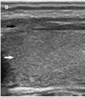
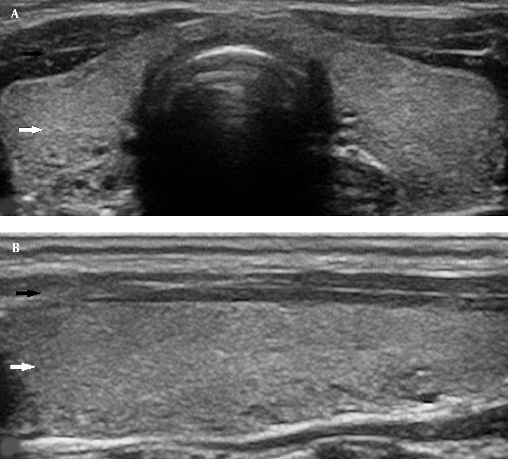
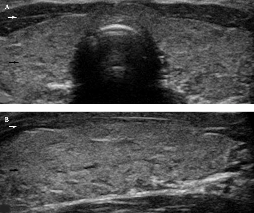
A thyroid US was performed by a board-certified radiologist using a high-resolution US system equipped with a 5-12 MHz linear array transducer (Aplio 500, Toshiba Medical System, Otawara, Japan). The children were categorized into two groups based on the thyroid parenchymal appearance on the US. The first group (group 1) was composed of subjects with a normal homogeneous echo pattern of the TG similar to that of the surrounding connective tissue (Figure 1). The second group (group 2) consisted of subjects with a heterogeneous irregular echo pattern of the TG with echogenicity similar or lower to the neck muscles (Figure 2).
Thyroid function tests included thyroid-stimulating hormone (TSH), free triiodothyronine (fT3), free thyroxine (fT4), thyroglobulin antibody (Tg-Ab), and thyroperoxidase antibody (TPO-Ab). These laboratory tests were reported as near to the day of US observation as possible. The TSH, fT3, and fT4 values were divided into three levels as follows: (1) low TSH (< 0.54 mIU/L), normal TSH (0.54 - 4.53 mIU/L), and elevated TSH (> 4.53 mIU/L); (2) low fT3 (< 2.3 ng/L), normal fT3 (2.3 - 5.2 ng/L), and elevated fT3 (> 5.2 ng/L); and (3) low fT4 (< 0.7 ng/dL), normal fT4 (0.7 - 1.48 ng/dL), and elevated fT4 (> 1.48 ng/dL).
The Tg-Ab and TPO-Ab values were categorized into four groups as follows: (1) positive Tg-Ab (> 4.11 IU/mL) and negative Tg-Ab (< 4.11 IU/mL); (2) positive TPO-Ab (> 5.61 IU/mL) and negative TPO-Ab (< 5.61 IU/mL); (3) both Tg-Ab and TPO-Ab positivity; and (4) both Tg-Ab and TPO-Ab negativity.
3.3. Statistical Analysis
Continuous variables were summarized as mean ± standard deviation, and categorical variables were presented as numbers and percentages. The student’s t-test was performed to compare the continuous data, and the chi-square test was used to compare categorical variables between the two groups. Significant correlations were determined using Pearson’s correlation coefficient.
A stepwise multiple linear regression analysis was performed to identify which thyroid function test was most closely correlated with the TG echo pattern. The diagnostic values of thyroid US assessments, including sensitivity, specificity, positive predictive value (PPV), and negative predictive value (NPV), in predicting thyroid function tests were calculated. The statistical analysis was performed using SPSS software (version 20.0). P-values ≤ 0.05 and ≤ 0.001 were regarded as significant and highly significant, respectively.
4. Results
4.1. Demographic Data
Group 1 consisted of 70 (30.6%) male and 159 (69.4%) female children aged within the range of 5-18 years (mean age: 11.88 ± 3.75 years). Group 2 included 44 (21.5%) male and 161 (78.5%) female children aged within the range of 6-18 years (mean age: 14.48 ± 3.08 years).
4.2. Thyroid Function Tests in Groups
In children with a homogeneous echo pattern of the TG (group 1), TSH was normal, low, and high in 87.8% (210/229), 1.7% (4/229), and 10.5% (24/229) of the subjects, respectively. The fT3 and fT4 levels were normal in all the participants of group 1. In children with the heterogeneous echo pattern of the TG (group 2), TSH was normal, low, and high in 61.5% (126/205), 8.8% (18/205), and 29.8% (61/205) of the subjects, respectively. The fT3 levels were normal, low, and high in 94.6% (194/205), 1.5% (3/205), and 3.9% (8/205) of the participants, respectively. The fT4 values were normal, low, and high in 92.7% (190/205), 2.0% (4/205), and 5.4% (11/205) of the participants, respectively.
In group 1, Tg-Ab levels were negative and positive in 83.4% (191/229) and 16.6% (38/229) of the subjects, respectively. TPO-Ab values were negative and positive in 90.8% (208/229) and 9.2% (21/229) of the subjects, respectively. Among children in group 2, Tg-Ab levels were negative and positive in 31.7% (65/205) and 68.3% (140/205) of the participants, respectively. TPO-Ab values were negative and positive in 40.5% (83/205) and 59.5% (122/205) of the participants, respectively. Only 8.3% (19/229) and 56.1% (115/205) of children in groups 1 and 2 showed both Tg-Ab and TPO-Ab positivity, respectively. About 82.5% (189/229) and 28.3% (58/205) of subjects in groups 1 and 2 showed both Tg-Ab and TPO-Ab negativity, respectively. Table 1 shows a summary of TSH, fT3, fT4, Tg-Ab, and TPO-Ab values among male and female children with the homogeneous and heterogeneous echo patterns of the TG.
| Variables | Group 1 | Group 2 | P-Value b, c | ||||
|---|---|---|---|---|---|---|---|
| F | M | T | F | M | T | ||
| TSH | |||||||
| Normal | 138 (60.3) | 63 (27.5) | 201 (87.8) | 100 (48.8) | 26 (12.7) | 126 (61.5) | < 0.001 d |
| Low | 4 (1.7) | 0 | 4 (1.7) | 13 (6.3) | 5 (2.4) | 18 (8.8) | < 0.001 e |
| High | 17 (7.4) | 7 (3.1) | 24 (10.5) | 48 (23.4) | 13 (6.3) | 61 (29.8) | < 0.001 f |
| fT3 | |||||||
| Normal | 159 (69.4) | 70 (30.6) | 229 (100) | 152 (74.1) | 42 (20.5) | 194 (94.6) | < 0.001 d |
| Low | 0 | 0 | 0 | 2 (1.0) | 1 (0.5) | 3 (1.5) | = 0.001 e |
| High | 0 | 0 | 0 | 7 (3.4) | 1 (0.5) | 8 (3.9) | < 0.001 f |
| fT4 | |||||||
| Normal | 159 (69.4) | 70 (30.6) | 229 (100) | 151 (73.7) | 39 (19.0) | 190 (92.7) | < 0.001 d |
| Low | 0 | 0 | 0 | 2 (1.0) | 2 (1.0) | 4 (2.0) | < 0.001 e |
| High | 0 | 0 | 0 | 8 (3.9) | 3 (1.5) | 11 (5.4) | < 0.001 |
| Tg-Ab | < 0.001 d; < 0.001 e; < 0.001 f | ||||||
| Negative | 128 (55.9) | 63 (27.5) | 191 (83.4) | 45 (21.9) | 20 (9.8) | 65 (31.7) | |
| Positive | 31 (13.5) | 7 (3.1) | 38 (16.6) | 116 (56.6) | 24 (11.7) | 140 (68.3) | |
| TPO-Ab | < 0.001 d; < 0.001 e; < 0.001 f | ||||||
| Negative | 141 (61.6) | 67 (29.3) | 208 (90.8) | 63 (30.8) | 20 (9.8) | 83 (40.5) | |
| Positive | 18 (7.9) | 3 (1.3) | 21 (9.2) | 98 (47.8) | 24 (11.7) | 122 (59.5) | |
| Tg-Ab and TPO-Ab | < 0.001 d; < 0.001 e; < 0.001 f | ||||||
| Negative | 127 (55.6) | 62 (27.1) | 189 (82.5) | 39 (19.0) | 19 (9.3) | 58 (28.3) | |
| Positive | 17 (7.4) | 2 (0.9) | 19 (8.3) | 92 (44.9) | 23 (11.2) | 115 (56.1) | |
Abbreviations: n, number of patients; F, female; M, male; T, total group 1, homogeneous thyroid echo pattern; Group 2, heterogeneous thyroid echo pattern; TSH, thyroid-stimulating hormone; fT3, free triiodothyronine; fT4, free thyroxine; Tg-Ab, thyroglobulin antibody; TPO-Ab, thyroperoxidase antibody.
a Values are expressed as No. (%).
b P-values obtained using student’s t-test.
c P ≤ 0.001 considered highly significant.
d P-values to compare levels between female children.
e P-values to compare levels between male children.
f P-values to compare levels in the total study population.
4.3. Correlation
The mean age was significantly different between groups 1 and 2 (11.88 vs. 14.48 years; P < 0.0001). The percentage of female subjects was significantly higher in group 2 than in group 1 (78.5 vs. 69.4%; P = 0.031). The levels of TSH, fT3, fT4, Tg-Ab, and TPO-Ab were significantly different between the two groups (P < 0.0001 for all). The multiple stepwise regression analysis revealed that TSH, fT4, Tg-Ab, and TPO-Ab were most closely correlated with the TG echo pattern (Table 2).
| Predictors | Unstandardized Coefficients | Standardized Coefficient | t | 95.0% CI for β | R | Adjusted R-Squared | SEE | ||
|---|---|---|---|---|---|---|---|---|---|
| β | SE | β | Lower Bound | Upper Bound | |||||
| Model 1 [F (1, 432) = 172.960; P < 0.0001; R2 = 0.286] | 0.535 | 0.284 | 0.423 | ||||||
| TPO-Ab | 0.284 | 0.022 | 0.535 | 13.15 | 0.242 | 0.326 | |||
| Model 2 [F (2, 431) = 101.743; P < 0.0001; R2 = 0.321] | 0.566 | 0.318 | 0.413 | ||||||
| TPO-Ab | 0.171 | 0.043 | 0.323 | 5.36 | 0.109 | 0.234 | |||
| Tg-Ab | 0.143 | 0.032 | 0.283 | 4.70 | 0.083 | 0.203 | |||
| Model 3 [F (3, 430) = 79.823; P < 0.0001; R2 = 0.358] | 0.598 | 0.353 | 0.402 | ||||||
| TPO-Ab | 0.146 | 0.031 | 0.275 | 4.64 | 0.084 | 0.208 | |||
| Tg-Ab | 0.150 | 0.030 | 0.295 | 5.03 | 0.091 | 0.208 | |||
| TSH | 0.123 | 0.025 | 0.196 | 4.98 | 0.074 | 0.171 | |||
| Model 4 [F (4, 429) = 65.156; P < 0.0001; R2 = 0.378] | 0.615 | 0.372 | 0.396 | ||||||
| TPO-Ab | 0.143 | 0.031 | 0.269 | 4.60 | 0.082 | 0.204 | |||
| Tg-Ab | 0.152 | 0.029 | 0.300 | 5.20 | 0.095 | 0.210 | |||
| TSH | 0.108 | 0.025 | 0.172 | 4.37 | 0.059 | 0.156 | |||
| fT4 | 0.220 | 0.059 | 0.144 | 3.74 | 0.104 | 0.336 | |||
Abbreviations: SEE, standard error of estimate; SE, standard error; CI, confidence interval; TPO-Ab, thyroperoxidase antibody; Tg-Ab, thyroglobulin antibody; TSH, thyroid-stimulating hormone; fT4, free thyroxine.
a P ≤ 0.001 considered highly significant.
All thyroid function tests were within normal ranges in 73.4% (168/229) and 14.6% (30/205) of children with homogeneous and heterogeneous echo patterns, respectively. The Tg-Ab and TPO-Ab positivity with normal TSH, fT3, and fT4 levels were observed in 5.7% (13/229) and 35.1% (72/205) of the participants in groups 1 and 2, respectively. These differences reached a statistical significance (P < 0.0001).
Homogeneous TG on the US had 73.8% sensitivity in predicting normal TSH and 100% sensitivity in predicting normal fT3 and fT4. The sensitivity values of homogeneous TG for predicting negative Tg-Ab and TPO-Ab were 78.7 and 85.3%, respectively. Table 3 shows the sensitivity, specificity, PPV, and NPV of sonographic TG patterns in predicting the thyroid function tests.
| Thyroid Function Test | Sensitivity (%) | Specificity (%) | PPV (%) | NPV (%) |
|---|---|---|---|---|
| TSH | 73.8 | 61.5 | 38.5 | 87.8 |
| fT3 | 100 | 45.9 | 5.4 | 100 |
| fT4 | 100 | 45.3 | 7.3 | 100 |
| Tg-Ab | 78.7 | 25.4 | 68.3 | 83.4 |
| TPO-Ab | 85.3 | 28.5 | 59.5 | 90.8 |
Abbreviations: PPV, positive predictive value; NPV, negative predictive value; TSH, thyroid-stimulating hormone; fT3, free triiodothyronine; fT4, free thyroxine; Tg-Ab, thyroglobulin antibody; TPO-Ab, thyroperoxidase antibody.
5. Discussion
A thyroid US is a safe, low-cost, easily accessible, and first-line imaging method for the evaluation of the TG parenchyma in children and adolescents (1, 2). A thyroid US can easily distinguish normal and homogeneous from heterogeneous parenchyma (2, 3). The association between TG echogenicity and thyroid function has been well studied in the general adult population (6-10). This study reported an association between normal and heterogeneous TG echo patterns with thyroid function tests in a large sample of children.
The present study firstly aimed to examine the relationship between thyroid parenchymal echogenicity and thyroid function. The second aim was to assess the validity of the thyroid US in predicting thyroid function parameters in the pediatric population.
Diffuse thyroid diseases, including Hashimoto’s thyroiditis and Graves’s disease, are commonly presented with heterogeneous and irregular thyroid parenchyma on the US due to diffuse lymphocytic infiltration, follicular degeneration, and reduction of colloid contents (4, 11). The correlation between the heterogeneous thyroid echo pattern and abnormal thyroid function has been reported in numerous studies with a wide age range of participants (4, 6-8).
Park et al. reported that TSH, T3, and fT4 were within abnormal limits in 64.2, 47.4, and 54.7% of patients with decreased thyroid parenchymal echogenicity, respectively (11). Additionally, 59% and 67.6% of the investigated patients showed positive levels of Tg-Ab and TPO-Ab, respectively (11). In another study performed by Trimboli et al., 78.4% of the adult patients with heterogeneous echo patterns had elevated TSH, and 76.3% had positive thyroid antibodies (12). Schiemann et al. (4) and Vejbjerg et al. (13) observed a significant association between hypoechogenicity or irregular echo patterns on the thyroid US and increased TSH levels. Previous studies have shown a high correlation between decreased thyroid echogenicity and Tg-Ab/TPO-Ab positivity (9, 10). A recent study conducted by Jeong et al. showed a significant association between decreased thyroid echogenicity and thyroid function tests in children with Hashimoto’s thyroiditis (5).
The current study compared the homogeneous and heterogeneous thyroid echo patterns with thyroid function in a large sample of pediatric subjects. In the present study, Tg-Ab and TPO-Ab levels were positive in 68.3 and 59.5% of the children with heterogeneous echo patterns, respectively. Both antibodies were positive in 56.1% of the investigated patients. These results are in line with the results of previous studies on adult patients that prove the presence of genetic susceptibility to autoimmune thyroiditis (9, 10). Since thyroid hormone levels at presentation might vary between euthyroidism and hypothyroidism or hyperthyroidism in children with thyroid disorders, thyroid antibody positivity is an important marker in the diagnosis of thyroid disorders and matches well with the heterogeneous appearance on the thyroid US. However, it does not match in nearly 40% of both pediatric and adult patients, which is in concordance with the results of previous studies indicating the presence of heterogeneous thyroid echogenicity before the appearance of thyroid antibodies (9, 10).
The TSH, fT3, and fT4 levels were out of normal ranges in 38.6, 5.4, and 7.4% of children with heterogeneous thyroid parenchyma, respectively. The reported levels in pediatric patients differed remarkably from the adult series because children with autoimmune thyroiditis are usually detected in the initial phase when thyroid function is usually preserved (5). Unlike adult patients, younger patients show a weaker association between heterogeneous thyroid echo patterns and high thyroid hormone levels that might be partly attributable to the fact that children are diagnosed earlier in the euthyroid stage of autoimmune thyroiditis (5, 8). Heterogeneous thyroid parenchyma on the US in pediatric patients with normal thyroid hormone profiles should be regarded as an early sign of thyroid failure.
A limited number of studies have reported the association between normal homogeneous thyroid parenchyma and thyroid function tests in adults (6, 13, 14). Pedersen et al. reported that TSH levels were within abnormal limits in 9%, and TPO-Ab levels were positive in 10.2% of the patients with normal thyroid parenchyma (6). Vejbjerg et al. observed Tg-Ab and TPO-Ab positivity in 9.6% and 11% of subjects with normal thyroid echogenicity, respectively (13). In the present study, TSH levels were abnormal in 12.2% of the subjects; however, Tg-Ab and TPO-Ab levels were positive in 16.6 and 9.2% of children with normal thyroid parenchyma, respectively. The present study results are in concordance with previous literature data (6, 13).
In a study performed by Tam et al., 86.1% of adult patients with homogeneous echo patterns had normal TSH levels, and 93.4% of the subjects had negative thyroid antibodies (14). All thyroid tests were within normal ranges in 77.6% of the cases (14). In the current study, Tg-Ab and TPO-Ab levels were negative in 83.4 and 90.8% of children with normal thyroid parenchyma, respectively. The TSH levels were normal in 87.8% of the subjects; nevertheless, fT3 and fT4 levels were normal in all participants with homogeneous TG. In the present study, all thyroid function tests were normal in 73.4% of children with homogeneous thyroid parenchyma, similar to the results reported by Tam et al. (14).
Nordmeyer et al. reported that a thyroid US is useful for excluding diffuse thyroid diseases with an 84% ratio (15). Moreover, Trimboli et al. observed that a thyroid US had 90% sensitivity in predicting negative thyroid hormones and normal TSH levels (12). These results are comparable to the present study results. The current study showed that a normal thyroid US had 73.8% sensitivity in predicting normal TSH and 100% sensitivity in predicting normal fT3 and fT4. The sensitivity values in predicting negative Tg-Ab and TPO-Ab were 78.7 and 85.3%, respectively. These results confırm that the homogeneous echo pattern of the TG is able to predict euthyroid pediatric subjects with a high degree of precision; however, it does not in approximately 20% of cases. This piece of evidence might suggest that clinicians should also consider thyroid function examination in a diagnostic work-up in children with a normal thyroid US. On the other hand, all the thyroid function tests were within normal ranges in 14.6% of children with the heterogeneous echo patterns of the TG. These findings strengthen the role of the thyroid US in the diagnosis of thyroid disorders in apparently healthy children.
The current study had some limitations that should be addressed. Firstly, the present study was a retrospective, single-center study that included a limited number of children. Secondly, due to the lack of appropriate data, the results were compared to the results of similar studies on adults. Thirdly, the US assessments were undertaken at variable durations after thyroid function testing. The subjectivity of the US assessment has also been regarded as another limitation of the study. The US evaluations were performed by a single radiologist; therefore, inter-observer variability should be taken into account in future prospective multicenter studies.
5.1. Conclusion
In conclusion, the current study investigated the association of homogeneous and heterogeneous thyroid echo patterns with thyroid function in a large sample of children. The obtained results also confirmed the value of a thyroid US in predicting thyroid function in the pediatric population.


