


1. Introduction
Foreign bodies (FBs) of the external auditory canal (EAC) are common among pediatric patients in the emergency department of otorhinolaryngology. Clinicians have tried many ways to treat these patients, including using irrigation setup, forceps, cerumen curettes, and syringes (1, 2). However, removing spherical FBs in EAC, such as round beads and beans, is still challenging. General anesthesia may be required for some of these pediatric patients (3). In this study, we presented a simple and novel 3-dimensional (3D)-printing tool and successfully treated a pediatric patient with a spherical FB in EAC.
2. Case Presentation
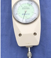
To remove a spherical FB in EAC, we designed a stape - like 3D attractor by Mimics version 21 (Materialise, Germany; Figure 1). The hemispherical portion of the attractor was 3 mm in diameter and could fit small and round FBs. The attractor could be tightly attached to a spherical FB in EAC with the superglue. The handle portion could be caught on with an ear hook. The model printing was performed with a 3D printer (HORI X400, Beijing, China) with nylon (FS 3300PA; Figure 2). To measure its tensile capacity, the dynamometer read 25 N before the handle portion was broken (Figure 3).

The 3-dimensional model of the attractor designed to remove spherical foreign bodies in the external auditory canal.

The 3-dimensional–printed attractor with a hemispherical portion of 3 mm in diameter.

The 3-dimensional–printed attractor can be tightly attached to the bead with the superglue, and the dynamometer reads 25 N before the handle portion is broken.
A 2-year-old child presented with a history of a plastic bead in the right EAC. The child was asymptomatic, and the diagnosis was confirmed by otoscopy. To remove the EAC FB, the child was required to sit still. The clinician then performed the procedures as follows: The clinician held the 3D-printed attractor with an ear hook, glued on the bottom of the hemispherical portion of the attractor, and carefully attached the hemispherical portion of the attractor to the surface of the bead in the EAC with the ear hook under an otoscope (Figure 4A). Then, the clinician waited for 10 minutes until the glue dried. Finally, the clinician pulled out the bead (7 mm) with the ear hook again under no anesthesia (Figure 4B). The child had no complaints during the procedures. The child received no further treatment, and no complications developed afterward.

A, The clinician tries to remove the bead in the EAC with an ear hook; B, The attractor is firmly stuck to the bead.
3. Discussion
EAC is divided into an outer cartilaginous and inner bony portion by a narrow isthmus. Manipulation of the FB embedded in the inner portion of EAC may cause pain and bleeding. The size, shape, and nature of FB determine the timing and procedures for removal. The electronic, botanical, and penetrating FBs warrant urgent removal, while removing other EAC FBs can be deferred, such as round beads and rubber. It was reported that complication incidences for smooth-surfaced objects were considerably higher than those for irregularly shaped objects (4, 5). The appropriate instruments are essential for the successful extraction of EAC FBs. Most FBs can be removed using commonly available instruments, such as irrigation setup, alligator/bayonet forceps, and cerumen curette (1, 2, 6). However, removing smooth-surfaced and spherical FBs requires finer instruments than those commonly used in the emergency department. The right-angled hooks, suction tube, super glue, and syringes have all been introduced to remove such FBs (7-9). However, the results are not satisfactory. We assumed that these clinicians might not have effective tools. Accordingly, we developed a simple, novel, and effective 3D-printing tool to remove EAC FBs.
The 3D-printing technique was first reported by Charles Hull in 1984 (10). Since then, it has gained much interest in the medical world. 3D-printed objects can be used to practice medical procedures and study complex cases. In this study, we printed a tiny tool to treat patients with EAC FBs. Many materials (such as biomaterials and nanomaterials) have been explored for 3D printability (11). We chose nylon for 3 main reasons. First, it does no more harm to EAC and can be easily removed if left inside. Second, we could stick it and some spherical FBs with superglue. Third, it is cost-effective and can sustain drag force up to 25 N, which is powerful enough to remove FBs from EAC. Further study is needed to confirm its effectiveness.
