1. Background
Chorioamnionitis is one of the common forms of placental inflammation which could induce preterm birth (1). Based on different diagnostic methods, chorioamnionitis can be divided into clinical and histological types. However, the incidence of the latter was confirmed to be twice that of the former (2). Although histological chorioamnionitis (HCA) usually exhibits no clinical symptoms, its influence on the fetus should not be ignored because it may induce inflammation in the fetus, further involving multiple organs of a preterm infant (3, 4). Scholars have claimed that preterm infants with HCA, delivered at less than 34 weeks of gestational age, have a poor prognosis (5, 6) and display short- and long-term complications of brain injury (7-10), posing a heavy burden on society and families. Additionally, the effects of prenatal anabolic steroids (AS) on the neurological system of preterm infants have gained much attention from scholars (11-15).
2. Objectives
Consequently, we aimed to investigate the correlation of HCA and prenatal AS administration with brain injury in premature infants (BIPI) at 30 - 34 weeks. This may provide a basis for the early diagnosis and treatment of this disease.
3. Methods
3.1. Research Objectives
We studied the effect of placental tissue chorioamnionitis and prenatal hormone use on brain damage in preterm infants less than 34 weeks of age. Furthermore, we evaluated the predictive value of inflammatory indicators in predicting the occurrence of brain damage in preterm infants with intrauterine infection. We studied the clinical data of preterm infants admitted to the Neonatal Ward of Yinchuan Maternal and Child Healthcare Hospital between January 2017 and January 2021.
The inclusion criteria were as follows: (1) Infants born in our hospital with a gestational age of 30 - 34 weeks; (2) infants whose mothers underwent placental pathological examination with the informed consent of the family members; and (3) infants whose mothers underwent the examination of B-ultrasound and magnetic resonance imaging (MRI) after the delivery.
The exclusion criteria entailed: (1) Infants who had intrauterine hypoxia and birth asphyxia indicated by the monitoring of prenatal fetal heart; (2) infants with congenital nervous system malformation, such as congenital hydrocephalus; (3) infants with severe perinatal complications, such as hypoglycemia encephalopathy and bilirubin encephalopathy; (4) infants with blood system diseases, such as moderate and severe anemia, abnormal coagulation, polycythemia, and hyperviscosity; (5) infants with diseases that induced abnormal hemodynamics, such as abnormal blood pressure, hypercapnia, hypothermia, severe and complicated congenital heart disease, heart failure, and respiratory failure; and (6) infants with genetic metabolic diseases.
3.2. Data Collection
The collected data comprised (1) general data, including gender, nationality, birth weight, and gestational age; (2) results of pathological examination of the placental tissue of mothers; (3) use of steroid hormones by mothers before delivery; (4) white blood cell (WBC) count, procalcitonin (PCT), and C-reactive protein (CRP) levels in the venous blood of preterm infants within 2 h of birth.
3.3. Histopathological Examination of the Placenta
Histological chorioamnionitis is defined as the amniotic membrane of the mother’s placenta showing neutrophil infiltration in the chorion and parietal decidua. The samples were collected during delivery and sent for inspection within 24 h of delivery. The intact placenta was fixed in 4% neutral formaldehyde for 48 h. The placental mass, umbilical cord length, adhesion, blood vessel number, the appearance of the mother’s placenta, and the fetus’s face were routinely evaluated. The placenta was sectioned parallelly at an interval of 1 cm. Routine samples were taken from 10 tissue blocks, including the whole placenta, fetal membrane, and umbilical cord (three fetal membranes, three umbilical cords, two maternal faces, and two fetal faces in placental essence). More samples were collected from abnormal parts during general observation. All specimens were embedded in paraffin, and tissue sections of 4 µm thickness were made. Next, the samples were stained with hematoxylin-eosin, and neutrophil infiltration was observed using an optical microscope. The diagnostic criterion of HCA (6) was neutrophil infiltration ≥ 5 HP under a microscope.
3.4. Steroid Usage
According to the American College of Obstetricians and Gynecologists Guidelines for Premature Birth (2016 version), administering an intramuscular injection of AS to pregnant women at week 23 - 34, who were likely to give birth in a week, could promote fetal lung maturity (16). Specifically, an intramuscular injection of betamethasone/dexamethasone at 12 mg/d was administered for two days. An entire course of AS was considered as the AS+ group, while AS without the entire course was grouped as the AS- group.
3.5. Diagnosis Criterion for Brain Injury
Cranial B-ultrasound was performed between day 0 to day 3 and day 7 after the infant’s birth with an interval of 2 weeks. MRI examination was completed at the corrected gestational age of 36 - 42 weeks. Based on the expert consensus of the Neonatal Professional Committee of the Chinese Medical Doctor Association (17), brain injury was determined as patients with high-risk factors of brain injury accompanied by clinical symptoms, such as central apnea, depression, change of consciousness, convulsion, abnormal muscle tone, or abnormal primitive reflex. The early brain injury of premature infants can be diagnosed combined with the following abnormalities in the head B-ultrasound or head MRI: (1) Brain edema; (2) intracranial hemorrhages, such as periventricular-intraventricular hemorrhage, subarachnoid hemorrhage, and cerebral parenchyma hemorrhage; (3) periventricular leukomalacia (PVL); and (4) other abnormalities, such as cerebral infarction.
3.6. Procalcitonin and C-reactive Protein Assessment
Blood samples were collected from newborns within 2 h of birth before administering antibiotics. The PCT levels of venous blood were assessed using electrochemiluminescence immunoassay by ELECSYS2010 all-active immunity analyzer. In contrast, the level of CRP in venous blood was measured using immunoturbidimetry (MAGE800 analyzer, Beckman Company, USA).
3.7. Grouping
According to the results of placental pathology and prenatal steroid use, the enrolled infants were divided into the following groups: HCA+ AS+, HCA+ AS-, HCA- AS+, and HCA- AS-.
3.8. Observation Index
The correlation of placental inflammation and steroid hormone usage with brain injury was observed in premature infants. Moreover, the clinical value of inflammatory indicators was explored to predict brain injury in preterm infants with intrauterine infection.
3.9. Statistical Analysis
SPSS 22.0 software was used for data analysis. The data conforming to normal distribution were expressed as mean ± standard deviation (X ± S). The comparisons between two independent sample groups were conducted by t-test, while comparison between multiple groups was conducted by single-factor analysis of variance. The variables with non-normal distribution were expressed as the median and interquartile range (25% - 75%). The Kruskal-Wallis test was used for comparison between multiple groups. Count data were compared between groups by the χ2 test. The ROC curve was plotted, and the area under the curve was calculated to determine the critical value. P < 0.05 indicated statistical significance.
4. Results
4.1. Comparison of the General Data Among All Four Preterm Infant Groups
We included 339 of 3,892 preterm infants treated during the study in the present investigation, including 96 girls and 243 boys. The mean gestational age was 32.51 ± 1.43 weeks, and the mean birth weight was 1979 ± 330 g. The number of cases in the HCA+ AS+, HCA+ AS-, HCA- AS+, and HCA- AS- groups was 93, 120, 81, and 45, respectively. No statistical differences were observed in the gestational age, birth weight, gender, and delivery mode between the four groups (P > 0.05) (Appendix 1).
4.2. Comparison of Brain Injury Between the Preterm Infants of Each Group
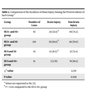
Of the 339 preterm infants, 216 showed HCA in placental pathology examination, with the incidence of brain injury being 29.1%. No HCA was observed in the placental pathological examination in the remaining 123 pregnant women, with the incidence of brain injury significantly lower than that of the HCA+ group (χ2 = 5.713, P < 0.05). In the current study, 171 cases of pregnant women were given enough steroids before delivery, and the incidence of brain injury was lower than that of the AS group. There is a statistical difference between AS+ and AS- groups (χ2 = 4.368, P < 0.05) (Table 1). Among 75 preterm infants with brain injury, 24 cases belonged to the HCA+ AS+ group, 36 to the HCA+ AS- group, three to the HCA- AS+ group, and 12 to the HCA- AS- group. The incidence of brain injury was statistically different in the four groups (χ2 = 4.738, P < 0.05). Furthermore, the incidence of brain injury in the HCA+ AS+, HCA+ AS-, and HCA- AS- groups was significantly higher than in the HCA- AS+ group (χ2 = 6.105, P = 0.013; χ2 = 9.086, P = 0.003; χ2 = 4.848, P = 0.047, respectively), while no significant difference was observed between the other groups (P > 0.05) (Table 2).
| Group | Number of Cases | Brain Injury | Non-brain Injury | χ2/F | P-Value |
|---|---|---|---|---|---|
| HCA+ group | 216 | 63 (29.1) | 153 (70.9) | 5.713 | 0.017 |
| HCA-group | 123 | 12 (9.8) | 111 (90.2) | ||
| AS+ group | 171 | 24 (14.0) | 147 (86.0) | 4.368 | 0.037 |
| AS-group | 168 | 51 (30.3) | 117 (39.7) |
a Values are expressed as No. (%).
a Values are expressed as No. (%).
b P < 0.05 compared to the HCA- AS+ group
4.3. Comparison of Infection Indices Between the Preterm Infants of Each Group
The early PCT levels of preterm infants were significantly different in each group (Z = 15.498, P < 0.05). The intra-group comparison revealed that the PCT level of preterm infants in the HCA+ AS- group was higher than the HCA- AS- group (P < 0.01). The difference in WBC count and CRP level between various groups was not statistically significant (P > 0.05) (Appendix 2).
4.4. Value of Early Procalcitonin Levels for Predicting Brain Injury in Preterm Infants with Chorioamnionitis
The ROC curve analysis of serum PCT after 2 h of admission showed that PCT could be used as a predictor of brain injury with statistical significance (P < 0.01) during intrauterine infection. The area under the curve for PCT was found to be 0.731, which predicted chorioamnionitis in preterm infants (Appendix 3). According to the Youden index, the critical value of PCT level within 2 h of the infant’s birth was 2.213 ng/mL, while the sensitivity and specificity were 70.7% and 80.2%, respectively. The serum PCT level of 2.213 ng/mL on admission provided a certain reference value for judging brain injury in preterm infants with intrauterine infection.
5. Discussion
Brain injury in premature infants is the primary cause of cerebral palsy and cognitive dysfunction in preterm infants, posing a severe threat to their physical and mental health. However, its causes are complex, and as a link between the mother and the fetus, the placenta is closely related to the development of the fetal nervous system.
Some researchers claim that the occurrence of chorioamnionitis in the maternal placenta is one of the risk factors for BIPI (18). Upon maternal placenta developing HCA, we found the incidence of BIPI to be 29.1%, which was 2.96 times higher than the incidence of brain injury in the HCA- group and was in agreement with the study of Guillen et al. (19). Fukuda et al. (20) observed that after the occurrence of placental HCA, reduced placental blood flow could cause a significant decrease in the fetal cerebral blood flow. As a result, the susceptibility of neurons to hypoxic injury increases, stimulating a vascular inflammatory cascade response, which in turn exacerbates the injury to the brain parenchyma (21). Moreover, HCA can activate the microglia by causing fetal inflammatory response syndrome, leading to the production of a large number of inflammatory cytokines and injury to the central nerve cells, axonal degeneration, and destruction of the immature blood-brain barrier. The final consequence is BIPI. Meanwhile, inflammatory factors raise the activity of caspase and a large number of cytotoxic substances, such as excitatory amino acids, that cause brain cell apoptosis (22). When HCA occurs in the placenta, BIPI may eventually occur in various ways. In this study, the incidence of BIPI was 30.3% in mothers who did not use AS before delivery, which was significantly higher than that observed in the AS+ group. This suggested that the prenatal use of steroid hormones could effectively prevent the occurrence of BIPI, which was consistent with other studies (23). The PVL is a common type of BIPI. Some researchers believe that the application of AS can significantly reduce the time of mechanical ventilation and the incidence of PVL, while AS can act as a protective factor against PVL in premature infants (24).
Many studies evaluate the correlation between AS and BIPI, and a line of evidence has confirmed that the prenatal use of AS can reduce BIPI occurrence. Consequently, the influence of placental inflammation on BIPI is included when studying the relationship between AS and BIPI. In this study, the subjects were divided into HCA+ AS+, HCA+ AS-, HCA- AS+, and HCA- AS- groups based on the results of the placental pathological examination and prenatal steroid hormone use. The incidence of brain injury in the HCA+ AS+, HCA+ AS-, and HCA- AS- groups was significantly higher than in the HCA- AS+ group. Compared to the HCA+ AS- group, the incidence of BIPI in the HCA- AS+ group was lower than in the HCA- AS- group, which indicated that the adequate use of AS before delivery could reduce the incidence of BIPI regardless of placenta inflammation. This may be related to the pharmacological action of AS, which can promote the maturation of fetal brain tissue (25), stabilize cerebral capillaries and blood pressure hemodynamics (26, 27), reduce basal metabolism, inhibit brain injury mediated by hypoperfusion (28), inhibit toxic organic cytokines, and reduce nerve cell apoptosis (29).
Intrauterine infection in preterm infants stimulates the production of inflammatory cells and secretion of large amounts of inflammatory cytokines, both of which directly or indirectly trigger ischemia and hypoxia in nerve cells, affecting the development of the nervous system in preterm infants (29-31). Procalcitonin is an inflammation-related factor synthesized rapidly and is free from the effects of hormones. When inflammation occurs, PCT levels are significantly elevated. However, after the inflammation is controlled, PCT levels decline rapidly, which can be used as an indicator to assess the inflammatory response (32). Few relevant studies have been conducted at home and abroad to investigate the importance of using PCT as a monitoring indicator to predict the occurrence of BIPI with HCA and for early guidance in diagnosing and treating brain injury. In the present research, no statistical difference was observed in the WBC and CRP levels of different groups. However, PCT levels exhibited differences between the groups, where the HCA+ AS- group showed higher PCT levels than the HCA- AS- group. To evaluate the occurrence of BIPI with intrauterine infection, ROC curves were plotted, and it was found that the area under the ROC for PCT levels within 2 h was 0.731 postnatally. According to the Youden index, the critical value of PCT within 2 h was 2.213 ng/mL, while the sensitivity and specificity were 70.7% and 80.2%, respectively. In other words, when the PCT level was 2.213 ng/mL on admission, it showed a certain reference value for judging whether premature infants with intrauterine infection had a brain injury.
In summary, BIPI is associated with intrauterine infection in the mother and the lack of an entire course of steroid hormones before delivery. It is more likely to occur in the presence of both. Therefore, AS should be emphasized in clinical work, while routine placental pathology can be recommended for mothers at 28 - 30 weeks of gestational age to avoid a missed diagnosis of HCA, reducing the incidence of BIPI and improving the prognosis. The serum PCT levels after 2 h of birth can provide a laboratory basis for the early monitoring of BIPI. We found that a value of 2.213 ng/mL on admission bears a certain reference value for determining brain injury occurrence in preterm infants with intrauterine infection.
