


1. Background
Neonatal infections have nonspecific manifestations, and in some cases, it is difficult to diagnose the source of the infection. One of the causes of infection in a neonate is urinary tract infection (UTI). Diagnosis of UTI is given based on the urine culture (1, 2). A definite diagnosis of UTI cannot be made based on urine analysis before the urine culture results are ready. Screening tests such as positive nitrite or gram staining sometimes show false-positive results, as it has been already observed in cases with negative urine culture, especially in neonates (3, 4).
On the other hand, the leukocyte esterase dipstick test is not accurate in diagnosing pyuria in febrile patients (4), and a negative leukocyte esterase test cannot rule out pyuria; so this test cannot detect cases of infection without microscopic examination and urine culture as far as it may increase the chance of false negatives (5, 6). In previous literature, it has been shown that the presence of less than 10 white blood cells (WBCs) in a simple urine test indicates normal urine. Although, in some cases, the presence of 15 to 20 WBCs has also been associated with negative urine culture in general, there is no definite number that is highly sensitive and specific to diagnose infection in neonates, and due to the underdevelopment of immune response in them, we may not see reliable pyuria (4, 7).
Urinary tract infection in neonates includes a wide range of symptoms; for instance, growth retardation, polyuria, oliguria, jaundice, etc. (8, 9). Since urosepsis manifestations are nonspecific in neonates, urine culture is requested before the first dose of antibiotics, and urinary tract malformations are investigated during the treatment course in neonates with UTI (2). Although neonates with structural disorders of the urinary system are at higher risk of UTIs than normal infants, the relationship between pyuria and urinary tract abnormality has not yet been established. However, the association between positive urinary culture and structural abnormalities of the urinary system has already been detected (10, 11).
2. Objectives
This study investigated the relationship between simple urine tests and renal abnormality.
3. Methods
This study was performed as a cross-sectional-analytical evaluation on 100 neonates with UTIs in Tehran Children’s Medical Center, and the relationship between renal anomalies and simple urine tests was investigated.
3.1. Study Design
Neonates who were hospitalized for various reasons and were diagnosed with UTIs were selected. Urine samples were taken from all neonates by urine bag and catheterization or suprapubic in a sterile manner for urine culture. More than 1,000 colonies in the catheter urine sample or even one count in the suprapubic sample were considered positive. Counts of WBCs, which were more than ten, bacteriuria, positive nitrite, and positive leukocyte esterase in early detection of UTI were considered valuable. None of the neonates with UTI had positive blood cultures in our study. Finally, neonates with positive urine culture were treated with antibiotics, and also renal and urinary tract ultrasound and VCUG were performed to diagnose renal abnormalities and urinary reflux. SPSS21 software was used to investigate the relationship between urinary indices and urinary tract anomalies. The alpha level has been set to 0.05, and P-values less than 0.05 were considered significant.
3.2. Inclusion Criteria
Neonates with UTIs admitted to the children’s Medical Center in 2016 - 2017 were included in this study.
3.3. Exclusion Criteria
The patients who had incomplete information were excluded from this study.
3.4. Statistical Analysis
Descriptive statistics were implemented by calculating the frequency, percentage, mean ± standard deviation (SD). To compare the qualitative data, the chi-square test was used for contingency with tables of cell counts more than 5, and Fisher’s exact test was used for contingency with tables of cell counts less than 5. For the comparison of the quantitative data, we used the Wilcoxon test. P < 0.05 was considered statistically significant.
3.5. Ethical Considerations
Since urine analysis, urine culture, sonography, and VCUG in neonates with UTI are necessary for the treatment of the disease; therefore, no additional action was taken on patients besides what should have been done in the process of the treatment. Patients’ information was recorded anonymously. The study was approved by the Ethics Committee of Tehran University of Medical Science with the following ethical code: IR.TUMS.CHMC.REC.1398.122.
4. Results
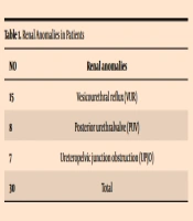
The mean age of neonates was 13.64 days. The mean gestational age was 37.17 weeks, and the mean weight was 3,310 grs. A total number of 30 patients had renal anomalies. The most common observed anomaly was urinary reflux (Table 1). The most common organism found in this study was Klebsiella (31%), which was associated with renal anomalies in 45% of cases, but none of the organisms were significantly associated with renal anomalies (P-value 0.987) (Table 2). There was a statistically significant correlation between fever with the renal anomaly (P-value 0.002). Comparison of urinalysis in the two groups with and without renal anomalies showed a statistically significant difference (Table 3). Pyuria (P-value = 0.003), bacteriuria (P-value = 0.016), nitrite positive (P-value = 0.001), and leukocyte esterase (P-value = 0.001) showed a statistically significant correlation with the renal anomaly. However, there was no statistically significant correlation between gender, cause of hospitalization, positive culture, CRP level, and type of organism with the renal anomaly.
| No. | Renal Anomalies |
|---|---|
| 15 | Vesicourethral reflux (VUR) |
| 8 | Posterior urethralvalve (PUV) |
| 7 | Ureteropelvic junction obstruction (UPJO) |
| 30 | Total |
Renal Anomalies in Patients
| Organism | Renal Anomalies | P-Values | |
|---|---|---|---|
| No | Yes | ||
| Enterobacter | 3 | 4 | 0.987 |
| Candida | 10 | 8 | |
| Escherichia coli | 10 | 9 | |
| Staphylococcus | 5 | 3 | |
| Klebsiella | 17 | 14 | |
| Serratia | 4 | 3 | |
| Enterococcus | 4 | 4 | |
| Total | 53 | 47 | |
The Organisms Were Significantly Associated with Renal Anomalies
| Index | Renal Anomalies | P-Value | |
|---|---|---|---|
| No | Yes | ||
| Pyuria | 0.003 | ||
| Yes | 7 | 19 | |
| No | 46 | 28 | |
| Bacteriuria | 0.016 | ||
| Few | 23 | 8 | |
| Many | 16 | 19 | |
| Negative | 14 | 20 | |
| Nitrite | 0.001 | ||
| Positive | 10 | 24 | |
| Negative | 43 | 23 | |
| Leukocyte esterase | 0.001 | ||
| Positive | 4 | 21 | |
| Negative | 49 | 26 | |
| Reinfection | 0.694 | ||
| Positive | 27 | 22 | |
| Negative | 26 | 25 | |
Comparison of Urine Analysis in Two Groups with and Without Renal Anomalies
5. Discussion
The findings of this study showed that some indicators in urinalysis, including pyuria, bacteriuria, nitrite, and leukocyte esterase, have statistically significant correlations with a renal anomaly. Based on the results observed in this study, these indicators are important for reevaluating antibiotics and assessing renal anomalies accurately. However, the best choice of empirical treatment for UTIs in neonates is aminoglycosides (12). In the Crain and Gershel’s study, out of 32 positive urine cultures in infants younger than eight weeks of age with UTIs, 50% had normal urine analysis. Then more than half of the UTIs in neonates would have been missed based on urine analysis (13).
In the study of Falakaflaki et al., proteinuria and hematuria in neonates were associated with ureteropelvic junction and vesicoureteral reflux (14). Mohamed et al. found that the rate of UTIs in neonates with pyuria was 5.44 times higher than in other neonates (15). Eberechukwu et al.’s study showed the presence of hematuria, glucosuria, and ketonuria in the urine of the infants was not usual, and it should be investigated in terms of the underlying factor (16).
The most common anomaly observed in our study was urinary reflux, which is similar to other studies (15, 16). The most common organism found in this study was Klebsiella (31%), which was associated with renal anomalies in 45% of cases, but none of the organisms were significantly associated with renal anomalies. In the study of Cleper et al., Klebsiella was more likely to be associated with urinary reflux (17).
5.1. Conclusions
Based on the results observed in this study, some indicators related to UTIs in neonates, including pyuria, bacteriuria, nitrite, and leukocyte esterase, which are seen in simple urinalysis, have a statistically significant correlation with renal anomaly and need to reevaluate antibiotics and assess renal anomalies accurately.
