


1. Background
Cerebral palsy (CP) is a group of permanent disorders affecting the immature fetal or infant brain, characterized by non-progressive conditions that lead to activity limitations in movement and posture development. In addition to motor disorders, cerebral palsy is frequently accompanied by sensory, perceptual, cognitive, communication, and behavioral disorders, as well as epilepsy and secondary musculoskeletal problems (1, 2). Cerebral palsy results from lesions in the developing brain, with variations in the condition based on the timing of the lesion, clinical presentation, location, and severity (3).
Globally, the incidence of cerebral palsy, one of the most severe disabilities of childhood, varies between 1.5 and 3 per 1,000 live births (4), while in Turkey, the incidence is reported as 4.4 per 1,000 live births (5). The frequency of cerebral palsy is inversely related to gestational age and birth weight, with rates ranging from 90 cases per 1,000 newborns weighing less than 1,000 grams to 1.5 cases per 1,000 newborns weighing 2,500 grams or more (3). Although prematurity and low birth weight are the primary risk factors for cerebral palsy, numerous other factors are associated with or may increase the risk (6). Brain disorders in the immature brain can occur in utero, at birth, after the newborn period, or in early childhood. Brain injury, often resulting from hypoxia, infection, stroke, or hypotension, is typically accompanied by an inflammatory process. While there is no international consensus on the upper age limit for brain damage in defining cerebral palsy, injuries occurring up to the age of 2 are generally considered cerebral palsy (7).
The clinical classification of cerebral palsy is varied, with several different classification systems in use. Common classifications include those by Balf and Ingram (8) and Hagberg et al. (9). Additionally, the Surveillance of Cerebral Palsy in Europe (4) has recommended classifying CP into spastic, ataxic, and dyskinetic types. Balf and Ingram classified CP into categories such as diplegic, hemiplegic, tetraplegic, ataxic, dyskinetic, and mixed, based on the location and severity of neurological symptoms (8). Hagberg et al.’s classification, on the other hand, categorizes CP into spasticity, dyskinetic syndromes, and ataxia (9).
The diagnosis of cerebral palsy is primarily clinical, but international guidelines recommend the use of neuroimaging to evaluate the underlying cause. Approximately 10% of patients with cerebral palsy have normal neuroimaging results. The need for further neurometabolic testing to exclude other diagnoses should be considered on an individual basis. Determining the diagnosis and underlying cause is crucial for making informed decisions regarding treatment and prognosis. The diagnosis of cerebral palsy is based on identifying and classifying the movement disorder. In some cases, a standardized assessment of general movements can detect cerebral palsy as early as five months of age. However, a definitive diagnosis is typically made during the second year of life, when consistent symptoms are present and neuroimaging can be performed (7). The management of cerebral palsy aims to enhance functional capacity and independence while preventing secondary complications. Management strategies include physical and occupational therapies, the use of mechanical aids, orthopedic interventions, and the medical and surgical treatment of comorbid conditions. Advances in neonatal care have contributed to a decrease in the prevalence of cerebral palsy, and improvements in early diagnosis have had a promising impact on outcomes. Early intervention programs are a critical component of cerebral palsy management, as they address the condition as early as possible and support early brain neuroplasticity, which can lead to better long-term outcomes (10).
2. Objectives
The aim of this study is to determine the clinical features of patients with cerebral palsy, as well as to identify the risk factors and MRI findings associated with different types of cerebral palsy.
3. Methods
3.1. Study Type and Study Group
This study retrospectively evaluated the medical records of 202 children diagnosed with cerebral palsy at the Bursa Uludağ University Faculty of Medicine, Child Neurology Clinic, between January 2017 and June 2020. The study population represents the Bursa metropolitan region, which is located in the Southern Marmara region of Turkey. Bursa is the fourth most populous city in the country, with a population of 3,194,720 (11). The study protocol was approved by the Bursa Uludağ University Clinical Research Ethics Committee (2020-21/3).
3.2. Data Collection and Definitions
Demographic data, including age, gender, consanguinity, maternal age, and other relevant factors, were collected retrospectively. Information on possible relevant abnormalities during pregnancy, birth, and the neonatal period was also gathered, including infection history, drug and toxic exposure, pre-eclampsia, placental anomalies, chronic maternal diseases, polyhydramnios, oligohydramnios, asphyxia, resuscitation history, meconium aspiration, breech presentation, multiple pregnancies, intrauterine growth restriction, prematurity, postmaturity, birth weight, neonatal sepsis, meningitis, convulsions, hypoglycemia, hyperbilirubinemia, cerebral hemorrhage/infarction, and trauma or surgical interventions.
Additionally, data on comorbidities such as intellectual disability, epilepsy, behavioral disorders, gastrointestinal dysfunction, visual impairment, hearing impairment, language and speech disorders, orthopedic problems, and sleep disturbances were collected, along with information on the types of cerebral palsy (spastic, dyskinetic, ataxic, and mixed) as recorded in the patients' medical files. The diagnosis of cerebral palsy was made by the attending neuropediatric physician.
Cranial MRI data were available for all 202 cases. The MRI findings were categorized into various patterns, including malformations (nervous system developmental anomalies, cysts, calcifications, etc.), white matter damage of immaturity (periventricular leukomalacia, periventricular hemorrhage), focal infarction/hemorrhage, cortical-subcortical damage (multicystic encephalomalacia, other cortical damage), basal ganglia-thalamus damage, miscellaneous (diffuse atrophy, encephalomalacia), and other pathologies (cerebellar atrophy, subdural hemorrhage, etc.), with some cases showing normal MRI results.
The severity of cerebral palsy was determined using the Gross Motor Function Classification System (GMFCS), which classifies motor impairments in the lower extremities into five levels. The GMFCS ranges from level I (patients can walk and run with balance and speed limitations) to level V (independent mobility is impossible) (12).
3.3. Inclusion/Exclusion Criteria
Patients between the ages of 2 and 18 who were followed up with a diagnosis of cerebral palsy were included in the study. Exclusion criteria were applied to patients with progressive motor disorders, hypotonia as the sole clinical feature, or isolated spinal neural tube defects. Additionally, patients whose brain damage developed after 28 days postnatally and those whose medical records were inaccessible were also excluded from the study.
3.4. Statistical Analysis
Statistical analyses were conducted using the SPSS software (IBM SPSS Statistics for Windows, Version 28). The conformity of continuous variables to a normal distribution was assessed using the Shapiro-Wilk test. Variables that were normally distributed are presented as mean and standard deviation, while those that did not follow a normal distribution are presented as median, minimum, and maximum values. Based on the results of the normality test, the Mann-Whitney U test and Kruskal-Wallis test were used for comparisons between groups. Categorical variables were compared between groups using the Pearson chi-square test and the Fisher-Freeman-Halton test. A P-value of less than 0.05 was considered statistically significant.
4. Results
The study cohort consisted of 202 patients with a mean age of 8.02 years (SD 4.54, range 1.8 - 18.2 years), with a predominance of males (60.4%, n = 122; females 39.6%, n = 80). Consanguinity was present in 18.3% (n = 37) of the children’s parents. Regarding maternal pregnancies, 75 cases (37.1%) were first pregnancies, 58 cases (28.7%) were second pregnancies, and 69 cases (34.1%) involved three or more pregnancies. Maternal age at birth was below 20 years in 6 cases (3%) and above 35 years in 18 cases (8.9%). The majority of patients were born preterm (56.4%, n = 114), and 11.9% (n = 24) were from multiple births. Delivery methods included vaginal delivery in 49% (n = 99) of cases, planned cesarean section (CS) in 13.9% (n = 28), and emergency CS in 37.1% (n = 75). Birth weights were below 1000g in 23 cases (11.4%), between 1000-1500g in 34 cases (16.8%), between 1500 - 2500g in 54 cases (26.7%), between 2500 - 4000g in 89 cases (44.1%), and above 4000g in two cases (1%). Resuscitation at birth was required in 63 cases (31.2%), and 156 cases (77.2%) had neonatal hospitalization, with an average hospitalization period of 44.9 days (range 1 - 210 days).
Prenatal risk factors were identified in 51 cases (25.2%), including placental abnormalities in 15 cases, preeclampsia-eclampsia in 11 cases, polyhydramnios in 8 cases, oligohydramnios in 6 cases, a maternal history of chronic disease in 6 cases, hypertension during pregnancy in 5 cases, gestational diabetes in 4 cases, and infections during pregnancy in 5 cases. Perinatal/natal risk factors were present in 156 cases (77.2%), including prematurity in 114 cases, asphyxia and the need for resuscitation in 63 cases, multiple pregnancies in 22 cases, meconium aspiration in 14 cases, intrauterine growth retardation in 12 cases, premature rupture of membranes in 6 cases, and cord entanglement in 5 cases. Postnatal risk factors were present in 143 cases (70.8%), including neonatal sepsis/meningitis in 101 cases, neonatal convulsions in 37 cases, hyperbilirubinemia in 35 cases (with 24 cases receiving phototherapy and 3 cases requiring exchange transfusion), cerebral hemorrhage/infarction in 33 cases, neonatal hypoglycemia in 12 cases, and postnatal surgery in 6 cases (Table 1).
| Variables | Values |
|---|---|
| Age (y) | 8.02 ± 4.54 |
| Sex | |
| Male | 122 |
| Female | 80 |
| Consanguinity | 37/202 (18.3) |
| Maternal age at birth (y) | |
| < 20 | 6 (3) |
| 20 - 35 | 178 (88.1) |
| > 35 | 18 (8.9) |
| Number of pregnancies | |
| First | 75 (37.1) |
| Second | 58 (28.7) |
| Third or more | 69 (34.1) |
| Delivery types | |
| Vaginal delivery | 99 (49) |
| Planned C/S | 28 (13.9) |
| Emergency C/S | 75 (37.1) |
| Gestational age(week) | |
| < 28 | 26 (12.9) |
| 28 - 32 | 37 (18.3) |
| 33 - 37 | 51 (25.2) |
| 38 - 42 | 85 (42.1) |
| > 42 | 3 (1.5) |
| Birth weights (g) | |
| < 1000 | 23 (11.4) |
| 1000 - 1500 | 34 (16.8) |
| 1500 - 2500 | 54 (26.7) |
| 2500 - 4000 | 89 (441) |
| > 4000 | 2 (1) |
| Resuscitation at birth | 63 (31.2) |
| Newborn hospitalization | 156 (77.2) |
| Etiological factors | |
| Prenatal | 51 (25.2) |
| Placental abnormalities | 15 |
| Preeclampsia-eclampsia | 11 |
| Polyhydramnios | 8 |
| Oligohydramnios | 6 |
| Maternal chronic diseases | 6 |
| Hypertension in pregnancy | 5 |
| Gestational diabetes | 4 |
| Infection in pregnancy | 5 |
| Perinatal/natal | 156 (77.2) |
| Prematurity | 114 |
| Asphyxia/resuscitation | 63 |
| Multiple pregnancy | 22 |
| Meconium aspiration | 14 |
| Intrauterine growth retardation | 12 |
| Premature rupture of membranes | 6 |
| Cord entanglement | 5 |
| Postnatal | 143 (70.8) |
| Neonatal sepsis/menengitis | 101 |
| Neonatal convulsion | 37 |
| Neonatal hyperbilirubinemia | 35 |
| Cerebral hemorrhage-infarction | 33 |
| Neonatal hypoglycemia | 12 |
| Postnatal surgery | 6 |
Clinical Features of Patients with Cerebral Palsy a
The majority of patients in the study had spastic cerebral palsy, accounting for 81.6% (n = 165) of the cases, with the subtypes distributed as follows: Diparesic in 35.6% (n = 72), tetraparesic in 31.7% (n = 64), and hemiparesic in 14.4% (n = 29). The dyskinetic subtype was present in 6.4% (n = 13) of the patients, the ataxic subtype in 3.5% (n = 7), and the mixed cerebral palsy subtype in 8.4% (n = 17).
According to the GMFCS, the distribution of patients was as follows: Seven patients (3.5%) were at level I, 60 patients (29.7%) at level II, 56 patients (27.7%) at level III, 45 patients (22.3%) at level IV, and 34 patients (16.8%) at level V.
Epilepsy was the most common comorbidity, affecting 50.5% (n = 102) of the patients. Other identified comorbidities included intellectual disability in 39.6% (n = 80), visual impairment in 28.2% (n = 57), gastrointestinal dysfunction in 26.7% (n = 54), orthopedic problems in 22.8% (n = 46), language and speech disorders in 21.3% (n = 43), hearing impairment in 7.4% (n = 15), behavioral disorders in 7.4% (n = 15), and sleep problems in 2.5% (n = 5).
Cranial MRI findings were categorized as follows: White matter damage of immaturity was observed in 73 cases (36.1%), cortical-subcortical damage in 42 cases (20.8%), miscellaneous findings in 25 cases (12.4%), focal infarction/hemorrhage in 21 cases (10.4%), basal ganglia-thalamus damage in 11 cases (5.4%), malformations in 9 cases (4.5%), cerebellar atrophy in 4 cases (2%), and normal findings in 17 cases (8.4%) (Table 2).
| Variables | Values |
|---|---|
| Cerebral palsy types | |
| Spastic | 165 (81.6) |
| Spastic tetraparesis | 64 (31.7) |
| Spastic diparesis | 72 (35.6) |
| Spastic hemiparesis | 29 (14.4) |
| Dyskinetic | 13 (6.4) |
| Choreoathetoid | 4 (2) |
| Dystonic | 9 (4.5) |
| Ataxic | 7 (3.5) |
| Mixt | 17 (8.4) |
| GMFCS | |
| Level I | 7 (3.5) |
| Level II | 60 (29.7) |
| Level III | 56 (27.7) |
| Level IV | 45 (22.3) |
| Level V | 34 (16.8) |
| Comorbidity | |
| Intellectual disability | 80 (39.6) |
| Epilepsy | 102 (50.5) |
| Behavioral disorders | 15 (7.4) |
| Gastrointestinal dysfunction | 54 (26.7) |
| Visual impairment | 57 (28.2) |
| Hearing impairment | 15 (7.4) |
| Language and speech disorders | 43 (21.3) |
| Orthopedic problems | 46 (22.8) |
| Sleep problems | 5 (2.5) |
| Cranial MRI findings | |
| White matter damage of immaturity | 73 (36.1) |
| Cortical-subcortical damage | 42 (20.8) |
| Miscellaneous | 25 (12.4) |
| Focal infarction/hemorrhage | 21 (10.4) |
| Basal ganglia-thalamus damage | 11 (5.4) |
| Malformation | 9 (4.5) |
| Cerebellar atrophy | 4 (2) |
| Normal | 17 (8.4) |
Clinical Features, Comorbidities and Cranial MR Findings in Patients with Cerebral Palsy a
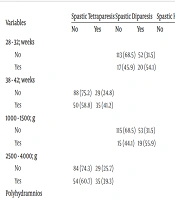
We examined the factors influencing the types of cerebral palsy individually (Tables 3 and 4). In patients with spastic diparesis, the following factors were significantly more common: Pregnancy between 28 - 32 weeks (P = 0.01), birth weight between 1000 - 1500 grams (P = 0.007), a history of oligohydramnios (P = 0.002), multiple pregnancies (P = 0.05), prematurity (P < 0.001), and neonatal sepsis (P = 0.001). Additionally, the group without comorbidities (P = 0.001) was larger among these patients compared to other types of cerebral palsy. Lower GMFCS levels were also significantly more prevalent in this group: GMFCS I (P = 0.009), GMFCS II (P < 0.001), and GMFCS II (P = 0.008). The most common MRI finding in patients with spastic diparesis was white matter damage of immaturity (P < 0.001).
| Variables | Spastic Tetraparesis | Spastic Diparesis | Spastic Hemiparesis | |||
|---|---|---|---|---|---|---|
| No | Yes | No | Yes | No | Yes | |
| 28 - 32; weeks | ||||||
| No | 113 (68.5) | 52 (31.5) | ||||
| Yes | 17 (45.9) | 20 (54.1) | ||||
| 38 - 42; weeks | ||||||
| No | 88 (75.2) | 29 (24.8) | ||||
| Yes | 50 (58.8) | 35 (41.2) | ||||
| 1000 - 1500; g | ||||||
| No | 115 (68.5) | 53 (31.5) | ||||
| Yes | 15 (44.1) | 19 (55.9) | ||||
| 2500 - 4000; g | ||||||
| No | 84 (74.3) | 29 (25.7) | ||||
| Yes | 54 (60.7) | 35 (39.3) | ||||
| Polyhydramnios | ||||||
| No | 136 (70.1) | 58 (29.9) | ||||
| Yes | 2 (25) | 6 (75) | ||||
| Oligohydramnios | ||||||
| No | 130 (66.3) | 66 (33.7) | ||||
| Yes | 0 (0) | 6 (100) | ||||
| Asphyxia/ resuscitation | ||||||
| No | 102 (73.4) | 37 (26.6) | ||||
| Yes | 36 (57.1) | 27 (42.9) | ||||
| Multiple pregnancy | ||||||
| No | 120 (66.7) | 60 (33.3) | ||||
| Yes | 10 (45.5) | 12 (54.5) | ||||
| Prematurity | ||||||
| No | 69 (78.4) | 19 (21.6) | ||||
| Yes | 61 (53.5) | 53 (46.5) | ||||
| Neonatal sepsis/menengitis | ||||||
| No | 76 (75.2) | 25 (24.8) | ||||
| Yes | 54 (53.5) | 47 (46.5) | ||||
| Neonatal convulsion | ||||||
| No | 119 (72.1) | 46 (27.9) | ||||
| Yes | 19 (51.4) | 18 (48.6) | ||||
| Intellectual disability | ||||||
| No | 95 (77.9) | 27 (22.1) | ||||
| Yes | 43 (53.8) | 37 (46.3) | ||||
| Epilepsy | ||||||
| No | 82 (82) | 18 (18) | ||||
| Yes | 56 (54.9) | 46 (45.1) | ||||
| Gastrointestinal dysfunction | ||||||
| No | 118 (79.7) | 30 (20.3) | ||||
| Yes | 20 (37) | 34 (63) | ||||
| No comorbidity | ||||||
| No | 110 (70.5) | 46 (29.5) | ||||
| Yes | 20 (43.5) | 26 (56.5) | ||||
| Orthopedic problems | ||||||
| No | 119 (76.3) | 37 (23.7) | ||||
| Yes | 19 (41.3) | 27 (58.7) | ||||
| GMFCS-1 | ||||||
| No | 129 (66.2) | 66 (33.8) | ||||
| Yes | 1 (14.3) | 6 (85.7) | ||||
| GMFCS-2 | ||||||
| No | 102 (71.8) | 40 (28.2) | 132 (93) | 10 (7) | ||
| Yes | 28 (46.7) | 32 (53.3) | 41 (68.3) | 19 (31.7) | ||
| GMFCS-3 | ||||||
| No | 102 (69.9) | 44 (30.1) | ||||
| Yes | 28 (50) | 28 (50) | ||||
| GMFCS-4 | ||||||
| No | 124 (79) | 33 (21) | ||||
| Yes | 14 (31.1) | 31 (68.9) | ||||
| GMFCS-5 | ||||||
| No | 124 (73.8) | 44 (26.2) | ||||
| Yes | 14 (41.2) | 20 (58.8) | ||||
| White-matter damage | ||||||
| No | 99 (76.7) | 30 (23.3) | ||||
| Yes | 31 (42.5) | 42 (57.5) | ||||
| Focal-infarction/ hemorrhage | ||||||
| No | 168 (92.8) | 13 (7.2) | ||||
| Yes | 5 (23.8) | 16 (76.2) | ||||
| Miscellaneous | ||||||
| No | 127 (71.8) | 50 (28.2) | ||||
| Yes | 11 (44) | 14 (56) | ||||
| Total | 138 (68.3) | 64 (31.7) | 130 (64.4) | 72 (35.6) | 173 (85.6) | 29 (14.4) |
Clinical Features, Risk Factors, Comorbidities and MR Findings with Spastic Cerebral Palsy Types a
| Variables | Choreoathetoid CP | Dystonic CP | Ataxic CP | Mixt CP | ||||
|---|---|---|---|---|---|---|---|---|
| No | Yes | No | Yes | No | Yes | No | Yes | |
| Vaginal delivery | ||||||||
| No | 102 (99) | 1 (1) | ||||||
| Yes | 91 (91.9) | 8 (8.1) | ||||||
| Hyperbilirubinemia | ||||||||
| No | 163 (97.6) | 4 (2.4) | ||||||
| Yes | 30 (85.7) | 5 (14.3) | ||||||
| Intellectual disability | ||||||||
| No | 121 (99.2) | 1 (0.8) | ||||||
| Yes | 74 (92.5) | 6 (7.5) | ||||||
| Epilepsy | ||||||||
| No | 97 (97) | 3 (3) | ||||||
| Yes | 88 (86.3) | 14 (13.7) | ||||||
| Gastrointestinaldysfunction | ||||||||
| No | 142 (95.9) | 6 (4.1) | ||||||
| Yes | 43 (79.6) | 11 (20.4) | ||||||
| Visual problems | ||||||||
| No | 143 (98.6) | 2 (1.4) | ||||||
| Yes | 52(91.2) | 5(8.8) | ||||||
| Language speech disorders | ||||||||
| No | 151 (95) | 8 (5) | ||||||
| Yes | 34 (79.1) | 9 (20.9) | ||||||
| GMFCS-3 | ||||||||
| No | 144 (98.6) | 2 (1.4) | ||||||
| Yes | 51 (91.1) | 5 (8.9) | ||||||
| GMFCS-5 | ||||||||
| No | 160 (95.2) | 8 (4.8) | ||||||
| Yes | 25 (73.5) | 9 (26.5) | ||||||
| Basal ganglia-thalamus damage | ||||||||
| No | 189 (99) | 2 (1) | 187 (97.9) | 4 (2.1) | ||||
| Yes | 9 (81.8) | 2 (18.2) | 6 (54.5) | 5 (45.5) | ||||
| Malformation | ||||||||
| No | 188 (97.4) | 5 (2.6) | ||||||
| Yes | 7 (77.8) | 2 (22.2) | ||||||
| Cerebellar atrophy | ||||||||
| No | 193 (97.5) | 5 (2.5) | ||||||
| Yes | 2 (50) | 2 (50) | ||||||
| Miscellaneous | ||||||||
| No | 168 (94.9) | 9 (5.1) | ||||||
| Yes | 17 (68) | 8 (32) | ||||||
| Total | 198 (98) | 4 (2) | 193 (95.5) | 9 (4.5) | 195 (96.5) | 7 (3.5) | 185 (91.6) | 17 (8.4) |
Clinical Features, Risk Factors, Comorbidities and MR Findings with Other Cerebral Palsy Types (Choreoathetoid, Dystonic, Ataxic, Mixt CP) a
For patients with spastic tetraparesis, the factors that were significantly more common included pregnancy between 38 - 42 weeks (P = 0.013), birth weight between 2500 - 4000 grams (P = 0.038), a history of polyhydramnios during pregnancy (P = 0.013), a history of asphyxia/resuscitation at birth (P = 0.022), and a history of neonatal convulsions (P = 0.014). Regarding comorbidities, intellectual disability (P < 0.001), gastrointestinal dysfunction (P < 0.001), and orthopedic problems (P < 0.001) were significantly higher in these patients. Higher GMFCS levels, specifically GMFCS IV (P < 0.001) and GMFCS V (P < 0.001), were also more prevalent. The most common MRI finding in patients with spastic tetraparesis was miscellaneous findings (P = 0.005).
In patients with spastic hemiparesis, GMFCS-2 (P < 0.001) was significantly more common. The most frequent MRI finding in these patients was focal infarction/hemorrhage (P < 0.001).
Lastly, in patients with choreoathetotic-type cerebral palsy, the most common MRI finding was basal ganglia-thalamus damage (P = 0.015).
In patients with dystonic cerebral palsy, vaginal delivery (P = 0.015) and a history of hyperbilirubinemia (P = 0.009) were found to be significantly more common. The most frequent MRI finding in these patients was basal ganglia-thalamus damage (P < 0.001).
For patients with ataxic cerebral palsy, intellectual disability (P = 0.016) and visual impairment (P = 0.02) were more prevalent comorbidities. GMFCS level III (P = 0.019) was significantly higher in this group. The most common MRI findings in ataxic cerebral palsy were cerebellar damage (P = 0.006) and malformation (P = 0.033).
In patients with mixed-type cerebral palsy, epilepsy (P = 0.006), gastrointestinal dysfunction (P = 0.001), and language and speech disorders (P = 0.003) were detected more frequently. GMFCS level V (P < 0.001) was also significantly higher in these patients. The most common MRI finding in patients with mixed-type cerebral palsy was miscellaneous findings (P < 0.001).
The Gross Motor Functional Classification System levels of the patients, categorized according to their cerebral palsy types, are provided in Table 5.
| Cerebral Palsy Types | Gross Motor Function Classification System | Total | ||||
|---|---|---|---|---|---|---|
| Level I | Level II | Level III | Level IV | Level V | ||
| Spastic tetraparesis | 0 (0) | 2 (3.1) b | 11 (17.2) b | 31 (48.4) b | 20 (31.3) b | 64 (100) |
| Spastic diparesis | 6 (8.3) b | 32 (44.4) b | 28 (38.9) b | 6 (8.3) b | 0 (0) b | 72 (100) |
| Spastic hemiparesis | 1 (3.4) | 19 (65.5) b | 8 (27.6) | 0 (0) b | 1 (3.4) b | 29 (100) |
| Choreoathetoid CP | 0 (0) | 2 (50) | 1 (25) | 0 (0) | 1 (25) | 4 (100) |
| Dystonic CP | 0 (0) | 3 (33.3) | 1 (11.1) | 2 (22.2) | 3 (33,3) | 9 (100) |
| Ataxic CP | 0 (0) | 2(28.6) | 5 (71.4) b | 0 (0%) | 0 (0%) | 7 (100) |
| Mixt CP | 0 (0) | 0 (0) b | 2 (11.8) | 6 (35.3) | 9 (52.9) b | 17 (100) |
| Total | 7 | 60 | 56 | 45 | 34 | 202 |
Gross Motor Functional Scale According to Cerebral Palsy Types a
5. Discussion
Cerebral palsy is one of the most common motor disorders in childhood (4). This study aimed to describe the clinical characteristics, risk factors, comorbidities, MRI findings, and factors associated with different types of CP among patients in our region.
Consistent with the literature, our study showed a male predominance (122 males to 80 females, with a ratio of 1.52) (13-16). This male dominance suggests a biological predisposition for boys to develop CP, and identifying the underlying causes may provide valuable insights into the etiology of CP.
The clinical features of cerebral palsy vary between developing and developed countries. In developing countries, spastic tetraparesis is typically the most common type of cerebral palsy, whereas spastic diparesis is more common in developed countries. This difference likely reflects the higher survival rate of premature infants in high-income countries due to better prenatal and postnatal care (17). However, the situation in developed countries is evolving; recent literature indicates that spastic hemiparesis has become the most common subtype in these regions (18).
In our study, spastic cerebral palsy was the most common type, with spastic diparesis being the most frequent subtype. This finding may be attributed to the fact that the study was conducted in a developed region of our country, where patients generally have better socioeconomic status and greater access to healthcare services. Comparative studies from various regions highlight these differences. For instance, Prabha et al. reported in North India that spastic tetraparesis (47.8%) was the most common, followed by spastic diparesis (28.3%), spastic hemiparesis (11.6%), extrapyramidal cerebral palsy (6.52%), and ataxic/hypotonic types (5.7%) (19). Similarly, Chaudhary et al. in Nepal found that spastic tetraparesis (44.4%) was most common, followed by spastic diparesis (34.9%) and spastic hemiparesis (19%) (16). Conversely, Blasco et al. in Spain reported that spastic hemiparesis (62.1%) was the most common subtype, followed by spastic diparesis (19%) and spastic tetraparesis (15.5%) (20).
The differences between developing and developed countries in CP prevalence reflect variations in etiology and population characteristics. In developing countries, where perinatal (e.g., asphyxia, sepsis) and postnatal risk factors (e.g., meningitis, jaundice, trauma) constitute the majority of cases, the etiological landscape differs markedly from that in developed countries, where prematurity is the leading risk factor (17). In our study, most patients were born preterm (56.4%, n = 114) and had low birth weights (54.9%, n = 111). This is likely due to the developmental level of our study region. Other significant etiological risk factors identified included asphyxia/resuscitation (31.2%, n = 63), neonatal sepsis/meningitis (50%, n = 101), and neonatal hospitalization (77.2%, n = 156), with a mean hospitalization period of 44.9 days. The majority of our patients were premature, low-birth-weight infants who required resuscitation at birth and experienced prolonged hospitalizations, often being treated for suspected neonatal sepsis and meningitis. Children with such a medical history—especially those born preterm or who faced significant prenatal or postnatal challenges—are often identified as "at risk for cerebral palsy" or other developmental issues. For these patients, primary care physicians, in collaboration with newborn follow-up programs and therapists, can use validated assessment tools, which require special training, to monitor and evaluate their development more closely (1).
Comorbidities associated with cerebral palsy significantly impact the individual's overall health and quality of life, influencing their ability to participate in various aspects of life. Gincota Bufteac et al. reported that 59% of individuals with CP had intellectual disabilities and 50% had epilepsy (21). Similarly, Basaran et al. found that 58.1% of CP patients had speech problems, 52.4% had intellectual disabilities, 37.6% had visual impairments, and 29.5% had active epilepsy (14). In our study, epilepsy (50.5%) was the most common comorbidity, followed by intellectual disability (39.6%) and visual impairment (28.2%).
A multisystemic approach is required for managing cerebral palsy, addressing not only motor impairments but also the wide range of comorbidities that often accompany the condition. Such an approach can help improve the overall health and quality of life for individuals with cerebral palsy. In line with the literature, our study found that lower GMFCS levels were significantly more prevalent in patients with spastic diparesis (GMFCS I, II, and III), spastic hemiparesis (GMFCS 2), and ataxic cerebral palsy (GMFCS III). Higher GMFCS levels were significantly more common in patients with spastic tetraparesis (GMFCS IV and V) and mixed-type cerebral palsy (GMFCS 5) (Table 5). Prabha et al. similarly reported that spastic diparesis and spastic hemiparesis were significantly associated with lower GMFCS levels (I - III), while spastic tetraparesis and extrapyramidal cerebral palsy were more commonly related to severe activity limitations (IV and V) (19). Towsley et al. also found that spastic tetraparesis mainly occurs as severe cerebral palsy with high GMFCS IV and V levels, while children with spastic diparesis are mostly ambulatory (GMFCS I - III) (22). These findings highlight the relationship between cerebral palsy subtypes and functional outcomes, further underscoring the need for tailored therapeutic approaches based on the specific needs of each patient.
Cranial MRI is the most recommended neuroimaging modality for assessing cerebral palsy. In the European Cerebral Palsy Group study, cranial MRI findings were abnormal in 88.3% of patients, with the most common findings being white matter damage due to immaturity (42.5%), basal ganglia involvement (12.8%), and cortical-subcortical damage (9.4%) (23). White matter damage due to immaturity is the most frequently reported pathology in the literature (13, 24-26).
In our study, cranial MRI findings were abnormal in 91.6% of the patients. The most common pathology identified was white matter damage due to immaturity (36.1%), followed by cortical-subcortical damage (20.8%), consistent with the literature. Specific MRI findings correlated with different types of cerebral palsy in our study: White matter damage of immaturity was the most common finding in patients with spastic diparesis; miscellaneous findings were most common in patients with spastic tetraparesis; focal infarction/hemorrhage was the predominant finding in patients with spastic hemiparesis; basal ganglia-thalamus damage was most frequently observed in patients with choreoathetotic and dystonic cerebral palsy; cerebellar damage and malformation were the most common findings in patients with ataxic cerebral palsy; and miscellaneous findings were also most common in patients with mixed-type cerebral palsy.
These results highlight the importance of cranial MRI in not only confirming the diagnosis of cerebral palsy but also in identifying specific patterns of brain injury that correlate with different clinical subtypes, which can be valuable for prognosis and tailored treatment planning.
5.1. The Implications of This Study
White matter damage of immaturity was most frequently associated with spastic diparesis. The key risk factors for these patients included prematurity, low birth weight, oligohydramnios, multiple pregnancies, and neonatal sepsis/meningitis. Notably, the absence of comorbidities was more common in this group compared to other cerebral palsy subtypes. Patients with spastic diparesis also exhibited lower GMFCS levels (GMFCS I, II, and III), indicating better motor function.
Miscellaneous MRI findings were most frequently associated with spastic tetraparesis. The primary risk factors for these patients included term birth, normal birth weight, polyhydramnios, asphyxia/resuscitation, and a history of neonatal convulsions. Common comorbidities in this group were epilepsy, intellectual disability, gastrointestinal dysfunction, and orthopedic problems. High GMFCS levels (GMFCS IV and V) were also significantly more prevalent in patients with spastic tetraparesis, reflecting more severe motor impairments.
Miscellaneous findings were also commonly associated with mixed-type cerebral palsy. In this group, epilepsy, gastrointestinal dysfunction, and language and speech disorders were frequent comorbidities. High GMFCS levels (GMFCS V) were significantly more common, indicating severe motor impairments. Focal infarction/hemorrhage was most often associated with spastic hemiparesis, and these patients commonly presented with a lower GMFCS level (GMFCS II), indicating relatively better motor function. Basal ganglia-thalamus damage was most frequently linked with choreoathetotic and dystonic cerebral palsy. In patients with dystonic cerebral palsy, a history of vaginal delivery and hyperbilirubinemia was more commonly identified.
Cerebellar damage and malformations were most frequently associated with ataxic cerebral palsy. Common comorbidities in this group included intellectual disability and visual impairment, and GMFCS level III was significantly more prevalent, indicating moderate motor impairments.
These findings underscore the diverse presentations and underlying risk factors associated with different cerebral palsy subtypes, highlighting the importance of personalized management approaches based on specific clinical and radiological features.
5.2. Limitations
The most important limitations of our study include its retrospective design and the fact that it was conducted as a single-center study, which may limit the generalizability of the findings. Additionally, the absence of a control group is a significant limitation. To strengthen our conclusions and improve our ability to establish causality in the results, future research should focus on planning prospective, multicenter, case-control studies. Such studies would provide a more robust understanding of the factors associated with cerebral palsy and enhance the generalizability of the findings.
5.3. Conclusions
Cerebral palsy has a diverse range of underlying etiologies, risk factors, and types. Accurately determining the type of cerebral palsy, its underlying causes, the severity of the disease, and any accompanying comorbidities is crucial for effective treatment planning and prognosis prediction. Cerebral palsy is a multifaceted condition, and its management becomes more challenging when considering the various secondary complications that often accompany it. This disease significantly impacts the quality of life for both patients and their families, making life more difficult and necessitating urgent intervention to address preventable risk factors.
In this study, it was determined that MRI findings can serve as valuable indicators for identifying different types of cerebral palsy. Additionally, the study revealed that risk factors, disease severity, and accompanying comorbidities differ according to the specific type of cerebral palsy. Therefore, it is essential that each patient with cerebral palsy be evaluated on an individual basis, with a treatment approach tailored to their specific etiology, risk factors, and comorbidities. This individualized approach can help optimize treatment outcomes and improve the quality of life for those affected by cerebral palsy.
