1. Introduction
Henoch-schonlein purpura (HSP) is a systemic small vessel vasculitis which usually presents with petechial and purpuric rashes on the lower extremities and buttocks with possible renal, gastrointestinal and joint involvement. The presentations are mainly due to the deposition of immunoglobulin A (IgA) into the vessel walls. Although this phenomenon is believed to be associated with preceding group A streptococcal or mycoplasmal infection, its main cause remains unknown (1).
2. Case Presentation
Considering the quite infrequent presentations of this disease, up to now, children whose genital system has been affected were reported as cases with scrotal involvement (2). Nonetheless, some rare cases of HSP with penile involvement as the first presentation of the disease have been reported to date. In this report, we present the clinical history of nine HSP cases that presented with penile skin involvement.
2.1. Case Series
According to our records, 168 patients with the diagnosis of HSP were hospitalized in our center during the past three years. Among them, 100 (59.5%) patients were male and 68 (40.5%) female. Totally, 33 (33%) of male patients had involvement of the genitalia. Among those with involvement of genitalia, nine patients, 9% of all hospitalized male patients, referred with penile skin involvement.
2.1.1. Case 1
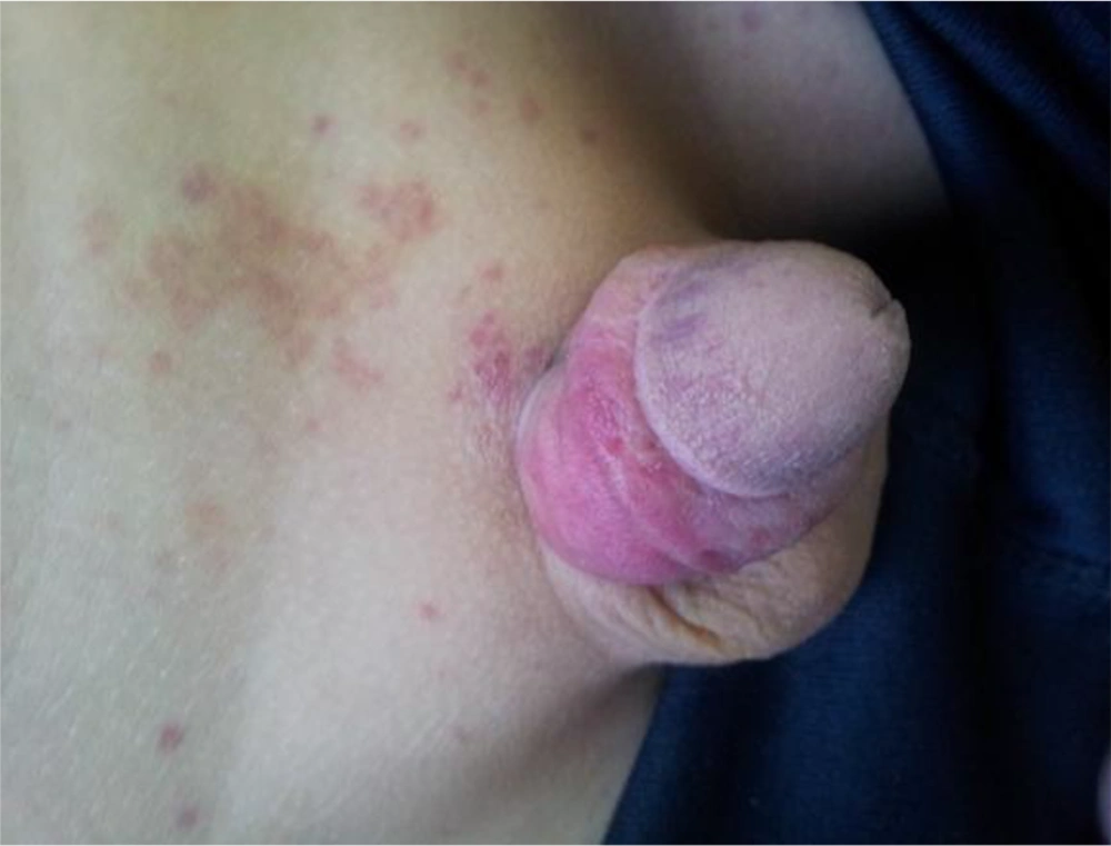
We were consulted urgently to visit a three-year-old boy suspected for HSP who was admitted to the rheumatology ward. His symptoms started eight days before admission as left ankle arthritis and malaise followed by purpuric rashes on lower extremities and buttocks with gradual involvement of upper limbs. Thirty six hours prior to admission, the patient’s penis was involved by a homogenous erythematous inflammatory rash with subsequent tenderness, ecchymosis of the prepuce, and indurations and no voiding difficulty (Figure 1). No history of upper respiratory tract infection was given and the patient had no complaint of abdominal pain or bowel habit change. He was irritated but not febrile.
Patient’s laboratory findings revealed normal electrolytes and biochemistry panel, whereas serum IgE level of 126 IU/mL (range 0.19 - 16.9) and IgA level of 2000 mg/dL (range 22 - 159) were identified; however, IgG and IgM were within normal limits. Moreover, erythrocyte sedimentation rate (ESR) was 21 millimeters (mm). No proteinuria or any other abnormality was detected in urinalysis. Penile doppler ultrasonography (DUS) showed no abnormality in blood flow and furthermore, scrotal ultrasonography revealed normal testes. Finally, the result of skin biopsy taken from a single rash confirmed the diagnosis: leukocytoclastic vasculitis in the resolving phase.
Subsequently, oral prednisolone (2 mg/kg) was administered and the patient developed significant healing in both generalized rash and penile inflammation within six hours, and the symptoms abated after 54 hours. Steroid therapy was continued for 96 hours and the patient was discharged from hospital teaching his parents how to taper the corticosteroid in the following eight days. Afterward, no complication or relapse was reported by parents in the follow up visits.
2.1.2. Case 2
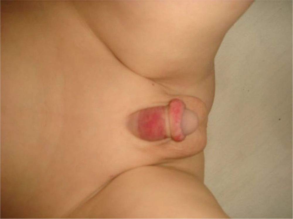
A 4.5-year-old boy was visited at rheumatology ward who had developed palpable petechial and purpuric rashes that first appeared on the lower part of his right leg and spread on the entire lower extremities and buttocks soon after. In addition, 4-limb-edema and then left extremities’ painfulness occurred the day after. Histories of upper respiratory tract infection and mild abdominal pain without diarrhea or hematochezia were given. On physical examination, he was a normotensive, uncircumcised and non-ill boy whose hands and calves had mild edema with a mild tenderness of the left quadriceps muscle. Moreover, erythema and edema was observed on the shaft and prepuce of his penis which occurred on the third day of admission (Figure 2).
In laboratory findings, WBC count of 8700/mL (Polymorphonuclear cells = 46%, Lymphocytes = 50%) and ESR level of 25 mm were detected on the admission day. Three days later, WBC increased to 10900/mL (P = 64%, L = 30%, Eos = 1.1%) and ESR decreased to 17. Serum electrolytes and biochemistry panel were found normal, anti-nuclear antibody (ANA) was negative. Serum immunoglobulins were within normal limits: IgA 92 mg/dL (range 22 - 159), IgM 97 mg/dL (range 47 - 200) and IgG 1125 mg/dL (range 441 - 1135). Urinalysis was negative for protein and glucose, however, RBC count of 10 - 12 per high power field (HPF) and WBC count of 1 - 2 per HPF were detected, which increased to 12 - 15 RBC per HPF the day after. Also, no growth was reported in urine culture, while abdominal and scrotal ultrasonography in addition to penile DUS were normal.
On the fourth day of hospital stay, the patient developed purpuric rash on left ear and periorbital edema. Because of the severe systemic symptoms such as periorbital edema, myalgia and prepuce edema, low dose prednisolone twice daily (0.5 mg per kg/day) was commenced. Dramatic response was observed as all symptoms (except for mild arthralgia of the right knee joint) relieved and the patient was discharged from the hospital two days after initiating the steroid therapy. On the day before discharge, RBC count decreased to 5 - 6 per HPF in urinalysis. Pathological study confirmed the diagnosis: leukocytoclastic vasculitis. Three and six month follow-up showed complete relief.
2.1.3. Case 3
The patient was a 4.5-year-old boy who was hospitalized at rheumatologic ward, with a subsequent request of urologic consult. He had a positive history for upper respiratory tract infection, which was followed by abdominal pain, upper and lower extremities pain, and edema with difficulty in weight bearing. The presentation was palpable lesions on calves, buttocks, penis, and scrotal region with a diameter of one millimeter to five centimeters. Moreover, the lesions were edematous, painless and non-itching. Physical examination revealed normal genitourinary, gastrointestinal, and neuromuscular systems. However, examination of joints revealed 2+ edema of both ankles. Laboratory data showed leukocytosis (WBC: 13800 with 50% polymorphonuclear cells), 3 - 5 RBC per HPF in urinalysis (U/A), while the other tests were normal. Skin biopsy revealed vasculopathic changes through dermis. Moreover, immune-fluorescence study discovered positive IgA count (2+) with negative IgM and C3, and the diagnosis was leukocytoclastic vaculitis. Abdominopelvic sonography revealed deactivated small basal mesenteric lymph nodes. The abdominal pain abated during hospital stay. Considering the lack of neurological, renal, and gastrointestinal manifestations, he was followed as an outpatient.
2.1.4. Case 4
An 8-year-old boy was hospitalized at rheumatologic ward with developing itching lesions. The lesions had appeared from 15 days ago firstly on the upper extremities that subsequently spread to lower limbs. He complained of right ankle and knee pain. Moreover, he had edematous extremities since four days ago and could not bear his weight. Also, he had one episode of fever, diarrhea and abdominal pain, without any history of urinary or respiratory tract infection. Moreover, his drug history was indicative of taking prednisolone due to allergic reactions. Physical examination revealed anterior neck lymphadenopathy (0.5 × 0.5 cm), mild bilateral wheezing of lungs and maculopapular rashes on upper and lower limbs, especially on the left arm and buttocks. Genital examination showed testicular pain as well as penile and scrotal edema.
Laboratory data showed leukocytosis: WBC 10840 with 70% Polymorphonuclear cells), microcytic and hypochromic anemia (Hb 8, MCV 55, MCH 17), anisocytosis, eliptocytosis as well as presence of oval cells and helmet cells in peripheral blood smear (PBS). C-reactive protein (CRP) level was high (100). Vacuities markers were positive for ANA (5). Urinalysis was positive for RBC (3 - 5 per HPF), stool examination was negative. Biochemistry profile and abdominopelvic sonography was normal. Immunofluorescence study revealed positive IgA (296 mg/dL), IgE (736 mg/dL) and negative IgG and IgM. In serology, anti-streptolysin O (ASO) antibody was negative. Skin biopsy revealed leukocytoclastic vasculitis with sedimentation of IgA through papillary dermis. The patient was managed with administration of oral prednisolone, the symptoms gradually abated and the medication was tapered. During follow-up periods, we did not find any abnormal finding in physical examination or laboratory tests.
2.1.5. Case 5
A 5-year-old boy was hospitalized at rheumatologic ward because of the scattered painful macular rash on the back and distal lower extremities 10 days prior to admission. He complained of lower extremities edema and generalized erythematous maculopapular lesions on the back, buttocks and then upper extremities. He did not have any history of respiratory, urinary, or gastrointestinal infections. On physical examination, he had erythematous maculopapular rashes in an edematous background on lower extremities and buttocks. Also, the patient had obvious tenderness in penis and testes. Penile involvement was among his primary presentations; hence, he was hospitalized for probable testicular torsion at first. Regarding his skin manifestations, HSP was confirmed. One week after admission, lesions of the extremities became hemorrhagic. His past medical history was indicative of recurrent episodes of fever and urine color changes and one episode of dysentery about one month ago. There was no history of lymphadenopathy and arthritis. In drug history, he used dexamethasone and hydroxyzine because of episodes of allergic reactions in the past.
Laboratory data showed leukocytosis (WBC 11700 with 70% polymorphonuclears), high level of CRP (68), but normal biochemistry profile, ESR, hemoglobin and ASO levels. Urinalysis discovered 2 - 4 WBC and RBC per HPF; but urine culture was negative. Stool examination showed 3 - 4 WBC, despite the negative stool culture. Immunological study revealed upper limit IgA (63 mg/dL) and normal IgM, IgG and ANA. Chest X-ray and abdominopelvic sonography were also normal. At first he received hydrocortisone due to oral therapy intolerance, but after GI and skin recovery, the treatment was changed to oral prednisolone. Finally, he was discharged with complete recovery.
Two months after his admission on the outpatient follow-up visit, active U/A (2+ proteinuria) was revealed. His kidney biopsy showed crescentic glomerulonephritis. He received intravenous methylprednisolone pulse therapy (30 mg/day for three days) with maintained oral prednisolone (1 mg/kg/day) for three months and azathioprine (1 mg/kg/day) for six months. One year after his admission, the glomerulonephritis was completely resolved. During his next follow-up visits, HSP recurrence was evident due to the positive U/A (hematuria) and skin lesions. Accordingly, we monitored him as an outpatient with urinalysis and skin biopsy results. On the follow-up visit, the results of his previous skin biopsy showed leukocytoclastic vasculitis, but his new laboratory tests and physical examinations revealed normal findings.
2.1.6. Case 6
A 6-year-old male was admitted to the rheumatologic ward with complaints of petechial and purpuric lesions on his lower extremities especially calves with concomitant edema, tenderness of ankles, severe limitation of lower joints range of motion and inability to weight bearing. On genital examination, we discovered scrotal edema with penile tenderness. In past medical history, he had several episodes of epistaxis and upper respiratory tract infection and one episode of abdominal pain. There was no history of fever, diarrhea or vomiting.
Laboratory investigations revealed microcytic and hypochromic anemia (Hb 9.5, MCV 56, MCH 16), increased ESR (54) with normal other tests. immunofluorescence study of dermal lesions revealed positive fine granular IgA (+1) and trace granular IgM, with negative IgG and complement. Skin biopsy revealed leukocytoclastic vasculitis with neutrophil infiltration around dermal vessels. Chest X-ray, abdominopelvic sonography in addition to DUS of testes was normal. Finally, he was discharged with normal general state. He was routinely managed as an outpatient and remained symptomless in follow-up visits.
2.1.7. Case 7
An 8-year-old boy was admitted to rheumatology ward with complaints of abdominal tenderness, fever and right knee tenderness. On physical examination, skin lesions were observed as prominent purpuric, petechial and ecchymotic lesions, with generalized distribution in upper and lower limbs, in addition to focal lesions in trunk and face. His hips were in external rotation and abduction with significant reduction of range of motion. Examination of genitalia showed swelling and tenderness in penile shaft. He had an upper respiratory tract infection (URTI) a few days prior to admission. Laboratory tests showed normal blood cells count and normal level of urea and Cr with mildly elevated ESR (15) and CRP (5.5). U/A revealed 8 - 10 RBCs per HPF. Abdominal sonography was normal and skin biopsy revealed leukocytoclastic changes, thus the diagnosis of HSP was established with significant penile involvement. He was discharged after complete recovery of signs and symptoms. In follow-up visits, all examinations and tests were normal, but U/A was still active (RBC = 12).
2.1.8. Case 8
A 5-year-old boy was admitted to rheumatologic ward with complaints of fever, joints tenderness, myalgia and loss of appetite. Physical examinations revealed pain and swelling in left knee and left ankle. Also, generalized purpura was observed in upper and lower limbs, which did not disappear with focal pressure. The left ear was painful with discharge which indicated otitis media. Genitalia examination revealed swelling of penile bulb with scattered erythema and petechial lesions. Lab study on admission day showed U/A with 7 - 8 RBC per HPF and trace proteinuria, normal CBC, BUN, Cr and normal IgA. Chest and abdominopelvic X-rays were normal. Biopsy of skin indicated leukocytoclastic vasculitis; therefore, the diagnosis of HSP with penile involvement was established. He was discharged after one week of hospitalization with complete recovery. On routine follow-up visits, we did not detect any abnormal finding in his examinations or lab tests.
2.1.9. Case 9
A 4-year-old boy was admitted with joints tenderness and limbs swelling. Joints involvement began first in both wrists then both knees and both ankles were involved. However, the range of motion remained normal in all joints. He also had general abdominal tenderness with no nausea or vomiting. On physical examination, we observed a purpuric rash on distal parts of lower limbs, which did not disappear with pressure. Genitalia examinations revealed the purpuric rash on penile shaft and scrotal edema. He mentioned episodes of URTI two weeks before admission. In lab data, CBC, blood urea, Cr, ESR, and CRP were normal. Also, U/A was normal. Blood immunoglobulins showed a lightly elevated IgA (76 mg/dL). Chest-X-ray and abdominal sonography were normal. In admission course, left eyelid swelling occurred. Skin biopsy showed leukocytoclastic vasculitis: the diagnosis was HSP with prominent involvement of penis. After complete recovery, he was discharged and during his scheduled follow-up visits, he remained fine.
3. Discussion
Genital involvement is uncommon in HSP; but until now, some cases of penile involvement have been reported in HSP patients that received different treatment strategies. Table 1 summarizes some of the clinical aspects of our HSP patients who referred with penile involvement within the past three years.
| Patient | Age | Treatment Regimen | Presence of GIA Manifestation | Time Point of Penile Involvement | Clinically Important Presentations During Disease Course | Follow Up Visits | Important Lab Findings |
|---|---|---|---|---|---|---|---|
| 3 | Prednisolone | Negative | Simultaneous with skin rashes | - | Normal | IgA = 2000 mg/dL, IgE = 126 mg/dL, ESR = 21 | |
| 4.5 | Prednisolone | Mild abdominal pain | Three days after skin rashes | - | Normal | IgG = 1125 mg/dL, ESR = 25, 10 - 12 RBC per HPF on U/A | |
| 4.5 | Prednisolone | Abdominal pain Sonography: enlargement of deactivated lymph-nodes in basal mesenteric | Simultaneous with skin rashes | - | Normal | WBC = 13800, RBC = 3 - 5 per HPF on U/A | |
| 8 | Prednisolone | Abdominal pain and diarrhea | Simultaneous with skin rashes | - | Normal | Hb = 8 g/dL, MCV = 55 fl, CRP = 100, IgA = 296 mg/dL, IgE = 736 mg/dL, 3 - 5 RBC per HPF on U/A | |
| 5 | Prednisolone, hydrocortisone | Abdominal pain and WBCB: 3 - 4 in S/E | Penile involvement was prior to skin rashes | Penile tenderness was highly suspicious for penile torsion | Proteinuria, crescentic GNC, cematuria | WBC = 11700, CRP = 68, 3 - 4 WBC on S/E, 2 - 4 RBC and WBC on U/A | |
| 5 | Prednisolone | Negative | Simultaneous with skin rashes | - | Normal | Hb = 9.5 g/dL, MCV = 56 fl, ESR = 54 | |
| 4 | Ibuprofen, prednisolone | Abdominal pain | Simultaneous with skin rashes | - | Normal | ESR = 15, CRP = 5.5, 8 - 10 RBC per HPF on U/A | |
| 5 | Prednisolone | Negative | Simultaneous with skin rashes | - | Normal | 7 - 8 RBC per HPF on U/A | |
| 8 | NSAIDD, prednisolon, azathioprine | Abdominal pain | Simultaneous with skin rashes | - | Normal | IgA = 76 mg/dL |
a Abbreviations: A: gastrointestinal; B: White Blood Cell; C: glomerulonephritis; D: non-steroidal anti-inflammatory drugs.
Burrows et al. reported a 19-year-old male with palpable rash on his penis which appeared after a streptococcal pharyngitis and resolved after penicillin was administered (3). David et al. presented a HSP case with mid-shaft to glans involvement of purpuric and erythematous rash who was only managed by conservative approach (4). Moreover, Ferrara et al. reported a HSP patient with involvement of the penis which had interestingly started after intravenous corticosteroid and antibiotics therapy due to his primary osteomastoiditis which eventually relieved spontaneously (5). Considering the treatment method, on the contrary, Sandell et al. reported a boy with HSP who was treated with pulsed methylprednisolon in addition to oral steroids because of his penile involvement (6). Additionally, Pennesi and coworkers presented a case of HSP who was treated with oral corticosteroid because his penis was involved (7). Furthermore, Lind and colleagues observed a boy with HSP and priapism who was finally treated with aspiration of corporal bodies under caudal anesthesia (8). Another 37-year-old patient was reported by Sari et al. in whom priapism occurred in the background of HSP, but resulted in penile necrosis and prosthesis implant (9). In addition to the mentioned reports, some investigators have stated this manifestation within their society as well. For example, in 1990, Dawod and Akl reported a HSP case with swollen penis among their patients (10). Moreover, Mintzer and colleagues evaluating 155 HSP patients, found three patients with penile involvement (2). Finally, in 2007, Akl mentioned some cases of HSP with penile swelling in Middle East countries (11).
From a therapeutic point of view, administration of corticosteroids for the treatment of HSP is still controversial. On one hand, some believe that steroids cause a rapid response in the healing process so that the penis would be saved from the permanent sequel of the severe end-organ vasculitis. On the other hand, self-limiting course of HSP has prevented other physicians from administration of corticosteroids. It seems that steroids have been considered for more complicated circumstances such as renal, central nervous system (CNS) or intestinal involvements of HSP. Besides, the general beneficial effects of early administration of corticosteroids has been demonstrated by Weiss et al. through a systematic review (12).
In our first case, because of the severity of local inflammation and possibility of permanent end-organ damage and probable future impotence, we started oral corticosteroid which caused dramatic healing response. However, our second case did not need steroid therapy from urological point of view, even though prednisolone was administered for other reasons. To our knowledge, this is the first report in which only administration of 10 mg prednisolone (almost 0.5 to 1 mgr per kg according to the severity of symptoms), much lower than in other reports, was used for the treatment of severe penile vasculitis which resulted in dramatic healing response. We presented nine cases of HSP with penile involvement in order to indicate another rare aspect of HSP and its possible complications as well as its appropriate treatment.
Although rare, male patients with HSP may present with penile skin involvement indicated by erythema, ecchymosis, induration, or edema of the prepuce/shaft. Thus, physicians should consider HSP as the differential diagnosis of such presentation.

