1. Introduction
Kawasaki disease (KD) as a febrile vasculitis is the second common vasculitis of childhood after Henoch-Schönlein Purpura. Its prevalence is 239 cases per 100,000 children under 5 years old in Asian countries vs about 50 per 100,000 in USA (1-3). Eighty percent of affected children are under 4 years old (4) and the peak age of incidence is 9 - 11 months (2). KD is the most common cause of acquired heart disease of infancy and childhood. Without sufficient treatment, it may cause coronary dilation and aneurysm in 25% of patients (1, 5); subsequently stenosis, thrombosis, myocardial infarction and congestive heart failure may happen (6). Early diagnosis and treatment may improve the prognosis significantly. Diagnosing infantile Kawasaki (younger than one year) is difficult because of obscure symptoms (7); at the same time they are at the higher risk of coronary abnormalities (8-11). Other cardiovascular involvements such as pericardial effusion, mitral insufficiency, congestive heart failure, myocardial systolic dysfunction, and systemic vasculitis (iliac, renal, axillary and mesenteric arteries) were reported (1, 12-16). Even rare interpretation of peripheral gangrene and amputation had been detected due to peripheral vasculitis (17, 18). Herein we report 3 cases of peripheral gangrene as the presentation of KD.
2. Case Presentation
2.1. Case 1
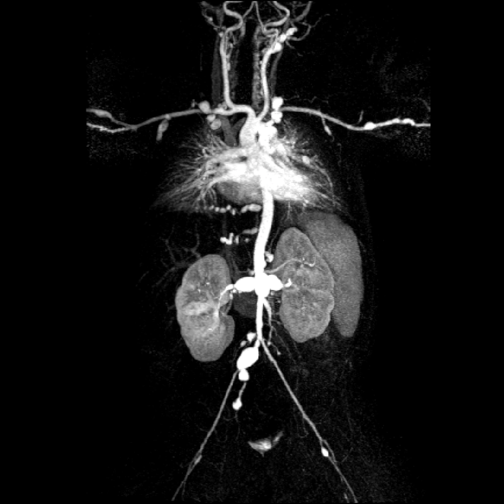
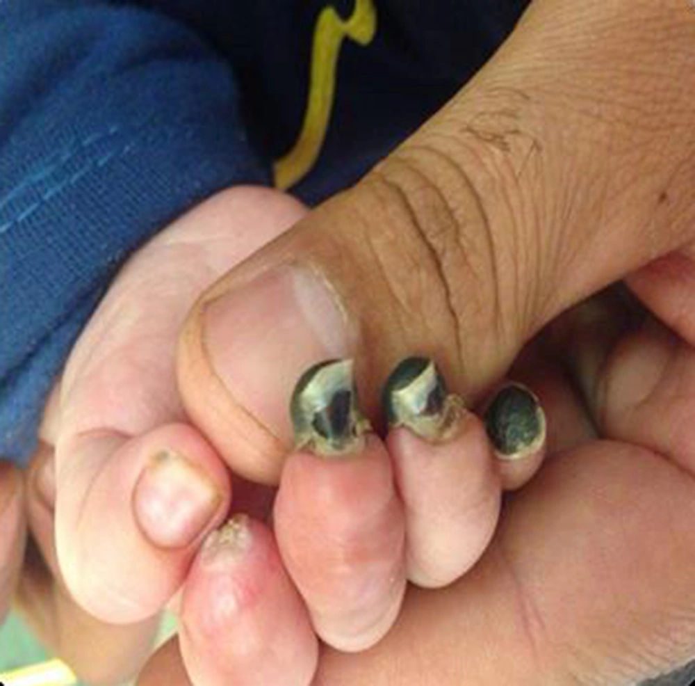
A 4-month-old boy presented with prolonged fever from since a month ago. Although sepsis work up and wide-spectrum anti-biotic therapy were done, there was no recovery. He was ill, febrile and severely anemic. In physical examination, pallor, hepato-splenomegaly and 3/6 systolic murmur were detected. He had peripheral gangrene on both sides lower and right side upper extremities. Peripheral pulses were undetectable. Lab data were as follow: WBC 14.1 × 10³ /mm3 (Neut 61%), Hb 7.1 g/dl, Plt 329 × 103 µL, CRP 201, ESR 74, Ammonia 1.2, Lactate = 27, negative cultures, sterile pyuria and normal electrolytes except hyponatremia (Na 129). Anti-phospholipid (apl) antibodies and anti-neutrophil cytoplasmic antibodies (ANCAs) were negative. Gallbladder wall thickness and edema with no hydrops, hepatomegaly and splenomegaly were detected in the abdominal sonography. Doppler sonography showed proximal stenosis in right side brachial artery and distal arteries of both lower extremities. Bone marrow aspiration was normal. Multiple aneurysms in the left coronary artery and other aortic branches were seen in the echocardiography, so magnetic resonance arteriography was done which showed multiple aneurysms in all coronary branches, both subclavian, external and internal iliac, bronchial, renal and mesenteric arteries (Figure 1). Because of KD as the diagnosis, treatment with high dose aspirin, methylprednisolone pulse therapy and IVIG initiated. Anti-coagulant therapy with heparin was done for peripheral gangrene and multiple arterial stenosis. Due to multiple aneurysms cyclophosphamide was added. The arterial dilations, aneurysms and organomegalies slightly improved. Unfortunately, peripheral gangrene remained and auto-amputation happened in distal phalanxes of the right hand and left foot (Figure 2). Follow-up of the patient after 1 year showed very mild improvement in the size of coronary aneurysms, but peripheral aneurysms were without improvement.
2.2. Case 2
Six-month-old girl, a known case of PHACE (Posterior fossa abnormalities, Hemangioma, Arterial lesions, Cardiac abnormalities/Aortic coarctation, and Eye abnormalities) syndrome was referred with 2-days history of fever, irritability and poor feeding. She had coryza and cough. At admission, she was febrile and irritable. Cardiac auscultation was abnormal due to aortic coarctation, but lung sounds were normal. There were no organomegalies. But the most important sign was distal cyanosis and scaling in hands and gangrene of the middle phalanx of the right hand, finger 3. Lab tests were normal: WBC 13.4 × 10³ /mm3 (Neut 45.1%), Hb 9.5 g/dl, Plt 470 × 103 µL, CRP 0.5, ESR 9, negative cultures and sterile pyuria. Pleocytosis (22.77 × 10³ /mm3), thrombocytosis (705 × 103 µL) and ESR rising (57) happened, during hospitalization. Apl antibodies and ANCAs were negative. Abdominal sonography and chest X-ray were normal. Due to dilation of descending aorta in echocardiography and angiography in addition to thrombocytosis and peripheral gangrene, Kawasaki disease was diagnosed. She was treated with aspirin, methylprednisolone, and IVIG, which terminated the fever. Peripheral cyanosis and gangrene bettered. In follow up after 6 months, dilatation of coronary improved relatively.
2.3. Case 3
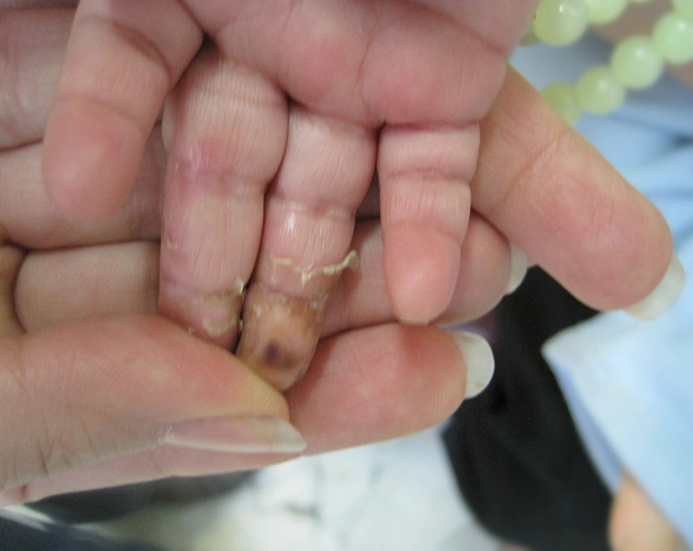
A 1-year-old boy was referred with high grade fever and intensifying peripheral gangrene. He was pretty ill, febrile and irritable. First he was admitted with a history of fever for 2 weeks, therefore sepsis work up and anti-biotic therapy initiated. Sucessively strawberry tongue, cervical lymphadenopathy and scaling appeared. Due to probability of Kawasaki disease, echocardiography was done which showed mild pericardial effusion and normal coronary arteries. Then he was treated with IVIG and aspirin. During the hospital stay, swelling and erythema of extremities happened. At the 6th day of admission, peripheral cyanosis and gangrene in distal phalanx of extremities happened. Because of continuous fever and peripheral gangrene, he was referred to our tertiary center (Figure 3). In second echocardiography, in addition to pericardial effusion, multiple aneurysms in RCA, LMCA, and LAD were detected. He had leukocytosis (32.73 × 10³ /mm3), anemia (Hb 10 g/dl), and severe thrombocytosis (1.344 × 10³ μL). In addition to aspirin and IVIG, methylprednisolone, azathioprine and heparin as an anti-coagulant were prescribed. Fever period ended and peripheral gangrene got better. Echocardiography after pulse of methylprednisolone showed decrease in size of aneurysms and no pericardial effusion. FANA level was 1/80 in speckled format, anti dsDNA level was 128 (normal range < 100) and complementary system evaluation was normal. In the acute phase of disease ANCAs and apl antibodies were negative. After 2 months, FANA and anti dsDNA became normal but there was no recovery in coronary aneurysms. He was admitted again. This time echocardiography findings were as follow: LMCA diameter 7.4 mm, RCA 4.3 mm, and LAD 5.2 mm. With continuation of aspirin, warfarin, and azathioprine (for 3 months), second pulse of methylprednisolone and IVIG and Infliximab were given. After a year cardiovascular angiography revealed: 4.5 mm diameter aneurysm in distal LMCA, LAD stenosis and non-appropriate tapering of RCA. His treatment continued with long-term use of low dose aspirin and warfarin for prevention of thrombosis.
3. Discussion
Gangrene is a rare presentation of Kawasaki disease that needs a high level of suspicion by physicians to discover the underlying disease. There are reports of peripheral gangrene due to KD from around the world, however it seems that the reports are less from Japan as a country from where most of the KD cases are reported (17). There is not a clear explanation why gangrene happens in KD, but some reasons have been emphasized as etiology such as arteritis, arteriospasm, thrombosis of inflamed vessels and decreased peripheral perfusion (17).
Most of the reported cases of gangrene due to KD, experienced a degree of sequel especially amputation of fingers, this has also occurred in our cases. However there is a report with no sequel from a 57 day infant in Korea in whom IVIG, dexamethasone, methotrexate and anticoagulant were administered (19). There is a wide range of different medications which are used in management of KD with gangrene. Some of the treatment agents that have been used are heparin, steroids, hydralazine, propranolol, warfarin, urokinase, PGE1, dipyridamole, nitroprusside, tissue plasminogen activator, prostacyclin, glyceryl trinitrate, nifedipin, and even caudal block (20). We additionally used cyclophosphamide due to multiple aneurysms in our first case and infliximab in case 3.
Regarding age of this complication, most of the reports present young age in infancy especially under seven months of age (20). But it is not a real clear cut for age as we have reported a one year old child with gangrene. There is also a report of a 22 year old man with Kawasaki whose disease was complicated by gangrene and amputation of fingers (21).
We reported three cases of peripheral gangrene to alert the physicians about this rare and important complication of KD, because sometimes this condition of a patient may be the first presentation of KD such as the infant we reported as our first case. Therefore, without a single guideline for managing peripheral gangrene of KD, more cases should be reported to understand the epidemiology of the disease and experimental studies should be done to evaluate the best medications for treatment of gangrene complicated KD.
Peripheral gangrene must be seen as an important sign of infantile Kawasaki disease. Early aggressive treatment after IVIG therapy with methylprednisolone pulse therapy and/or cytotoxic or biologic drugs can prevent permanent morbidities and sequels.


