


1. Background
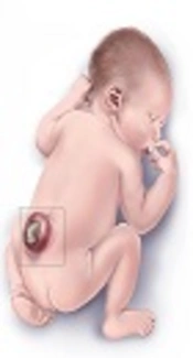
Spinal dysraphism is the most common congenital abnormality of the spinal cord and the most complex congenital condition with the incidence of 2.33 per 1000 births in Iran (1, 2). Most of these patients survive into adulthood. However, they are at risk of severe life-long disabilities, including paralysis, mental retardation, bowel and bladder dysfunction, and orthopedic disabilities (3-6). The majority of neonates born with spinal dysraphism have some degree of lower urinary tract and urodynamic dysfunction that may vary depending on the type, level, extent and completeness of the spinal lesion leading to long term sequelae of urinary tract infection, incontinence, and renal insufficiency (7). Most patients will ultimately require clean intermittent catheterization (CIC) and anticholinergic therapy (8, 9).
There is a worldwide consensus that identification of neonates born with spinal dysraphism who are at risk for upper urinary tract deterioration, using urodynamic studies, anticholinergic therapy and CIC may decrease the incidence of upper urinary tract deterioration and reduce the need for augmentation cystoplasty.In addition to that, neonates with urodynamic dysfunction, especially those with low bladder compliance and/or detrusor-sphincter dyssynergia, warrant close urological monitor in order to prevent urinary tract deterioration (10). Detrusor-sphincter dyssynergia, high leak point pressure and high intravesical filling pressure are good indicators of lower urinary tract dysfunction (11, 12).
2. Objectives
We performed the current study to find the incidence of upper urinary tract deterioration in Iranian newborns suffering from spinal dysraphism who were closely monitored for upper urinary tract deterioration using urodynamic studies, anticholinergic therapy and CIC.
3. Methods
In a prospective study, neonates born with the diagnosis of spinal dysraphism anomalies referred to the Children’s medical center, Tehran University of Medical Sciences, Iran in 2015 and 2016 were enrolled into the study.
3.1. Inclusion and Exclusion Criteria
Patients aged below 60 days and being diagnosed with spinal dysraphism including meningocele, myelomeningocele and spina bifida occulta were included in study. The diagnosis of spinal dysraphism was made with positive maternal serum or amniotic fluid alpha-fetoprotein and neural tube defects detected by fetal ultrasound at gestational age of 18 - 20 weeks. Neonates with history of surgical intervention, inability in performing urodynamic study and lack of adherence to treatments and follow-up were excluded from the study. Furthermore, three patients died due to their underlying disease and were excluded from study.
3.2. Ethics
All parents/guardians of the neonates included within this study signed the written informed consent prior to enrollment. This study was performed only after approval by the ethical committee of our center. No potential hazardous or high-risk evaluation or interventions were performed, diagnostic tests used during this study is recommended and performed by the majority of well-known international pediatric surgery centers with encouraging results.
3.3. Assessment and Follow-Up Protocol
All patients underwent surgical repair of spinal dysraphism by a same surgeon. Voiding cystourethrogram (VCUG) and urodynamic study were performed for all patients after surgery and based on the urodynamic study results, patients were classified into three groups, synergic group defined as coordination of detrusor muscles of the bladder and the urethral sphincter muscle, dyssynergic group recognized as dyscoordination of detrusor muscles of the bladder and the urethral sphincter muscle and complete denervation group characterized as no muscular and bioelectrical activity in urethral sphincter in any stage.
Our objective was to recognize high-risk groups (dyssynergic and denervation pattern) and offer prompt treatments such as CIC and anticholinergic therapy (oxybutynin 0.25 mg/kg/day per os, ExirPharma Co., Iran). Parents of patients in the low-risk group (synergic) were educated about urinary symptoms and prompt recruitment. Moreover, in patients with leak point pressure more than 40 cmH2O or detrusor sphincter dyssynergia (DSD) treatment using anticholinergics and CIC was started. DSD was characterized by increased electromyography sphincter activity during a detrusor contraction and by either a ‘‘spinning-top’’ configuration of the proximal urethra or a narrowing of the external sphincter area on VCUG.
All patients were followed with urine culture every three months, post voiding residual volume measurement every six months, renal/bladder ultrasound a year later. In cases of vesicourethral reflux or vesical hypertonicity VCUG was performed after a year. In all cases the second urodynamic study was performed nine months after the first study to detect the conversion of synergic to dyssynergic group. In dyssynergic group the aim was to evaluate efficacy of the therapeutic interventions. In patients with dyssynergia and vesicourethral reflux or vesical hypertonicity, dimercaptosuccinic acid (DMSA) scan was done initially and every twelve months. In case a symptomatic episode and febrile urinary tract infection (UTI) occurred, the scintigraphy was performed six months after the episode. Percutaneous vesicostomy was performed in cases of worsening upper urinary tract damage detected by hydronephrosis in ultrasound or defect in DMSA scan despite medical and CIC treatments. UTI was diagnosed if there were signs and symptoms of infection including fever of more than 38.5° C, change in continence pattern or change in the color or odor of urine accompanying with a positive urine culture defined as more than 105 colony-forming units/mL. The presence of urinary leukocytes was considered significant if more than five white blood cells/high power field were observed on microscopic examination of centrifuged urine. After each episode of UTI, ultrasound and DMSA scan was performed to assess the damage to kidneys. Defects on renal DMSA were recognized as decrease in relative renal function with a difference more than10% and/or hypocapturing area scarring or irregular distribution. The hydronephrosis was considered as pelvic diameter more than 15 mm.
3.4. Statistical Analysis
Data analysis was performed using statistical software (SPSS for Windows, Version 22.0, SPSS Inc., Chicago, IL, USA). Qualitative and quantitative variables were evaluated with the Chi-square and independent Student t-tests, respectively. Univariate survival analysis was performed with log-rank test, and multivariate survival analysis was performed with Cox proportional hazards regression model. Statistical significance was defined by P ≤ 0.05.
4. Results
During the study period, 55 neonates with age range of 1 - 60 days and the diagnosis of spinal dysraphism were enrolled into the study. General characteristics of the population and risk factors are depicted in Table 1. Results of the primary and secondary urodynamic studies are demonstrated in Table 2.
| Demographics | No. (%) |
|---|---|
| Gender, male | 34 (61.8) |
| Birth weight, < 2500 g | 6 (10.9) |
| Gestational age, < 36 w | 8 (14.5) |
| Mother’s age, y | |
| < 20 | 1 (1.8) |
| > 35 | 8 (14.5) |
| Father’s age, y | |
| > 35 | 14 (25.5) |
| Child’s rank | |
| First | 32 (58.2) |
| Second | 11 (20) |
| Third | 12 (21.8) |
| Maternal diabetes mellitus | 3 (5.5) |
Demographic Data of the Patients
| Group | Initial Study | Follow-Up Study |
|---|---|---|
| Synergic | 15 (27.3) | 11 (20) |
| Dyssynergic | 28 (50.9) | 32 (58.2) |
| Complete denervation | 12 (21.8) | 12 (21.8) |
Distribution of Patients in Each Group Based on Initial and Follow Up Urodynamic Studiesa
Five (9.1%) patients of the dyssynergic and complete denervation group, who were treated using CIC, were complicated with severe febrile urinary tract infection which required in-patient care. Hydrocephalus was seen in 23 (42%) patients in whom ventriculoperitoneal shunt was implanted. Vesicourethral reflux was seen in 16 (29.1%) cases. There was no significant relation between gender and variables such as urodynamic pattern (P = 0.55), location of the lesion (P = 0.77), hydrocephalous (P = 0.66), vesicourethral reflux (P = 0.24) and upper urinary tract deterioration (P = 0.12).
The results of the second urodynamic study and reviewing its relation with other variables revealed that location of the lesion was not a predictor of urodynamic pattern (P = 0.42). Also, the relation between urodynamic pattern with vesicourethral reflux (P = 0.66) and hydrocephalous (P = 0.66) was not significant. However, upper urinary tract deterioration was significantly related to the type of urodynamic pattern (P = 0.02). The risk for upper urinary tract deterioration was highest in the dyssynergic group (7 out of 32 patients), while just one out of 12 patients in the complete denervation group and none in the synergic group showed upper urinary tract deterioration. There was no significant relation between location of the lesion and upper urinary tract deterioration (P = 0.75), whereas there was significant relation between location of the lesion and hydrocephalus (P = 0.05). The frequency of location of the lesions and hydrocephalous is demonstrated in Table 3.
| Location of the Lesion | Total Number | Hydrocephalus |
|---|---|---|
| Lumbosacral | 19 | 7 (37) |
| Lumbar | 9 | 3 (33) |
| Thoracolumbar | 7 | 5 (71) |
| Sub occipital | 6 | 1 (17) |
| Sacral | 5 | 5 (100) |
| Cervical | 5 | 1 (20) |
| Thoracic | 2 | 0 (0) |
| Cervicothoracic | 2 | 1 (50) |
Frequency of Location of the Lesions and Patients with Hydrocephalus Required Ventriculoperitoneal Shunt Implantation Based on the Location of the Lesiona
5. Discussion
Spinal dysraphism may result in chronic medical conditions that impact life of affected children (2). Genitourinary system involvement in spinal dysraphism is the most common cause of neurogenic bladder in newborns which can cause severe morbidity and mortality (7, 8). It causes upper urinary deterioration through vesicourethral reflux and pyelonephritis and subsequently can lead to end-stage renal disease (13). Positive outcome of preventive measures such as CIC and anticholinergic therapy has been shown in several studies (14). However, no consensus has been reached in terms of follow-up regimen and time of intervention (15-19).
In the present study, it was shown that even under intensive follow-up regimen and supportive care conversion of the synergic group to dyssynergic group was inevitable, 26% of the patients in the synergic group converted to the dyssynergic type which was considered as high-risk. Therefore, our results may accentuate the importance of serial urodynamic study and educational empowerment of parents about signs of voiding dysfunction since CIC and anti-cholinergic agents have been shown to reduce, prevent and reverse upper urinary tract deterioration. Moreover, with our risk stratification and intervention 9.1% of patients developed febrile UTI, and renal function deterioration was seen in 18% of patients in the high-risk group after one year of follow-up. In a study by Hopps et al. 84 patients with myelomeningocele and lipomeningocele under six months of age were followed for 10 years. Patients were risk stratified based on presence of hydronephrosis or clinical evidence of urinary retention and high-risk patients underwent urodynamic study and VCUG. In that study, 45% of patients in the low-risk group were converted to the high-risk patients with mean age of three years and renal function deterioration was seen in 1.2% of the patients (20). In a study by Sager et al. on infants with myelomeningocele, 28.8% had dyssynergic bladder and 30% showed abnormal DMSA scan (21). Torre et al. assessed 502 patients with spinal dysraphism of which 283 had myelomeningocele, 90 had caudal regression syndrome, and 129 had spinal lipoma. Renal impairment was observed in 19 with myelomeningocele, 11 with caudal regression syndrome, and two with spinal lipoma (22). In a systematic review performed by Venboehr et al. on 1,564 patients with spina bifida, renal impairment was seen in 290 patients (25.7%) and end-stage renal disease was seen in 12 (1.3%) (7).
In conclusion, it may be an acceptable choice to perform urodynamic studies as soon as possible in all newborns diagnosed with spinal dysraphism and categorize patients in low and high-risk groups. It may be suggested to use CIC, prophylactic antibiotics and anti-cholinergic medications in high-risk and intensive follow-up and educational empowering of parents concerning signs of voiding dysfunction in low-risk patients. It may also be recommend performing CIC in newborns with difficulty in voiding prior to urodynamic study which may result in better parent’s cooperation and acceptance.
Even though all patients obtained the required test before they turned one year of age, non-simultaneity in gathering each of the requested studies posed an important limitation for the present study. Residing in far distances and socio-economic constraint caused delay in consultation, laboratory and pharmacological interventions. The relatively small sample size relative to the number of variables analyzed and correlated posed another limitation to our study.
