1. Introduction
Larva migrans syndrome is a rare disease. Wide spectrum of the parasites can cause the larva migrans syndrome but the leading cause is considered to be the ascarid nematodes; so the disease caused by them can be referred to as the ascarid visceral larva migrans syndrome (ascarid LMS) (1). The human is infected by ingesting the embryonated eggs and the involvement of the different parts of body occurs as a result of the migration of the larvae through the vessels (2-4).
2. Case Presentation
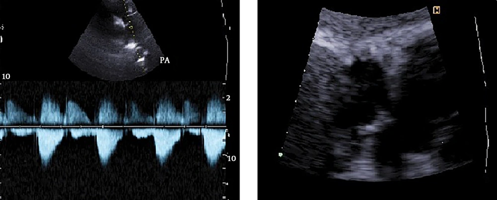
This 30-month-old girl, a resident of Qom, Iran was referred to our hospital due to a large ASD secundum and severe pulmonary valve stenosis. The parents stated that the child had no history of fever, abdominal pain, vomiting, diarrhea, cough, asthma, weight loss, fatigue or chest pain. Also there was no history of eating raw meat or geophagia. They had no pets and also no history of exposure to puppies. During her physical examination her vital signs were normal. There was no hepatomegaly. Electrocardiography showed normal sinus rhythm with right ventricle enlargement. Echocardiography revealed a large ASD secundum with left to right shunt associated with right atrium enlargement and right ventricle hypertrophy. We also noted a severe pulmonary valve stenosis with a pressure gradient of about 70 mmHg with no Tricuspid valve regurgitation and no pleural effusion (Figure 1). Surgical total correction for the cardiac lesions was decided as the treatment. The blood work-up including liver function tests were unremarkable, the patient did not have leukocytosis or eosinophilia and the liver function enzymes were not elevated.
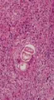
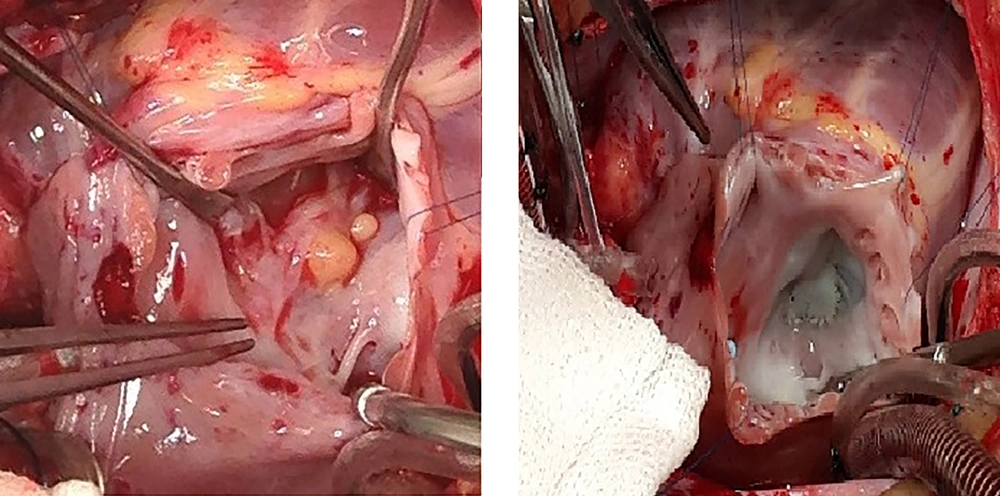
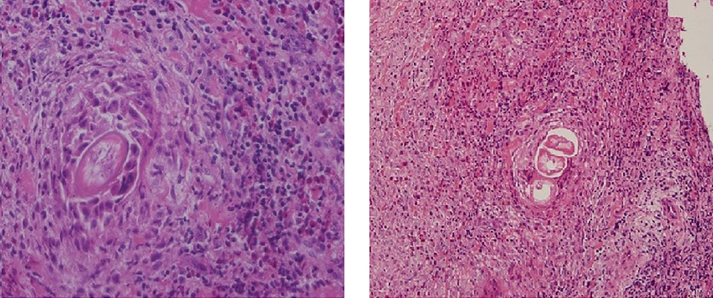
The patient underwent the surgery with median sternotomy approach. Following the cardiopulmonary bypass, cardioplegia and RA opening, we noted a large ASD secundum with normal drainage of pulmonary veins with incidental finding of an unusual mass on septal leaflet of the tricuspid valve in ventricular surface with the invasion to the myocardium. It was a well-defined mass with creamy to white colour that looked like lipoma or fibroma with much firmer texture. The size of the mass was about 7 to 8 millimeters and there was also a smaller satellite nodule near the mass with the same characteristics (Figure 2). Regarding the possibility of the cardiac tumors and its probable progression by considering the patient’s age, we decided to resect the mass totally and send it for further investigations. Finally ASD closure and pulmonary valve repair was done with bovine pericardial patch and the mass was resected completely and septal tricuspid valve was also repaired (Figure 2). The post-operative echocardiography was acceptable without any residual lesions. The histopathological findings for resected mass revealed a necrotic material, eosinophilis infiltration and larval elements that were surrounded by the muscle cells which were in favor of ascaridida infection (Figure 3). The diagnosis was double-checked and confirmed by department of parasitology of Tehran University of Medical Sciences, Tehran, Iran. Due to the direct pathology examination of the patient’s cardiac specimen, the definite diagnosis of infection caused by ascaridida was achieved. The post-operation blood work-up was normal and ophthalmological examinations did not reveal any ocular changes. The patient was referred to infectious disease clinic for medical treatment if necessary. At 12-month follow-up, the general state of the patient was good and echocardiographic findings demonstrated successful surgical outcome. For more exact determination of the larvae and by knowing its order, the titre of antibody (IgM and IgG) against Toxocariasis (ELISA method) was examined but the result was not conclusive. The remaining specimen was sent for PCR to determine the exact type of the larvae by DNA sequencing but unfortunately the amount of the remaining specimen was not enough and satisfying.
3. Discussion
The order ascaridida, consists of several families of parasitic round worms with three lips on the anterior end. Its important families include: The anisakidae, the ascarididae and the toxocaridae. Regarding the morphology of the larvae, our parasitologists propound the ascarididae and the toxocaridae as the probable cause of the infection but size (the larva was measured about 23 µm), scale and the morphology (closed lumen of the nematode section) were more compatible with the infection caused by the ascarididae.
Wide spectrum of the parasites can cause the larva migrans syndrome but the leading cause is considered to be the ascarid nematodes so the disease caused by them can be referred to as the ascarid visceral larva migrans syndrome (ascarid LMS) (1).
The embryonated eggs of the ascaridida worms are found in contaminated soil, vegetables and raw or undercooked meat. The human is infected by the incidental ingestion of the embryonated eggs. When the eggs reach the gastro intestinal tract, they hatch and second-stage larvae invade the intestinal mucosa and migrate through the blood and lymph vessels to the different parts of the body especially liver and lungs. As a result of the immune system activation and reaction to them, eosinophilia can occur (2-4) but as mentioned before, our patient lacked any eosinophilia before and after the surgery.
Cookston et al. observed the cardiac changes during infection with toxocara canis with cardiac involvement in mice. They reported that the myocardial lesion is developed by the infiltration of eosinophils and histocytes initially and progress to form granuloma with necrotic debris (5). In our case the mass was found on tricuspid septal valve with invasion to the myocardium and just like what Cookston et al. reported, the histopathological findings of the mass resected revealed a necrotic material, eosinophilis infiltration and larval elements. In 2016 Kuenzli et al. published a systematic review discussing Toxocara spp Infection with cardiac involvement. They reviewed 24 published cases and the cardiac entities described included myocarditis, pericarditis, and Loeffler’s endocarditis. The clinical presentation ranged from asymptomatic or mild disease to life threatening myocarditis/pericarditis with heart failure or cardiac tamponade, leading to death. They reported that only in 3 of the 9 cases, in whom histological analysis was performed, the granuloma of the parasite was detected (6).
We hypothetized that the endomyocarditis was probably caused by the direct migration of the larvae through the vessels and as it has reached the heart (more precisely right atrium) it is implanted on the septal leaflet of the tricuspid valve and involved the endocardium and myocardium of the adjacent areas.


