1. Background
Neonatal sepsis is a systemic infection in neonatal period. It is still a major cause of morbidity and mortality of newborns, especially in developing countries (1). Early diagnosis and treatment is essential to reduce morbidity and mortality (2). For this purpose some hematological findings can be helpful as blood culture is not available before 48 - 72 hours. Platelet indices; platelet count, PDW (platelet distribution width), and MPV (mean platelet volume) belong to this category of findings. Platelet count is important and thrombocytopenia is an early finding in neonatal sepsis (3). Although thrombocytopenia is seen in different types of neonatal sepsis it is reported to be more common in infections caused by Gram negative germs (2, 4) specially Klebsiella spp (5) ‘Enterobacter spp.’ (6) and fungal infections (3, 7). Some articles showed elevated PDW and MPV in septic neonates (3) and significant elevated level of MPV and even late return to normal level after treatment in Coagulase-negative staphylococci (8). If this becomes proven, regarding that the results of blood culture are available after 48 hours, this parameters can help to come to an early diagnosis of sepsis and even recognize the type of germs causing it (9). So this can be a clue for proper treatment.
2. Objectives
In this study, we evaluated platelet count, PDW, and MPV in neonatal sepsis caused by different types of germs. Comparing these findings the importance of them in early detection of the types of sepsis germs must be recognized.
3. Methods
In a prospective sectional study from September 2016 to March 2017, the septic neonates with positive blood culture who were referred to the Children’s Medical Center Hospital of Tehran were studied.
The neonates included in the study had at least one positive blood culture and two or more of the following symptoms: fever that would not drop with hydration of the patient, poor feeding, lethargy, irritability, tachypnea, and tachycardia. Excluded neonates were those without signs of infection, and a positive blood culture of a suspected contamination of sample and neonates with platelet disorders other than sepsis.
Blood culture was performed in 1 cc peripheral vessel sample in BACTEC medium with agar diffusion method. Coulter counters method was used for CBC (complete blood count). Platelets less than 150000 per microliter were considered as thrombocytopenia and more than 450000 as thrombocytosis. MPV up to 10 femtoliter was considered as being in normal limits and more than that as elevated. PDW higher than 17% was considered as significant.
After assessment the types of sepsis germs in positive blood cultures of the neonates, the ratios of thrombocytopenia, thrombocytosis, high MPV, and PDW in different germs were evaluated. Chi-squared test was used for comparison of qualitative variables. For comparison of quantitative variables, t-test, paired t-test, Fisher exact test and One-way analysis of variance (ANOVA) were applied. Normal distribution was calculated using Kolmogorov-Smirnov test. P value less than 0.05 was taken as significant.
The platelet indices measured were compared in different types of neonatal sepsis.
4. Results
In this study, 99 neonates admitted to Children’s Medical Center Hospital with positive blood culture were studied. Of these, 13 neonates were excluded as contaminated.
15 cases had early onset, 36 late onset sepsis and 35 nosocomial infection. The most common cause of blood infection in studied newborns was Gram-positive Staphylococcus epidermidis (22 cases, 25.5%).
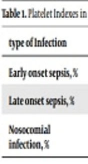
Thrombocytosis was seen in 15.2%, thrombocytopenia in 36.4%, elevated MPV in 25.6% and elevated PDW in 24.3% of included neonates. Thrombocytosis seen in 0% of early onset, 40% of late sepsis and 15.8% of nosocomial infections (P value 0.02) which was significant, thrombocytopenia existed in 33.3% of early, 40% of late sepsis and 50% of nosocomial infections (P value 0.52). Elevated MPV was seen in 85.7% of early, 69.2% of late sepsis and 73.5% of nosocomial infections (P value 0.16) and elevated PDW in 28.5% of early, 18.5% of late sepsis and 27.2% of nosocomial infections (P value 0.72) (Table 1). The ratio of platelet index for different germs in different types of neonatal sepsis was not estimated because of small number of cases in each group (Table 2).
| type of Infection | Thrombocytosis | P Value | Thrombocytopenia | P Value | High MPV | P Value | High PDW | P Value |
|---|---|---|---|---|---|---|---|---|
| Early onset sepsis, % | 0 | 0.02 | 33.3 | 0.52 | 85.7 | 0.16 | 28.5 | 0.72 |
| Late onset sepsis, % | 40 | 40 | 69.2 | 18.5 | ||||
| Nosocomial infection, % | 15.8 | 50 | 73.5 | 27.2 |
| Platelet Indexes | Early Onset Sepsis | Late Onset Sepsis | Nosocomial Infection | |||||||||
|---|---|---|---|---|---|---|---|---|---|---|---|---|
| G+ | G- | Fungal | P Value | G+ | G- | Fungal | P Value | G+ | G- | Fungal | P Value | |
| Thrombocytosis, % | 0 | 0 | 0 | UM | 29.5 | 17.6 | 0 | UM | 25 | 0 | 66.6 | 0.5 |
| Thrombocytopenia, % | 27.3 | 50 | 0 | UM | 11.8 | 41 | 0 | UM | 35 | 57.1 | 0 | 0.093 |
| High MPV, % | 9 | 33.3 | 0 | UM | 33.3 | 36.4 | 0 | UM | 15 | 36.4 | 33.3 | 0.89 |
| High PDW, % | 18 | 33.3 | 0 | UM | 0 | 45.5 | 0 | UM | 25 | 45.5 | 0 | 0.25 |
Abbreviation: UM, un measurable.
Thrombocytosis was seen in 34.2% of Gram-positive, 51.6 % of Gram-negative and 66.6% of fungal infections (P value = 0.26). Thrombocytopenia seen in 28.5% of Gram-positive, 16.6% of Gram-negative and 0% of fungal infections (P value = 0.62). Elevated MPV was seen in 19.5% of Gram-positive, 36% of Gram-negative and 33.3% of fungal infections (P value = 0.26), elevated PDW was in 15.2% of Gram-positive, 44% of Gram-negative and 0 % of fungal infections (P value = 0.026) (Table 3). The mean value of PDW in Gram-negative infections is not more than that of Gram-positive sepsis and fungal infections, i.e. 16.2%, 15.3 and 14.4% respectively (P value = 0.323).
| Types of Sepsis Agents | Thrombocytosis | P Value | Thrombocytopenia | P Value | High MPV | P Value | High PDW | P Value |
|---|---|---|---|---|---|---|---|---|
| Gram-negative sepsis, % | 51.6 | 0.26 | 16.6 | 0.62 | 36 | 0.26 | 44 | 0.26 |
| Gram-positive sepsis, % | 34.2 | 28.5 | 19.5 | 15.2 | ||||
| fungal infection, % | 66.6 | 0 | 33.3 | 0 |
5. Discussion
There are several hematological findings for early diagnosis of neonatal sepsis before availability of the blood culture results. Thrombocytopenia is one of these (3). Thrombocytopenia occurs in half of the cases with proven sepsis (10) and is used as a marker for screening of neonatal sepsis. It has been reported to be more common in association with some sepsis causing germs. This will be helpful to choose appropriate empirical therapy soon before availability of blood culture results. Ahmed et al. (11) reported to have seen thrombocytopenia more common in gram negative sepsis and that it had more prolonged duration in these cases than in Gram-positive sepsis. Some authors have even seen severe thrombocytopenia with Gram-negative sepsis (4). In addition, it correlated with the outcome of the patients especially in VLBW newborns (12). In a study Ahmad et al. (12) showed that thrombocytopenia was more common in fungal sepsis and Bhat et al. (13) reported high incidence of thrombocytopenia with Gram-negative and fungal infections. In addition prolonged thrombocytopenia, very low platelet levels, and need to platelet transfusion were poor prognostic factors. In a similar study by Alshorman Abdullah et al. thrombocytopenia was detected in 25% of infants with sepsis, but significant differences were not observed in Gram-positive and negative infections (14). All studies confirmed thrombocytopenia to be common in septic neonates, but can thrombocytopenia determine different types of neonatal sepsis germs, too?
Other platelet findings in diagnosis and predicting of sepsis are PDW and MPV (15). Elevated MPV (16) and PDW may be used as a marker for screening of sepsis. Choudhary et al. (17) reported that high MPV, high PDW and thrombocytopenia are more common in late onset than early onset sepsis. Although, Shaaban and Safwat (18) found MPV as a marker of early onset sepsis. Catal et al. (1) estimated that elevated MPV can be a diagnostic factor along with other findings and can even be utilized for following the course of the sepsis in neonates but, in line with our study, they didn’t find any correlation between elevated MPV and different kinds of sepsis germs. Akarsu et al. (19) also didn’t find any correlation between elevated MPV and PDW and different germs of sepsis, although we found that elevated PDW is more common in Gram-negative infections (P value = 0.026). Some authorss reported that septic patients with PDW levels greater than 18% have worse outcome (20). Ahmad and Waheed (3) did not accept this and introduced thrombocytopenia instead as the predictive factor in neonatal sepsis and found higher mortality rates in thrombocytopenic neonates.
5.1. Conclusions
Evaluating platelet indices in neonates with different sepsis germs we found that, although thrombocytosis may be a laboratory finding in septic neonates, it is not common in early onset infections (P value = 0.028). In addition, thrombocytopenia, elevated MPV or PDW can be laboratory findings for diagnosis of sepsis but are not specific for special types (namely early, late sepsis, nosocomial infection) of infection. Thrombocytosis, thrombocytopenia and elevated MPV are seen in Gram-positive, Gram-negative and fungal infections and they are not specific findings in determining different types of sepsis germs but PDW can be helpful because it was observed more common in Gram-negative infections in the present study (P value = 0.026), although the mean value of PDW in Gram-negative infections was not more than that in cases caused by other germs.
So, note that:
• In a young neonate with thrombocytosis sepsis is not the first thing to be considered because thrombocytosis is not common in early onset sepsis.
• In septic neonates pay attention to platelet indices, if PDW is over 17%, Gram-negative infections are more probable than Gram-positive and fungal infections. This is important for starting empiric therapy.
We admit that we had limitations in our study. The study was carried out in out-born hospital patients, so we had only few neonates with early onset sepsis. In addition, the number of studied neonates was limited. So, multicenter studies with larger sample size are necessary to come to more reliable conclusions.
