1. Background
Mechanical ventilation promotes alveolar derecruitment, atelectasis and increases physiological intrapulmonary shunt (1, 2). This can lead to impaired oxygen exchange. Positive end-expiratory pressure (PEEP) is used to counteract alveolar derecruitment and improve oxygen exchange. Literature data from human and animal studies show overall positive effect of PEEP on lung function (3-7). On the other hand, PEEP exerts negative hemodynamic effects. As distending pressure PEEP increases intrathoracic pressure, decreases venous return and cardiac output in normovolemic and hypovolemic patients (8). Depending on speed of titration and PEEP level these effects can be more or less clinically apparent (4, 7, 9). Slow PEEP titration up to 20 cmH2O during 15 minutes achieved recruitment, improved gas exchange and decreased intrapulmonary shunt without circulatory depression (4, 7).
2. Objectives
Based on data obtained from experimental model, we hypothesized that intraoperative preventive PEEP titration up to 11 cmH2O during 5 minutes can improve lung function without causing hypotension and bradycardia in preschool children with healthy lungs during general anesthesia for non-cardiothoracic surgery.
3. Methods
This was a prospective, randomized, two arms, unblinded, clinical trial in children undergoing general anesthesia for non-cardiothoracic surgery. Study was conducted at Institute for Mother and Child Health Care, Belgrade, Serbia between January 2017 and June 2017. Study was approved by Ethic Committee of Institute for Mother and Child Health Care (No 8/30, 2017.) and performed in accordance to criteria set by Declaration of Helsinki. All parents/legal guardians were informed about study protocol and children were enrolled only if parents/legal guardians gave informed consent. Study included 70 preschool children scheduled for general anesthesia for non-cardiothoracic surgery. They were randomly assigned into two groups; interventional (I group; n = 35) and control group (C group; n = 35). Inclusion criteria were: age 3 - 7 years, ASA I and II. Exclusion criteria were: current or recent (up to 4 weeks) upper airway infection, present cardiovascular and present respiratory comorbidity. All children were assessed preoperatively one day before surgery. Premedication and general anesthesia were the same in both groups. Children were premedicated with midazolam 0.1 mg/kg iv. Anesthesia induction was performed by thiopental 5 mg/kg iv, fentanyl 3 mcg/kg iv, sevoflurane 1 vol%, O2:air mixture 35%:65% and muscle paralysis with rocuronium 1 mg/kg. Trachea was intubated with proper sized tube. Anesthesia was maintained using sevoflurane 1.5 vol%/2 vol%, fentanyl 2 mcg/kg and rocuronium. Intravascular volume was maintained with Ringer lactate solution using 4:2:1 rule plus intraoperative losses (10). Both groups had the same initial ventilator settings (DatexOhmeda, Avance CS2, GE anesthesia machine) except for 20 minutes before the end of surgery. Initial ventilator settings were: PCV, Pinsp adjusted to achieve tidal volume (Vt) 6 - 8 mL/kg, respiratory rate adjusted to achieve end expiratory carbon dioxide (EtCO2) 35 mmHg - 45 mmHg, PEEP 3 cmH2O, air:oxygen 65%:35%. After intubation arterial line was placed and invasive monitoring of blood pressure set up. Monitoring during anesthesia was: hemoglobin oxygen saturation (SpO2), capnography and capnometry (EtCO2), invasive blood pressure (IBP), heart rate (HR), inspiratory pressure (Pins), peak airway pressure (Ppeak), mean airway pressure (Pmean) and dynamic lung compliance (Cdyn). We used monitoring incorporated in anesthesia machine (DatexOhmeda, Avance CS2, GE anesthesia machine). Arterial blood samples were analyzed immediately after obtaining the sample (ABL 90 Radiometer, Copenhagen, Denmark).
3.1. Allocation
Patients were randomly assigned in a 1:1 ratio into either interventional or control group. We used sealed envelope technique to allocate participants in groups. The box containing 70 envelopes with allocation instruction (I: interventional group; C: control group) was used to allocate patients randomly. Before first sampling of arterial blood anesthetist in charge pulled the envelope from the box and allocated patient to experimental or control group.
3.2. Intervention
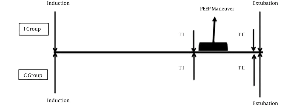
Twenty minutes before the end of anesthesia and surgery PEEP was increased in interventional group by 2 cmH2 on every 5 breaths up to 11 cmH2. Ventilation was maintained on PEEP 11 cmH2 for 2 minutes. Then, PEEP was decreased by 2 cmH2 for every 5 breaths to 5 cmH2O. Ventilation with PEEP of 5 cmH2O was unchanged until extubation in order to keep re-aerated alveoli open. Total time to perform the PEEP maneuver was 5 minutes. Control group was ventilated the whole time with baseline PEEP setting (3 cmH2O). PEEP 3 cmH2O is lowest PEEP level used in clinical practice and is associated with derecrutiment. As a part of study design, we kept it constant in control group to be able to differences in PaO2/FiO2 index and P(A-a)O2 gradient between two groups. Arterial blood was collected in the same two time points in both groups (Figure 1): TIME I- before PEEP titration, which was 20 minutes before the end of surgery and TIME II before extubation. Criteria for aborting the procedure were hypotension, bradycardia, desaturation below SpO2 94% and sudden rise in Ppeak. Based on previous published data hypotension was defined as SBP < 70 mmHg and MAP < 55 mmHg (11, 12). Bradycardia was defined as heart rate ≤ 60/min.
3.3. Study Outcomes
To evaluate oxygen exchange we used PaO2/FiO2 index and to evaluate changes in intrapulmonary shunt we used P(A-a)O2 gradient. Hemodynamic changes during PEEP titration were evaluated using MAP, SBP, and HR.
3.4. Statistical Analysis
Sample size was calculated using available literature data on PEEP effect on oxygen exchange (PaO2/FiO2) (13) with α = 0.05 and study power of 80%. The number needed was 35 participants per group. All data were analyzed using IBM-SPSS 20.0 (IBM Corp, New York, USA) statistical software. Results are presented as mean and standard deviation with CI 95% or proportions. To evaluate size effect PEEP titration produced, we calculated delta (Δ) and compared it between groups. To test the difference between groups Student’s t-test for independent samples and Mann-Whitney U test were used depending on data distribution. To test the difference within intervention group before and after PEEP titration paired sample t-test was used. To test the difference in SBP, MAP and HR in interventional group on different PEEP levels repeated measurements ANOVA was used. All tests were 2-tailed and P < 0.05 was considered statistically significant.
4. Results
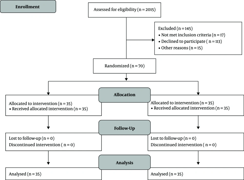
Between January and June 2017, 486 children were scheduled for surgery, 271 were younger than 3 and older than 7 years, 113 refused to participate, 10 had upper respiratory tract infection, 7 had present chronic heart and lung conditions and 15 didn’t show up at all on the day of surgery. All enrolled participants between January 2017 and June 2017 completed the study and were analyzed for outcomes (Figure 2). Baseline data of participants are presented in Table 1.
| Group | P Value | ||
|---|---|---|---|
| Interventional | Control | ||
| Age, y | 4.9 ± 1.5 | 5.1 ± 1.6 | 0.728b |
| Body weight, kg | 18.8 ± 5.2 | 19.4 ± 5.3 | 0.682b |
| Gender (male) | 19 (54) | 25 (71) | 0.138c |
| Comorbidity present | 14 (40) | 6 (17) | 0.034c |
| Type of surgery | 0.328c | ||
| Abdominal | 12 (34) | 10 (29) | |
| Urology | 7 (20) | 14 (40) | |
| Orthopedics | 7 (20) | 5 (14) | |
| Reconstructive surgery | 9 (26) | 6 (17) | |
| Procedure duration > 60 min | 20 (57) | 22 (63) | 0.238c |
aResults are presented as No. (%) or mean ± SD
bt-test
cχ2 test
4.1. Main Outcomes
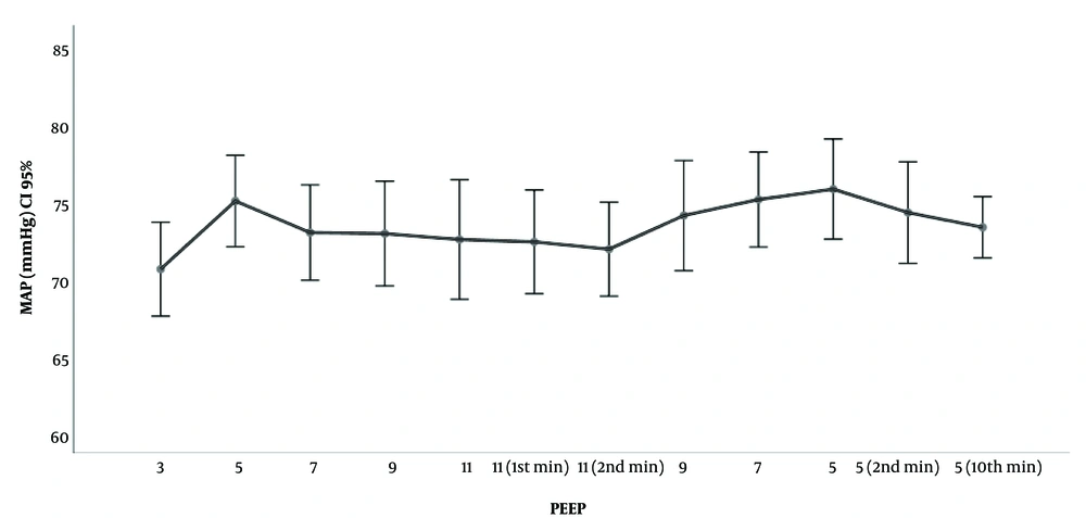
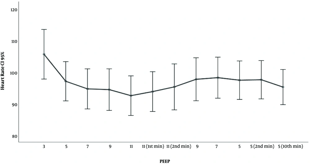
Differences in P(A-a)O2 and PaO2/FiO2 between and within groups in TIME I and II are presented in Table 2. There was significant difference between groups in TI (before PEEP maneuver) in both parameters. Independent t-test for delta between groups (Δ TI - TII) revealed that there was a statistically significant difference in magnitude of change in P(A-a)O2 and PaO2/FiO2 between groups which means that intervention had effect. Comparison within I group showed that there was significant difference in both parameters before and after PEEP titration (TI vs. TII). Both parameters improved after PEEP maneuver. Table 3 displays changes in SBP, DBP, MAP and HR during PEEP titration in interventional group. There were no episodes of hypotension or bradycardia during PEEP titration, all subjects remained hemodynamically stabile. Figure 3 displays changes in MAP on different PEEP levels. Figure 4 displays HR on different PEEP levels.
| Group | P Value | ||
|---|---|---|---|
| Interventional | Control | ||
| PaO2/FiO2 | |||
| TI (Before) | 522.0 (508.2 - 535.9) | 554.7 (547.3 - 562.2) | < 0.001b |
| TII (After) | 552.3 (545.2 - 559.4) | 554.2 (541.6 - 566.8) | 0.784b |
| P value (TI - TII) | < 0.001c | 0.870c | |
| Δ PaO2/FiO2 | -30.3 (-38.7 - -21.8) | 0.52 (-5.87 - 6.91) | < 0.001b |
| P(A-a)O2 | |||
| TI (Before) | 20.1 (15.5 - 24.7) | 8.7 (6.5 - 10.9) | < 0.001d |
| TII (After) | 13.3 (10.8 - 15.9) | 9.7 (6.1 - 13.4) | 0.230d |
| P value (TI - TII) | < 0.001c | 0.350c | |
| Δ P(A-a)O2 | 6.7 (4.1 - 9.4) | -1.0 (-3.2 - 1.1) | < 0.001b |
Abbreviation: Δ, difference (TI - TII)
aResults are presented as mean and 95% CI.
bIndependent samples t-test
cPaired samples t-test
dMann-Whitney U test
| Measurement | MAP | Systolic BP | Heart Rate |
|---|---|---|---|
| PEEP 3 vs. 5 | 70.6 (67.5 - 73.6) | 100 (96.3 - 103.7) | 105.6 (97.7 - 113.4) |
| PEEP 5 vs. 7 | 75 (72 - 77.9)b | 102.3 (99.3 - 105.3) | 97 (90.8 - 103.2)b |
| PEEP 7 vs. 9 | 72.9 (69.8 - 76)b | 100.5 (96.9 - 104.2)b | 94.6 (88.3 - 101)b |
| PEEP 9 vs. 11 | 72.9 (69.5 - 76.3) | 99.1 (94.7 - 103.6)b | 94.4 (87.8 - 101) |
| PEEP 11 vs. 11 in 1st minute | 72.5 (68.6 - 76.4) | 98.6 (93.8 - 103.5) | 92.5 (86.2 - 98.7)b |
| PEEP 11 in 1st minute vs. 11 in 2nd minute | 72.3 (69 - 75.7) | 97.5 (93.9 - 101.2) | 93.8 (87.5 - 100.1) |
| PEEP 11 vs. 9 | 71.8 (68.8 - 74.9) | 96.9 (93.5 - 100.2) | 95.3 (88 - 102.5) |
| PEEP 9 vs. 7 | 74 (70.5 - 77.6)b | 100.2 (96.3 - 104.1)b | 97.7 (90.9 - 104.4)b |
| PEEP 7 vs. 5 | 75.1 (72 - 78.1)b | 99.4 (96 - 102.8) | 98.2 (91.7 - 104.7) |
| PEEP 5 vs. 5 in 2nd minute | 75.7 (72.5 - 79) | 99.6 (95.9 - 103.3) | 97.4 (91.3 - 103.5) |
| PEEP 5 in 2nd minute vs. PEEP 5 in 10th minute | 74.2 (70.9 - 77.5) | 96.3 (89.9 - 102.6) | 97.5 (91.5 - 103.6) |
| PEEP 5 in 10th minute vs. PEEP 5 before extubation | 73.3 (71.3 - 75.3) | 97.7 (95.1 - 100.4) | 95.2 (89.6 - 100.8)b |
aResults are presented as mean and CI 95%.
bRepeated measurements ANOVA; significant difference (P < 0.05) between that measurement and one before.
5. Discussion
For more than two decades PEEP was used to improve lung function on mechanically ventilated patients. Proposed values that should prevent perioperative deterioration of lung function are 7 - 15 cmH2O (14). Most of data regarding intraoperative preventive use of PEEP was pooled from adult studies and extrapolated to pediatric population. The aim of this study was to evaluate slow stepwise up and down PEEP titration effect from 5 - 11 cmH2O on intrapulmonary shunt, oxygenation and hemodynamics in preschool children in general anesthesia with muscle paralysis for non-cardiothoracic surgery. As markers of lung function we used alveolar-arterial gradient (P(A-a)O2) and oxygen exchange index (PaO2/FiO2). P(A-a)O2 is simple and reliable indicator of physiological shunt alterations in cardiorespiratory stable patients on constant FiO2. PaO2/FiO2 has been widely used to evaluate oxygen exchange and it is modified with PEEP and respiratory condition of the patient (15). Due to simplicity of bedside calculation of both parameters, absence of cardiorespiratory comorbidity in study population and ventilation with constant FiO2, we used those parameters to evaluate PEEP titration effect. In our study effect produced by PEEP maneuver was beneficial for patients in interventional group. P(A-a)O2 decreased within interventional group and compared to control. Improvement in P(A-a)O2 was a consequence of decrease in intrapulmonary shunt caused by applied PEEP maneuver. Similar results were presented in studies on adult patients and laboratory animals (4, 16). Ambrosio et al. observed decrease in P(A-a)O2 with increasing PEEP from 5 - 20 cmH2O (4). In pediatric patients, titration of PEEP decreased P(A-a)O2 1 hour after laparscopic surgery compered to children ventilated without PEEP (17). Positive pressure ventilation with PEEP 10 cmH2O also caused significant decrease of P(A-a)O2 in patients after coronary artery bypass grafting (16). The difference of mentioned studies on humans regarding our study was the study sample. We investigated effect on cardiorespiratory healthy preschool children. Nevertheless, in both studies authors mitigated conditions that could limit the use of P(A-a)O2 gradient. All samples in their study were obtained when patients were cardiorespiratory stable and on constant FiO2, similar to conditions in our study. Therefore we believe this is comparable to our study. As marker of oxygen exchange we investigated changes in PaO2/FiO2 index since it is influenced by PEEP. PEEP titration improved oxygen exchange within interventional group and compared to control group. Improvement in PaO2/FiO2 index was also demonstrated on isolated experimental lung when PEEP 5 and 10 was applied (5). In pediatric patients with ARDS, stepwise down PEEP titration from 25 cmH2O to “open lung PEEP” led to improvement in PaO2/FiO2 index (13). These results are similar to ours. On the other hand, we cannot ignore that sometimes PEEP application fails to improve PaO2/FiO2 index. In experimental model application of PEEP 5 - 7 failed to improve PaO2/FiO2 index but it did slow down raise of elastance that was used as marker of derecruitment (18). In pediatric patients after cardiopulmonary bypass (CBP) adding PEEP 8 cmH2O didn’t cause improvement in PaO2/FiO2 index compared to PEEP 0 (19). Our study group consisted of children with healthy lungs and atelectasis developed was consequence of mechanical ventilation only. In contrast, study population in the mentioned study was patients with injured lungs. Adding PEEP up to 11 cmH2O in our study was enough to improve oxygen exchange in healthy lungs but adding PEEP 8 wasn’t enough to improve oxygenation in injured lungs. Indeed, PEEP modifies PaO2/FiO2 index and the same index value can be obtained under different respiratory conditions and PEEP levels (20). Oxygenation changes when PEEP is applied better correlate with aerated lung parts than with poorly aerated or collapsed areas (21). So, the effect that PEEP produces regarding oxygenation depends not only on PEEP level but also on initial lung condition. Therefore, when evaluating lung function, especially in terms of lung recruitment, oxygenation exchange should not be used as the only marker.
Application of PEEP can have negative hemodynamic effect, regardless of its beneficial effect on lung function. To estimate its impact on hemodynamics we used MAP, SBP and HR. Besides the fact that they are easy to obtain, those parameters are advised as obligatory monitoring in every day practice. PEEP increases intrathoracic pressure. As a consequence, venous return decreases, right ventricular (RV) afterload increases, ventricular compliance and contractility decrease (8, 22). Increase in RV afterload leads to left shifting of septum, decrease in left ventricular end-diastolic diameter and left ventricular end-diastolic area (23). In hypovolemic and normovolemic patients, PEEP reduces cardiac output due to decreased venous return (22). Above mentioned changes in cardiac performance may or may not be clinically significant (23). During our PEEP trial there was not a single case of hypotension or/and bradycardia, as invasive blood pressure and ECG monitoring showed. All patients remained hemodynamically stable on each PEEP level. This is in accordance with previously published data. In systematic review Algera et al. analyzed hemodynamic effect of low PEEP (med 0, IQR 0 - 5) vs. high PEEP (med 10, IQR 3 - 20) in experimental animals with uninjured lungs. In 2 trials, application of high PEEP didn’t cause hypotension and bradycardia, 1 trial documented lower blood pressure (24). In study by Ambrosio et al. during 15 minutes PEEP titration up to 20 cmH2O cardiac output, cardiac index, systemic and pulmonary vascular resistance were significantly lower at PEEP of 20 cmH2O but this wasn’t clinically significant. Animals in both groups remained hemodynamically stable with no differences in HR and MAP within and between groups (4). Although this is experimental study, it is similar to ours in some points: uninjured lung, level of applied PEEP and the slow PEEP titration. Slow titration of PEEP up to 15, when compared to fast PEEP titration up to 10 and 20, causes less circulatory depression (7). In clinical setting, in hemodynamically stable children after repair of congenital cardiac defect, PEEP 4 - 12 cmH2O caused fall of cardiac index, but this reduction wasn’t clinically significant (25, 26). Several studies used MAP and HR to evaluate hemodynamic effect of PEEP in hemodynamically stable patients. Although MAP and HR can be reduced up to 5% with PEEP 10, this was not recognized as bradycardia or hypotension (27, 28). In their review article, Berger and Takala discussed impact of PEEP on hemodynamics, more precisely, on mean systemic filling pressure (MSFP) and venous return as major factors whose changes, induced by PEEP, influence CO. They pointed out controversy in experimental research, where some studies document unchanged venous return and CO or decreased CO with PEEP 10 - 20 cmH2O (22). Few years previously authors investigated impact of lower PEEP levels 5 - 10 cmH2O with stable airway pressure and found no effect on MSFP, venous return or resistance to venous return (29). None of CO determinants (and hemodynamics) was changed. As authors speculated, reason for these different results is probably a consequence of ventilator strategy with small pressures and tidal volumes. This is similar to our study since we used protective ventilation strategy, as mentioned earlier.
Limitation to our study is its open label design. We are aware that if the anesthesiologist who performed PEEP titration was not involved in data collection, and the observer who collected the data was unaware of the study groups, the results would be more convincing.
5.1. Conclusions
Our investigation confirmed hypothesis of study that preventive slow stepwise up and down PEEP titration form of 5 - 11 cmH2O improves oxygen exchange and decreases intrapulmonary shunt without compromising hemodynamics in preschool children in general anesthesia. Applied PEEP levels are sufficient to improve lung function and safe to use in everyday practice in children without cardiorespiratory comorbidity.


