Chemistry
Melting points were determined with a MEL-TEMP model 1202D. FT-IR spectra were recorded with a Bruker Tensor 27 spectrometer as KBR disks. The MS spectra were recorded using Agilent 7000-3Q mass spectrometer at an electron impact mode with an ionization voltage of 70 eV. 1H-NMR and 13C-NMR spectra were recorded with Bruker Spectrospin Avance 300 and 75 MHz spectrometers. CDCl3 and DMSO were used as solvents, while TMS was utilized as an internal standard for chemical characterizations. All chemical shifts were reported as δ (ppm) and coupling constants (J) are given in Hz. Thin-layer chromatography was performed with glass-backed plates (20 × 20 cm2, 500 μ) using silica gel (Merk Kieselgel 60 HF254, Art. 7739). The chemical reagents used for the synthesis were purchased from Merck and Sigma-Aldrich.
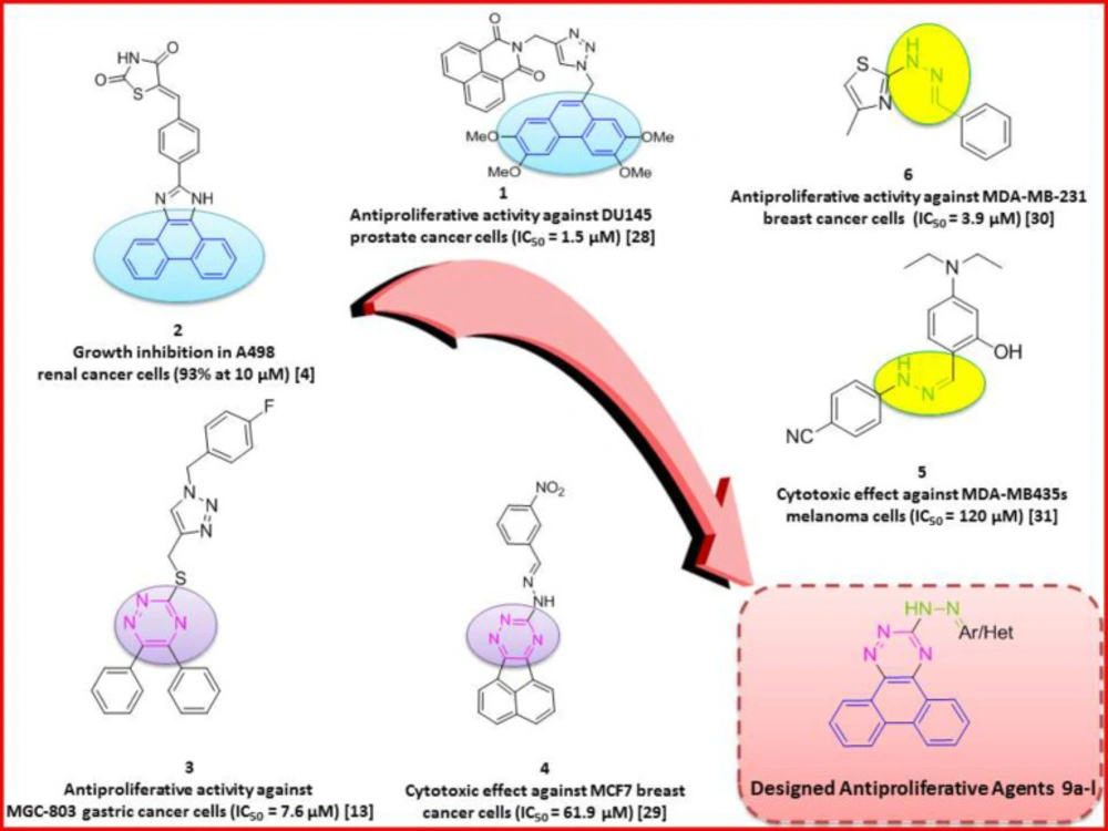
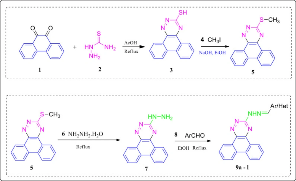
Synthesis of phenanthro(9,10-e)(1,2,4)triazine 3-thiol (3)
To a solution of Phenanthrene 9, 10 dione (1) (10 mmol) and acetic acid (40 mL), thiosemicarbazide (2) (20 mmol) was added and refluxed for 4 h. The reaction progression was monitored using TLC. The precipitated product was filtered and washed with enough amounts of cold water and ethanol mixture. Orange solid; Yield: 95%; mp: 122-124 °C; 1H-NMR (300 MHz, CDCl3): δH 14.65 ppm (s, 1H, NH), 8.55 (m, 2H, Ar-H), 8.15 (m, 5H, Ar-H), 7.55 (m, 9H, Ar-H), 6.72 (s, 1H, SH); IR (KBr, cm -1) υ max: 3413 (-NH-), 3145 (ArCH), 1596 (Ar N=N), 1500 (-C=N-), 1481 (Ar C=C); MS (EI) m/z (%): 263 (M+, 40), 235 (89), 176 (100).
Synthesis of 3-(methylthio)phenanthro(9, 10-e)(1,2,4)triazine (5)
Phenanthrotriazine thiol (3) (10 mmol) was added to the mixture of NaOH (460 mg, 11.5 mmol) in warm ethanol (50 mL) then the excess amount of methyl iodide (4) (15 mmol, 1 mL) was added after the temperature was decreased. The reaction mixture was stirred at room temperature for 3 h. The reaction progress was monitored using TLC. The resulting yellow suspension was filtered through a filtration paper and then washed with enough amounts of cold ethanol. Yellow solid; Yield: 78%; mp: 157-160 °C; 1H-NMR (300 MHz, CDCl3): δH 14.65 ppm (s, 1H, NH), 8.55 (m, 2H, Ar H), 8.15 (m, 5H,Ar H), 7.55 (m, 9H, Ar H), 6.72 (s, 1H, SH); IR (KBr, cm -1) υ max:2927 (ArCH), 1605 (Ar N=N), 1503 (-C=N-), 1482 (Ar C=C); MS (EI) m/z (%): 263 (M+, 40), 235 (89), 176 (100).
3-(2-benzylidene hydrazinyl)phenanthro(9, 10-e)(1,2,4)triazine(7)
Methyl thiol (5) (2 mmol) was dissolved in 2-propanol and refluxed. Excess amounts of hydrazine hydrate (6) (5 mL) were added and the mixture was refluxed overnight and the reaction progression was monitored using TLC. The resulting yellow suspension was filtered through a filtration paper and then washed with enough amounts of cold water.
Yellow solid; Yield: 77%; IR (KBr, cm -1) υ max: 3444 (-NH-), 3039 (ArCH), 1556 (Ar N=N), 1523 (-C=N-), 1447 (Ar C=C); MS (EI) m/z (%): 261 (M+, 57), 286 (79), 190 (100).
General procedure for the synthesis of hydrazones (9a-l)
Hydrazine (7) (0.5 mmol) was dissolved in warm ethanol (20 mL). The appropriate aryl aldehyde (8) (0.5 mmol) was added and the reaction mixture was refluxed for 3 h and the reaction progression was monitored using TLC. The resulting yellow suspension was filtered through a filtration paper and then washed with enough amounts of cold ethanol.
3-(2-benzylidenehydrazinyl)phenanthro [9,10-e][1,2,4]triazine (9a)
Yellow solid; Yield: 73%; mp: 286-290 °C; 1H-NMR (300 MHz, DMSO): δH9.14-9.17 (m, 2H, Ar H),8.83-8.86 (m, 2H, Ar H), 8.40 (s, 1H, =CH), 7.96-7.99 (d, J = 9 Hz, 1H, Ar H),7.81-7.89 (m, 5H, Ar H), 7.44-7.54 (m, 3H, Ar H); IR (KBr, cm -1) υ max: 3419 (-NH-), 2983 (ArCH), 1588 (Ar N=N), 1529 (-C=N-), 1447 (Ar C=C); MS (EI) m/z (%): 349 (M+, 24), 272 (28), 218 (100), 190 (96).
3-(2-(2-methoxybenzylidene)hydrazinyl)phenanthro[9,10-e][1,2,4]triazine (9b)
Yellow solid; Yield: 76%; mp: 252-254 °C; 1H-NMR (300 MHz, DMSO): δH9.12-9.16 (m, 2H, Ar H),8.77-8.82 (m, 2H, Ar H), 8.75 (s, 1H, =CH),8.04-8.07 (d, J = 9Hz, 1H, Ar H),7.93-7.97 (m, 1H, Ar H), 7.78-7.83 (m, 3H, Ar H), 7.40-7.45 (t, J = 9Hz, 1H, ArH), 7.07-7.15 (m, 2H, Ar H), 3.90 (s, 3H, CH3); IR (KBr, cm -1) υ max: 3445 (-NH-), 2977 (ArCH), 1584 (Ar N=N), 1522 (-C=N-), 1447 (Ar C=C); MS (EI) m/z (%): 379 (M+,29), 272 (14), 246 (37), 218 (100), 190 (76).
3-(2-(4-methoxybenzylidene)hydrazinyl)phenanthro[9,10-e][1,2,4]triazine (9c)
Yellow solid; Yield: 76%; mp: 288-292 °C; 1H-NMR (300 MHz, DMSO): δH9.14-9.16 (m, 2H, Ar H),8.83-8.85 (m, 2H, Ar H), 8.34 (s, 1H, =CH),7.94-7.99 (m, 1H, Ar H),7.76-7.83 (m, 5H, Ar H), 7.06-7.08 (d, J = 6 Hz, 2H, Ar H), 3.83 (s, 3H, CH3); IR (KBr, cm -1) υ max: 3445 (-NH-), 2989 (ArCH), 1611 (Ar N=N), 1526 (-C=N-), 1430(Ar C=C); MS (EI) m/z (%): 379 (M+, 12), 245 (27), 218 (100), 190 (55).
2-((2-(phenanthro[9,10-e][1,2,4]triazin-3-yl)hydrazono)methyl)phenol (9d)
Yellow solid; Yield: 61%; mp: 328-332 °C; 1H-NMR (300 MHz, DMSO): δH9.09-9.15 (m, 2H, Ar H),8.84-8.87 (m, 2H, Ar H), 8.56 (s, 1H, =CH),7.97-8.02 (t, J = 9 Hz, 1H, Ar H),7.81-7.91 (m, 3H, Ar H), 7.51-7.57 (d, J = 6 Hz, 1H, Ar H), 7.30-7.36 (t, J = 7.5 Hz, 1H, Ar H), 6.97-7.05 (m, 2H, Ar H); IR (KBr, cm -1) υ max: 2966 (ArCH), 1585 (Ar N=N), 1525 (-C=N-), 1430 (Ar C=C); MS (EI) m/z (%): 365 (M+, 45), 218 (100), 190 (92).
3-((2-(phenanthro[9,10-e][1,2,4]triazin-3-yl)hydrazono)methyl)phenol (9e)
Yellow solid; Yield: 64%; mp: 219-223 °C; 1H-NMR (300 MHz, DMSO): δH9.06-9.14 (m, 3H, Ar H),8.78 (s, 1H, =CH), 8.71-8.76 (m, 2H, Ar H),7.90-7.94 (t, J = 7.2 Hz, 2H, Ar H),7.72-7.80 (m, 5H, Ar H), 4.63 (s, 1H, NH); IR (KBr, cm -1) υ max: 3445 (-NH-), 3047 (ArCH), 1556 (Ar N=N), ~1530 (-C=N-), 1447 (Ar C=C); MS (EI) m/z (%): 365 (M+, 16), 286 (27), 272 (31), 218 (85), 190 (100).
3-((2-(phenanthro[9,10-e][1,2,4]triazin-3-yl)hydrazono)methyl)benzene-1,2-diol (9f)
Yellow solid; Yield: 57%; mp: 329-333 °C; 1H-NMR (300 MHz, DMSO): δH9.11-9.19 (m, 2H, Ar H),8.80-8.87 (m, 2H, Ar H), 8.52 (s, 1H, =CH),7.98-8.03 (t, J = 7.2 Hz, 1H, Ar H),7.81-7.90 (m, 3H, Ar H), 6.97-7.00 (d, J = 6 Hz, 1H, Ar H), 6.88-6.90 (d, J = 6 Hz, 1H, Ar H), 6.76-6.81 (t, J = 9 Hz, 1H, Ar H); IR (KBr, cm -1) υ max: 3495 (-NH-), 2851 (ArCH), 1596 (Ar N=N), 1525 (-C=N-), 1470 (Ar C=C); MS (EI) m/z (%): 381 (M+, 80), 364 (21), 218 (99), 190 (100).
3-(2-(2-nitrobenzylidene)hydrazinyl)phenanthro[9,10-e][1,2,4]triazine (9g)
Yellow solid; Yield: 70%; mp: 225-229 °C; 1H-NMR (300 MHz, DMSO): δH8.98-9.15 (m, 3H, Ar H),8.75 (s, 1H, =CH), 8.68-8.72 (m, 2H, Ar H),7.87-7.97 (m, 2H, Ar H),7.72-7.84 (m, 5H, Ar H), 4.61 (s, 1H, NH); IR (KBr, cm -1) υ max: 3445 (-NH-), 3038 (ArCH), 1643 (-NO2), 1556 (Ar N=N), 1524 (-C=N-), 1447 (Ar C=C); MS (EI) m/z (%): 395 (M+, <10), 261 (100), 233 (18), 190 (82).
3-(2-(3-nitrobenzylidene)hydrazinyl)phenanthro[9,10-e][1,2,4]triazine (9h)
Yellow solid; Yield: 57%; mp: 216-219 °C; 1H-NMR (300 MHz, DMSO): δH9.05-9.19 (m, 3H, Ar H),8.77 (s, 1H, =CH), 8.70-8.74 (m, 2H, Ar H),7.86-7.97 (t, J = 6.9 Hz, 2H, Ar H),7.74-7.83 (m, 5H, Ar H), 4.62 (s, 1H, NH); IR (KBr, cm -1) υ max: 3444 (-NH-), 3038 (ArCH), 1644 (NO2), 1556 (Ar N=N), 1523 (-C=N-), 1447 (Ar C=C); MS (EI) m/z (%): 261 (94), 190 (100), 175 (44), 205(21), 233(17).
3-(2-(4-nitrobenzylidene)hydrazinyl)phenanthro[9,10-e][1,2,4]triazine (9i)
Yellow solid; Yield: 58%; mp: 240-244 °C; 1H-NMR (300 MHz, DMSO): δH9.05-9.12 (m, 3H, Ar H),8.77 (s, 1H, =CH), 8.71-8.74 (m, 2H, Ar H), 7.89-7.94 (t, J = 7.2 Hz, 2H, Ar H),7.74-7.79 (m, 5H, Ar H), 4.63 (s,1H, NH); IR (KBr, cm -1) υ max: 3444 (-NH-), 3046 (ArCH), 1556 (Ar N=N), 1523 (-C=N-), 1447 (Ar C=C); MS (EI) m/z (%): 394 (M+, 25), 272 (34), 218 (79), 190 (100).
4-((2-(phenanthro[9,10-e][1,2,4]triazin-3-yl)hydrazono)methyl)benzonitrile (9j)
Yellow solid; Yield: 54%; mp: 231-234 °C; 1H-NMR (300 MHz, DMSO): δH9.03-9.11 (m, 3H, Ar H),8.76 (s, 1H, =CH), 8.71-8.75 (m, 2H, Ar H),7.87-7.93 (t, J = 9Hz, 2H, Ar H),7.72-7.78 (m, 5H, Ar H), 4.63 (s, 1H, NH); IR (KBr, cm -1) υ max: 3444 (-NH-), 3035 (ArCH), 2226 (CN), 1556 (Ar N=N), 1523 (-C=N-), 1447 (Ar C=C); MS (EI) m/z (%): 374 (M+, 28), 272 (35), 218 (81), 190 (100).
3-(2-((1H-pyrrol-2-yl)methylene)hydr -zinyl)phenanthro[9,10-e][1,2,4]triazine (9k)
Yellow solid; Yield: 63%; mp: 210-214 °C; 1H-NMR (300 MHz, DMSO): δH9.02-9.12 (m, 3H, Ar H),8.76 (s, 1H, =CH), 8.71-8.73 (m, 2H, Ar H),7.88-7.96 (t, J = 6.9 Hz, 1H, Ar H),7.74-7.85 (m, 5H, Ar H); IR (KBr, cm -1) υ max:3439 (-NH-), 2917 (ArCH), 1556 (Ar N=N), 1524 (-C=N-), 1447 (ArC=C); MS (EI) m/z (%): 338 (M+, <10), 261 (100), 190 (84), 176 (38).
3-(2-(thiophene-2-ylmethylene)hydraz-inyl)phenanthro[9,10-e][1,2,4]triazine (9l)
Yellow solid; Yield: 40%; mp: 228-231 °C; 1H-NMR (300 MHz, DMSO): δH9.07-9.11 (m, 3H, Ar H),8.75 (s, 1H, =CH), 8.66-8.73 (m, 2H, Ar H),7.87-7.92 (t, J = 8.1 Hz, 1H, Ar H),7.67-7.83 (m, 5H, Ar H), 4.61 (s, 1H, NH); IR (KBr, cm -1) υ max: 3445 (-NH-), 1556 (Ar N=N), 1524 (-C=N-), 1447 (Ar C=C); MS (EI) m/z (%): 355 (M+, 14), 246 (31), 218 (100), 190 (81).
Evaluation of the antiproliferative effect
Fetal bovine serum (FBS), phosphate-buffered saline (PBS), RPMI-1640, and trypsin were obtained from Biosera (Ringmer, UK). Penicillin/streptomycin and MTT (3-(4,5-dimethylthiazol-2-yl)-2,5-diphenyltetrazolium bromide) were from Sigma-Aldrich (Saint Louis, MO, USA) and Invitrogen (San Diego, CA, USA), respectively. Dimethyl sulfoxide and doxorubicin were purchased from Merck (Darmstadt, Germany) and EBEWE Pharma (Unterach, Austria), respectively. The cells were acquired from Iranian Biological Resource Center, Tehran, Iran (HT-29 (human colorectal adenocarcinoma), MCF-7 (human breast adenocarcinoma) and Vero (African green monkey kidney) cells) or National Cell Bank of Iran, Pasteur Institute, Tehran, Iran (MOLT-4 (lymphoblastic leukemia) cells).
MTT reduction assay was employed to assess the viability of cancer cells after being treated with synthesized derivatives as previously described (
34). The cells were plated in flat-bottom 96-well microplates, at densities of 3 × 10
4 cells/mL (100 μL in each well) and then incubated overnight at 37 °C. Three to four different concentrations of synthesized derivatives (in the range of 1 to 100 μM) were placed in triplicate wells and the plates were incubated for 72 h at 37 °C. The concentration of DMSO did not exceed 0.25% in each well. At the end of the incubation time, 80 μL of the media in each well was replaced with the same amount of growth medium without phenol red containing 0.5 mg/mL of MTT. Cells were further incubated at 37 °C for 4 h and then 80 μL of the solution of each well was removed. The addition of DMSO (200μL) to each well solubilized the formazan crystals formed inside the viable cells after 1-hour incubation followed by 30-minute shaking. The optical absorbance of the final solution was determined at 570 nm using a Bio-Tek multimode plate reader (Synergy HTX). The cell viability was calculated compared to untreated control cells by comparison of absorbance measurements. CurveExpert software (version 1.34 for Windows) was used to determine the IC
50 value for each compound. To confirm the results, every experiment was repeated 3 to 5 times.
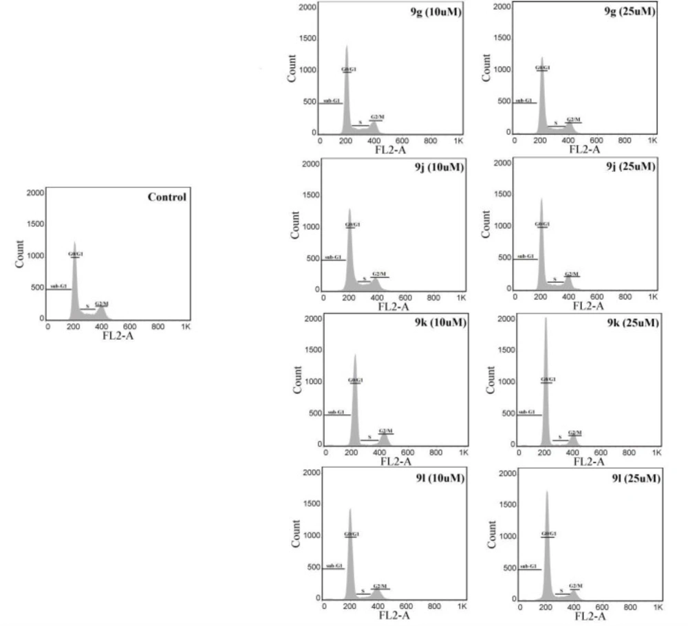
Cell Cycle Analysis
Alterations of the cell cycle were determined by the propidium iodide (PI)-RNase flow cytometric method (
35). MCF-7 cells were used to determine the percentage of cells in each phase of the cell cycle. In 6-well plates, 2 × 10
5 cells were seeded in each well and incubated at 37 °C for 48 h. Afterward, they were treated for 48 h with derivatives
9g,
9j,
9k and
9l at 10 and 25 μM. The cells were then collected after trypsinization, washed twice with PBS, and fixed by 5 mL ethanol 70% for at least 24 h at -20 °C. After fixation, MCF-7 cells were washed with PBS two times and stained with PI 20 µg/mL and RNase 200 µg/mL for 30 min in the dark at room temperature. A FACS Calibur flow cytometer (BD Biosciences) was used for the assays and 20,000 events were analyzed. The number of cells distributed in sub-G1, G0/G1, S, and G2/M were determined by CellQuest software (BD, USA).
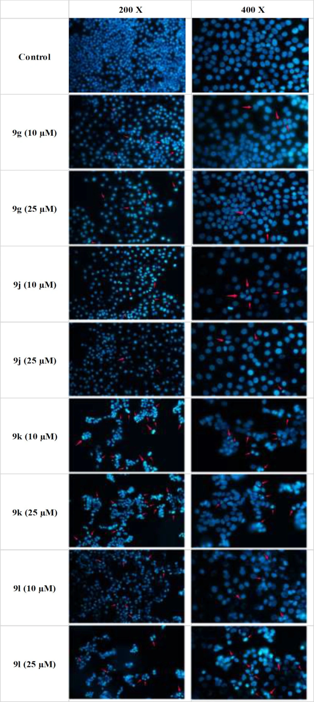
Hoechst 33258 staining
Hoechst 33258 was used as a DNA stain to detect apoptosis in cells. MCF-7 cells were seeded in a 6-well plate at a density of 5 × 10
4 cells/mL (2 mL per well) and incubated for 24 h. The whole medium was removed and different concentrations of test compounds
9g,
9j,
9k, and
9l diluted in 2 mL medium were placed in the different wells and incubated for 72 h. Afterward, the medium was removed again, 1 mL of 4% cold freshly prepared paraformaldehyde (PFA) was added, and cells were incubated for 20 min at room temperature. The cells were then washed 2 times with 1 ml PBS and incubated with 1 mL Hoechst 33258 2.5 µg/mL for 30 min in the dark at room temperature (
36,
37). In the end, the cells were washed with 1 mL PBS and imaged with a fluorescence microscope.
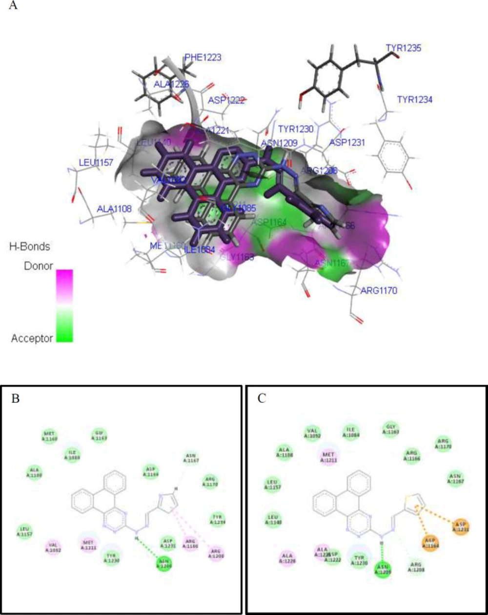
Docking analysis
The two most potent compounds (9k and 9l) were prepared using MarvinSketch 18.20.0 and geometry optimization was carried out by the steepest descent algorithm with MOPAC. The crystal structure of c-Met kinase receptor in complex with selective a c-Met inhibitor was obtained from the RCSB Protein Data Bank (PDB code 3ZZE). The molecular docking study was done with a GOLD software package using the GoldScore and ChemPLP scoring functions and default settings. The active site of protein was defined for the protein residues within 8 Å of the cognate ligand that accompanied the downloaded protein complexes. The cognate ligand was docked inside the 3ZZE to validate the docking algorithm using both scoring functions. Re-docking of the cognate ligand resulted in RMSD values of 0.854 Å and 1.01Å, for GoldScore and ChemPLP, respectively. Thus, the score value of the interactions between the ligands and c-Met kinase receptors with PDB code 3ZZE was calculated in terms of the GoldScore fitness function. The GoldScore fitness value for cognate ligand was calculated as 32.50. The molecular models of the docked compounds were visualized in the BIOVIA Discovery Studio software package and Pymol Software 1.7.4.4 Edu.