Effect of Zebularine in Comparison to Trichostatin A on the Intrinsic and Extrinsic Apoptotic Pathway, Cell Viability, and Apoptosis in Hepatocellular Carcinoma SK-Hep 1, Human Colorectal Cancer SW620, and Human Pancreatic Cancer PaCa-44 Cell Lines

authors:

avatar Masumeh Sanaei , avatar Fraidoon Kavoosi , *

Research Center for Non-communicable Diseases, Jahrom University of Medical Sciences, Jahrom, Iran.

how to cite: Sanaei M, Kavoosi F. Effect of Zebularine in Comparison to Trichostatin A on the Intrinsic and Extrinsic Apoptotic Pathway, Cell Viability, and Apoptosis in Hepatocellular Carcinoma SK-Hep 1, Human Colorectal Cancer SW620, and Human Pancreatic Cancer PaCa-44 Cell Lines. Iran J Pharm Res. 2021;20(3):e124295. https://doi.org/10.22037/ijpr.2021.115097.15196.

Abstract

Aberrant histone modifications or promoter region hypermethylation of tumor suppressor genes (TSGs) have been recognized as the important epigenetic molecular mechanism in cancer induction. The potential anticancer activities of histone deacetylase inhibitors (HDACIs) and DNA methyltransferase inhibitors (DNMTIs) have been investigated in recent years. The current study was assigned to investigate the effect of trichostatin A (HDACI) in comparison to zebularine (DNMTI) on the intrinsic pro-apoptotic (Bax, Bim, and Bak) and anti-apoptotic (Bcl-2, Mcl-1, and Bcl-xL) genes and extrinsic (DR4, DR5, FAS, FAS-L, and TRAIL genes) pathways, DNA methyltransferase 1, 3a, and 3b, histone deacetylase inhibitors 1, 2, and 3, cell viability, and apoptosis in hepatocellular carcinoma (HCC) SK-Hep 1, colorectal cancer SW620, and pancreatic cancer PaCa-44 cell lines. The SK-Hep 1, SW620, and PaCa-44 cells were cultured and treated with TSA and zebularine. To determine cell apoptosis, cell viability, and the relative gene expression level, flow cytometry assay, MTT assay, and qRT-PCR were done respectively. The result indicated that zebularine and TSA changed the expression level of the Bax, Bak, Bim Bcl-2, Bcl-xL, Mcl-1, DR4, DR5, FAS, FAS-L, TRAIL, DNA methyltransferase 1, 3a, and 3b, histone deacetylase inhibitors 1, 2, and 3 by which induced cell apoptosis and inhibit cell growth in all three cell lines. Concluding, TSA induced its role through both extrinsic and intrinsic apoptotic pathways in three cell lines, whereas, zebularine played its role via both pathways in the SK-Hep 1cell line, it had no significant effect on Bcl-2, Bcl-xL, and Mcl-1 gene expression in SW620 and PaCa-44 cell lines.

Introduction

Aberrant histone modifications or promoter region hypermethylation of tumor suppressor genes (TSGs) have been recognized as the important epigenetic molecular mechanisms in cancer induction and progression. These mechanisms can inactivate TSGs and cancer-related genes lead to cancer induction and progression. The N-terminal domains of all core histones are subject to diverse modifications, such as methylation, acetylation, phosphorylation, and ubiquitylation at certain residues (1). Acetylation is one of the most prominent posttranslational modifications affecting gene transcription and expression. Histone deacetylases (HDACs) and histone acetyltransferases (HATs) are responsible for the deacetylation and the acetylation of lysines of histone proteins respectively (2). The activity of HDACs condenses the chromatin structure of TSGs resulting in gene silencing and tumorigenesis. Previous analyses have divided HDACs into Class I HDACs (HDAC1, 2, 3, and 8), Class IIa HDACs (HDAC4, 5, 7, and 9), and the class III family of HDACs are also known as Sirtuins (3).

In addition to histone modification, DNA methylation, primarily at CpG dinucleotides, has long been identified to play an important role in controlling gene expression. Histone modifications and DNA methylation play a significant role in modulating chromatin structure which can control gene expression. DNA methyltransferases (DNMTs), are responsible for DNA methylation, the activity of these enzymes affects chromatin structure and gene expression (4). According to the structure and functions, DNMTs are divided into Dnmt1, Dnmt3a, Dnmt3b, and Dnmt3L (5). The potential anticancer activities of DNA methyltransferase inhibitors (DNMTIs) and HDAC inhibitors (HDACIs) have been investigated in recent years.HDACIs inhibit the activities of HDACs, resulting in an increase in the acetylation of the histone and an enhancement of the gene expression specially TSGs. DNMTIs are widely studied because DNA demethylation re-activates and up-regulates the expression of TSGs that are silenced by DNA hypermethylation. The combination of both HDACI and DNMTI has gained increasing interest as a possible targeted therapeutic cancer therapy (6).

In-vitro studies have demonstrated that DNMTIs (such as zebularine) and HDACIs (e.g. trichostatin A, TSA) can induce apoptosis through two molecular mechanisms comprising extrinsic and intrinsic apoptotic pathways (7, 8). The intrinsic pathway is controlled by the members of the BCL-2 family divided into three groups: promoters of apoptosis (BAX, BAK, and BOK); inhibitors of apoptosis (BCL-XL, BCL-2, MCL1, BCL-B, BCL-W, and A1); and regulatory BH3-only proteins (BIM, BAD, BID, BIK, BMF, NOXA, HrK, and PUMA) (9-11). The extrinsic pathway is triggered by the binding of death ligands to their death receptors (DRs) such as TRAIL-R1/DR4, TRAIL-R2/DR5, TRAIL-R3/DcR1, and TRAIL-R4/DcR2 (12). The current study was assigned to investigate the effect of TSA in comparison to zebularine on mitochondrial/intrinsic pro-apoptotic (Bax, Bim, and Bak) and anti-apoptotic (Bcl-2, Mcl-1, and Bcl-xL) genes and cytoplasmic/extrinsic (DR4, DR5, FAS, FAS-L, and TRAIL genes) pathways, DNA methyltransferase 1, 3a, and 3b, histone deacetylase inhibitors 1, 2, and 3, cell viability, and apoptosis in hepatocellular carcinoma SK-Hep 1, human colorectal cancer SW620, and human pancreatic cancer PaCa-44 cell lines.

Experimental

Materials

Hepatocellular carcinoma SK-Hep 1, human colorectal cancer cell lines SW620, and human pancreatic cancer PaCa-44 cell lines were purchased from the National Cell Bank of Iran-Pasteur Institute. The zebularine, TSA, and Dulbecco’s modified Eagle’s medium (DMEM) were obtained from Sigma (St. Louis, MO, USA) and dissolved in dimethyl sulfoxide (DMSO) to make a work stock solution. Further concentrations of the compounds, zebularine, and TSA were obtained by diluting the provided stock solution. Other necessary compounds comprising materials and various kits were purchased as provided for previous works (13, 14). The SK-Hep 1, SW620, and PaCa-44 cells were maintained in DMEM supplemented with fetal bovine serum 10% and antibiotics in a humidified atmosphere of 5% CO2 in air at 37 ℃. This work was approved by the Ethics Committee of Jahrom University of Medical science with a code number of IR.JUMS.REC.1399.079.

Cell culture and cell viability

The SK-Hep 1, SW620, and PaCa-44 cells were cultured in DMEM containing 10% FBS (Sigma) and 1% antibiotics which include 10,000 unit/mL penicillin G sodium(Sigma), 10,000 ug/mL streptomycin sulfate, and 25 ug/mL amphotericin B (Sigma) at 37 °C in 5% CO2 °C for 24 h and then seeded into 96-well plates (3 × 10⁵ cells per well). After 24 h, the medium was replaced with an experimental medium containing various concentrations of zebularine (0, 10, 25, 50, 75, 100, μM), and TSA (0, 0.5, 1, 2.5, 5, and 10 μM). The control groups were treated with DMSO, at a concentration of 0.05 %. After 24 h, the SK-Hep 1, SW620, and PaCa-44 cells, experimental and control, were investigated by MTT assay according to Standard protocols to determine cell viability. In this process, the MTT solution was added to each well for 4 h at 37 ℃ and then the MTT solution was changed by DMSO and shaken for 10 min to dissolve all of the crystals. Finally, the optical density was detected at a wavelength of 570 nM by a microplate reader. Each experiment was repeated three times (triplicates). IC50 was analyzed by GraphPad Prism Version 8.0. software. Non-linear regressions of log (inhibitor) vs response were selected for IC50 estimation. The bottom and top parameters were constrained to 0% and 100%.

Cell apoptosis assay

To determine SK-Hep 1, SW620, and PaCa-44 cells apoptosis, the cells were cultured at a density of 3 × 10⁵ cells/well and incubated overnight and then treated with zebularine and TSA, based on IC50 values demonstrated in Table 1, for 24 h. Subsequently, the cells were harvested by trypsinization, washed with cold PBS, and resuspended in Binding buffer (1x). Finally, Annexin-V-(FITC) and PI were used according to the protocol to determine the apoptotic cells by FACScan flow cytometry (Becton Dickinson, Heidelberg, Germany).

Real-time Quantitative Reverse Transcription Polymerase

Chain Reaction (qRT-PCR)

To determine the relative expression level of the Bax, Bak, Bim, Bcl-2, Bcl-xL, Mcl-1, DR4, DR5, FAS, FAS-L, TRAIL genes, DNA methyltransferase 1, 3a, and 3b, histone deac-etylase inhibitors 1, 2, and 3 qRT-PCR was done. The SK-Hep 1, SW620, and PaCa-44 cells were treated with zebularine and TSA, based on IC50 values demonstrated in Table 1, for 24 h, except control groups which were treated with DMSO only. Then qRT-PCR was done as our previous works (13, 14). The primer sequences are shown in Table 2 (15-26).

Results

Result of cell Cell viability by the MTT assay

The cell viability of the SK-Hep 1, SW620, and PaCa-44 cells treated with zebularine and TSA was investigated by MTT assay by which the activity of cellular enzymes reduced the tetrazolium salt MTT and produced a dark-blue formazan crystal. The crystal was dissolvable in DMSO to determine the viable cells. As shown in Figure 1, zebularine and TSA induced significant cell growth inhibition (****, P < 0.0001 and (**, P < 0.0027). IC50 values are shown in Table 1.

Result of cell apoptosis assay

To determine apoptosis, the SK-Hep 1, SW620, and PaCa-44 cells were treated with zebularine and TSA, based on IC50 values, for 24 h and then stained using annexin-V-(FITC) and PI to determine apoptotic cells in the early and late apoptosis stage. As indicated in Figures 24, both compounds induced cell apoptosis significantly (P < 0.001). Maximal and minimal apoptosis was seen in SK-Hep 1 cell treated with TSA and PaCa-44 cell treated with zebularine after 24 h of treatment respectively, Table 3.

Determination of genes expression

Effect of zebularine on the gene expression

The effect of zebularine on gene expression was evaluated by quantitative real-time RT-PCR analysis. The result indicated that zebularine up-regulated the expression of Bax, Bak, Bim, DR4, DR5, FAS, FAS-L, and TRAIL genes and down-regulated the expression of Bcl-2, Mcl-1, Bcl-xL, DNA methyltransferase 1, 3a, and 3b, histone deacetylase 1, 2, and 3 in SK-Hep 1cell line significantly. Additionally, zebularine up-regulated the expression of Bak, Bax, Bim, DR4, DR5, FAS, FAS-L, and TRAIL genes and down-regulated the expression of DNA methyltransferase 1, 3a, and 3b in SW620, and PaCa-44 cell lines significantly. It had no significant effect on Bcl-2, Bcl-xL, and Mcl-1 gene expression in SW620, and PaCa-44 cell lines (Figure 5).

Effect of TSA on the gene expression

The effect of TSA on gene expression was evaluated by quantitative real-time RT-PCR analysis. The result indicated that TSA up-regulated the expression of Bax, Bak, Bim, DR4, DR5, FAS, FAS-L, and TRAIL genes and down-regulated the expression of Bcl-2, Mcl-1, Bcl-xL, histone deacetylase 1, 2, and 3 in SK-Hep 1, SW620, and PaCa-44 cell lines significantly (Figure 6).

Discussion

The term “Apoptosis” derives from the Greek language and means trees shedding their leaves, the leaves “falling off” from trees, in autumn. Apoptosis is crucial for physiological processes, embryonic development, tissue homeostasis, and pathological diseases, such as cancer. Normally, it is triggered by two different molecular pathways comprising the death receptor (extrinsic) and mitochondrial (intrinsic) pathways, both of which converge on the activation of the cascade of caspases (27). HDACIs induce apoptosis through the activation of both pathways, intrinsic and extrinsic. They activate the extrinsic pathway via the upregulation of death receptors expression, reduction in c-FLIP, and upregulation of ligands such as TRAIL. Further, they activate the intrinsic pathway through the upregulation of several Bcl-2 family genes such as Bid, Bim, and Bmf (28). Similar to HDACIs, DNA demethylating drugs cause apoptosis via both extrinsic and intrinsic pathways (29).

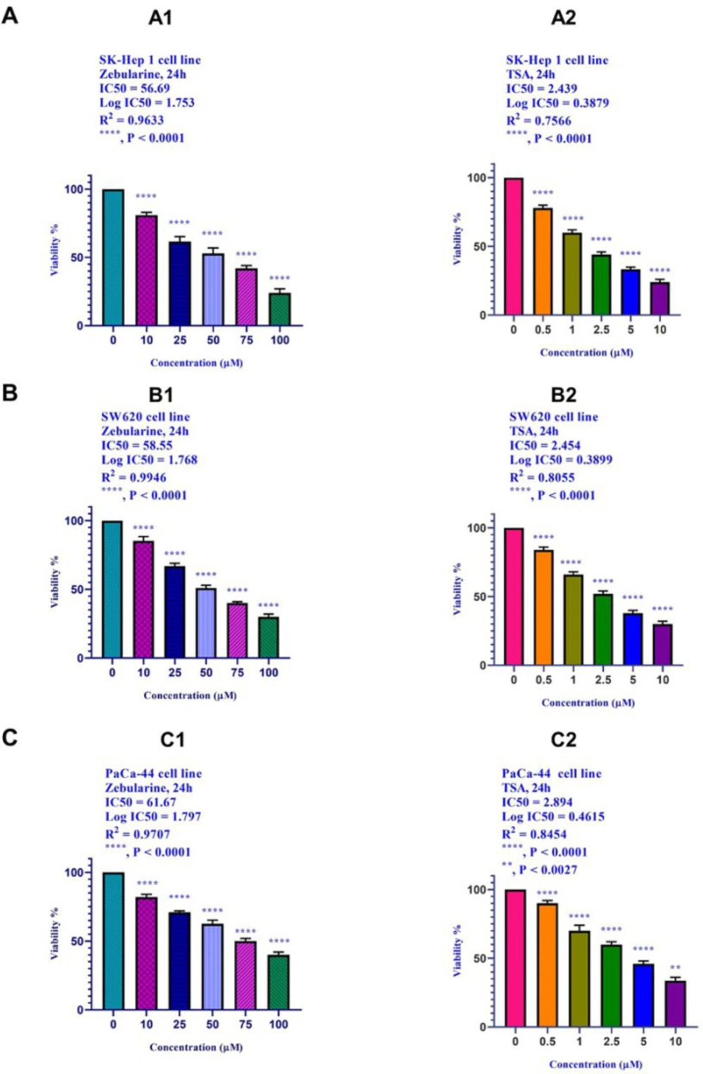
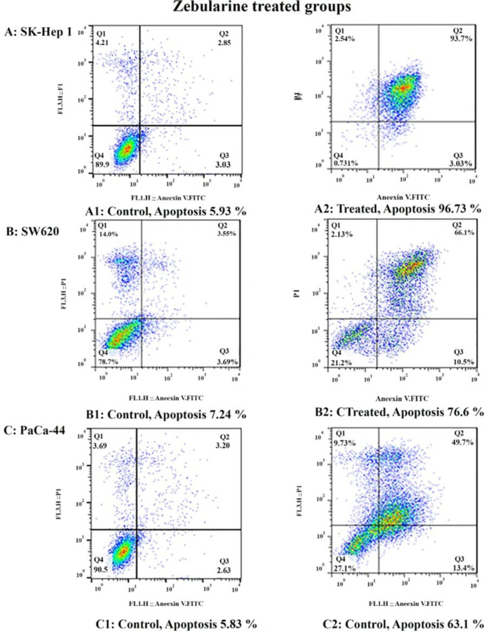
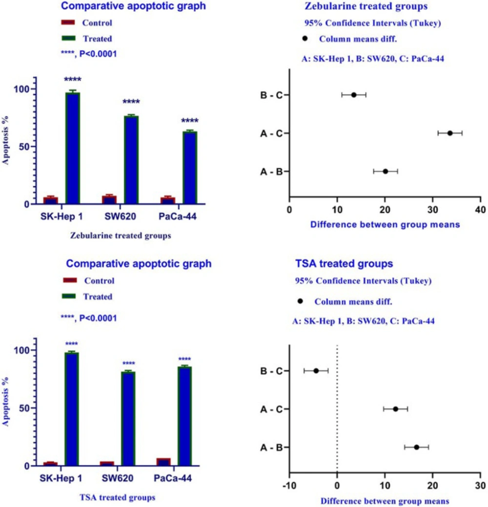
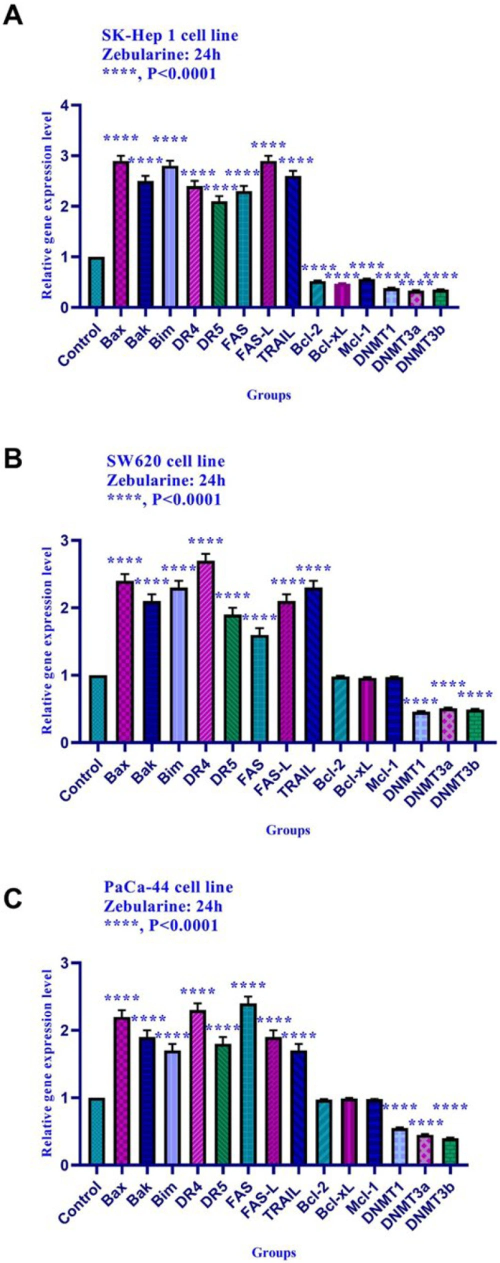
The results of current studies indicated that DNA methyltransferase inhibitor zebularine can induce apoptosis in hepatocellular carcinoma SK-Hep 1, human colorectal cancer SW620, and human PaCa-44 pancreatic cancer cell lines. It up-regulated the expression of the genes of the extrinsic apoptotic pathway and pro-apoptotic genes (as mentioned in the result part) and down-regulated the expression of the anti-apoptotic genes, DNMTs (1, 3a, and 3b), HDACs (1, 2, and 3) in SK-Hep 1 cell line significantly. Additionally, zebularine up-regulated the expression of the genes of the extrinsic apoptotic pathway and pro-apoptotic genes (as mentioned in the result part) and down-regulated the expression of DNMTs (1, 3a, and 3b), HDACs (1, 2, and 3) in SW620, and PaCa-44 cell lines significantly. We did not observe significant changes in the expression level of Bcl-2, Mcl-1, and Bcl-xL in SW620, and PaCa-44 cell lines treated with zebularine. Similarly, in-vitro studies have demonstrated that DNA demethylating agents zebularine and decitabine activate the mitochondrial apoptotic pathway in T cells (30). Similar to our findings, it has been reported that zebularine treatment induces cell-cycle arrest, and apoptosis in the HCC HepG2 cell line through the intrinsic pathway (31). It has been reported that DNA methyltransferase inhibitor 5-Aza-CdR down-regulated MCL-1 leads to apoptotic induction in AML cells (32). In recent studies, it was demonstrated that zebularine and 5-aza-dC down-regulate DNMT-1, DNMT-3a, and DNMT-3b in ovarian cancer cell lines (33). Several works have demonstrated that 5-Aza-CdR treatment decrease DNMT1 and DNMT3a mRNA expression resulting in a down-regulation of Bcl2 protein in colon cancer cell line HCT-116 (34). Experimental studies have shown that compounds with DNMTI activity such as 5-Aza-CdR generally induce tumor cell apoptosis by DNA demethylation in human cell lines A549, HepG2, Hep3B, A-498, and HCT-116 (35).

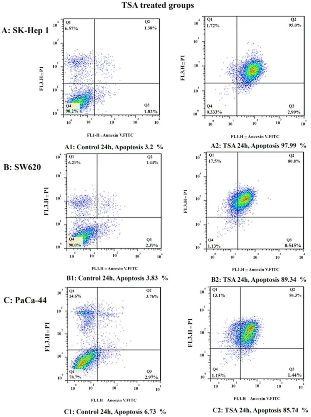
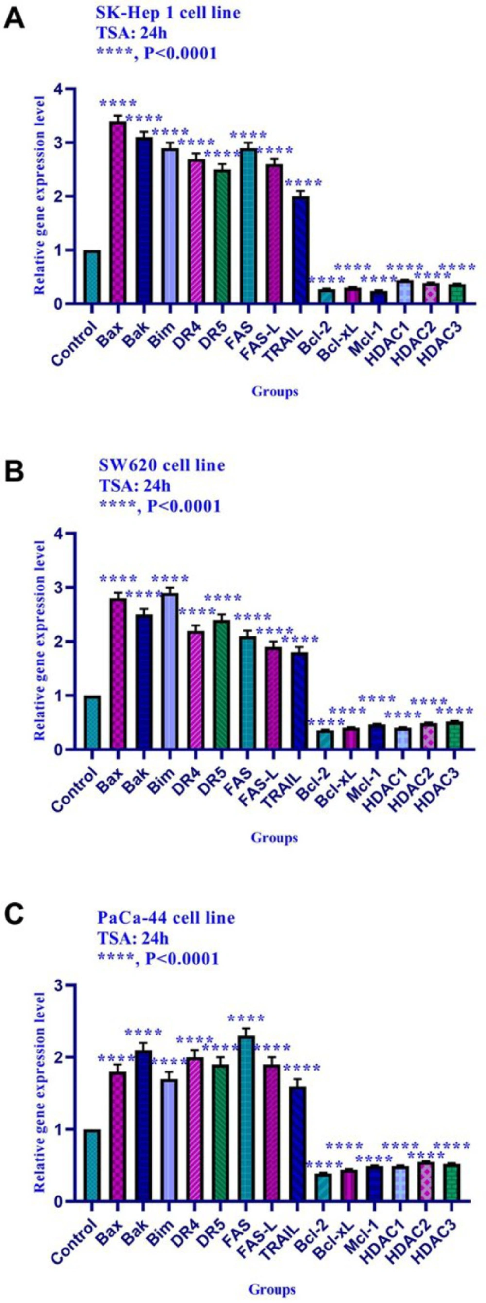
In the present work, we reported that histone deacetylase inhibitor TSA can induce apoptosis through both mitochondrial/intrinsic and cytoplasmic/extrinsic apoptotic pathways, in hepatocellular carcinoma SK-Hep 1, human colorectal cancer SW620, and human PaCa-44 pancreatic cancer cell lines through the activation of both extrinsic and intrinsic pathways, it up-regulated the expression of Bax, Bak, Bim, DR4, DR5, FAS, FAS-L, and TRAIL genes and down-regulated the expression of Bcl-2, Mcl-1, Bcl-xL, DNA methyltransferase 1, 3a, and 3b, histone deacetylase 1, 2, and 3 in all three cell lines significantly. Inconsistent with our result, other researchers have indicated that histone deacetylase inhibitor sodium butyrate decreases Bcl-XL protein levels by down-regulating its RNA expression in mesothelioma cell lines. Besides, depsipeptide, as an HDACI, decreases the expression of Bcl-2, Bcl-XL, and Mcl-1 in multiple myeloma cells (36). In human lymphatic endothelial cells (LEC), TSA-induced apoptosis by cytochrome c release contributed to activating caspases-3, caspases −7, and caspases −9 and the anti-apoptotic proteins down-regulation accompanied by up-regulation of p21, p27, and p53 (37). Resent in-vitro works have shown that TSA increases the ratio between the levels of expression of anti-apoptotic (BCL-w and BCL-xl) and pro-apoptotic (BIM)genes in pancreatic cancer cell lines CFPAC1, Miapaca2, HPAF, PSN1, Panc1 PC, Paca44, RT45P1, and T3M4 (38). Similar to our findings, other researchers have shown that HDACIs play their apoptotic roles through the extrinsic apoptotic pathway. It has been indicated that several HDACIs like SAHA, TSA, m-carboxycinnamic acid bishydroxamide (CBHA), LAQ824 (a cinnamic acid hydroxamate), and MS-275 can up-regulate the expression of TRIAL-R1 and TRIAL-R2 (39). In pancreatic cancer cell lines Panc1 and MiaPaCa2, treatment with TSA increase the expression of the TRAIL receptor 1 (DR5) (40). Additionally, HDACIs can induce apoptosis through activation of the death receptor pathway (TRAIL and Fas signaling pathways), the up-regulation of TRAIL, DR5, Fas, Fas-L in leukemic blasts (41).

Meanwhile, the extrinsic and intrinsic pathways are not the only molecular mechanisms of zebularine and TSA. The results of current studies indicated that DNA methyltransferase inhibitor zebularine and histone deacetylase inhibitor TSA can induce apoptosis through both mitochondrial/intrinsic and cytoplasmic/extrinsic apoptotic pathways, in hepatocellular carcinoma SK-Hep 1, human colorectal cancer SW620, and human PaCa-44 pancreatic cancer cell lines. Our finding demonstrated that zebularine induced significant apoptosis in all three cell lines. It up-regulated the expression of Bax, Bak, Bim, DR4, DR5, FAS, FAS-L, and TRAIL genes and down-regulated the expression of Bcl-2, Mcl-1, Bcl-xL, DNA methyltransferase 1, 3a, and 3b, histone deacetylase inhibitors 1, 2, and 3 in SK-Hep 1 cell line significantly. Additionally, zebularine up-regulated the expression of Bax, Bak, Bim, DR4, DR5, FAS, FAS-L, and TRAIL genes and down-regulated the expression of DNA methyltransferase 1, 3a, and 3b in SW620, and PaCa-44 cell lines significantly. It had no significant effect on Bcl-2, Mcl-1, and Bcl-xL gene expression in SW620, and PaCa-44 cell lines. Similarly, in-vitro studies have demonstrated that DNA demethylating agents zebularine and decitabine activate the mitochondrial apoptotic pathway in T cells (30). Similar to our findings, it has been reported that zebularine treatment induces cell-cycle arrest, and apoptosis in the HCC HepG2 cell line through the intrinsic pathway (31). It has been reported that DNA methyltransferase inhibitor 5-Aza-CdR down-regulated MCL-1 leads to apoptotic induction in AML cells (32). In recent studies, it was demonstrated that zebularine and 5-aza-dC down-regulate DNMT-1, DNMT-3a, and DNMT-3b in ovarian cancer cell lines (33). Several works have demonstrated that 5-Aza-CdR treatment decrease DNMT1 and DNMT3a mRNA expression resulting in a down-regulation of Bcl2 protein in colon cancer cell line HCT-116 (34). Experimental studies have shown that compounds with DNMTI activity such as 5-Aza-CdR generally induce tumor cell apoptosis by DNA demethylation in human cell lines A549, HepG2, Hep3B, A-498, and HCT-116 (35). As mentioned in the result section, TSA induced significant apoptosis in all three cell lines, SK-Hep 1, SW620, and PaCa-44. It up-regulated the expression of Bak, Bax, Bim, DR4, DR5, FAS, FAS-L, and TRAIL genes and down-regulated the expression of Bcl-2, Bcl-xL, Mcl-1, histone deacetylase inhibitors 1, 2, and 3 in these cell lines significantly. Inconsistent with our result, other researchers have indicated that histone deacetylase inhibitor sodium butyrate decreases Bcl-XL protein levels by down-regulating its RNA expression in mesothelioma cell lines. Besides, depsipeptide, as an HDACI, decreases the expression of Bcl-2, Bcl-XL, and Mcl-1 in multiple myeloma cells (36). In human lymphatic endothelial cells (LEC), TSA-induced apoptosis by cytochrome c release contributed to activating caspases-3, caspases −7, and caspases −9 and the anti-apoptotic proteins down-regulation accompanied by up-regulation of p21, p27, and p53 (37). Resent in-vitro works have shown that TSA increases the ratio between the levels of expression of anti-apoptotic (BCL-w and BCL-xl) and pro-apoptotic (BIM)genes in pancreatic cancer cell lines CFPAC1, Miapaca2, HPAF, PSN1, Panc1 PC, Paca44, RT45P1, and T3M4 (38). Similar to our findings, other researchers have shown that HDACIs play their apoptotic roles through the extrinsic apoptotic pathway. It has been indicated that several HDACIs like SAHA, TSA, m-carboxycinnamic acid bishydroxamide (CBHA), LAQ824 (a cinnamic acid hydroxamate), and MS-275 can up-regulate the expression of TRIAL-R1 and TRIAL-R2 (39). In pancreatic cancer cell lines Panc1 and MiaPaCa2, treatment with TSA increase the expression of the TRAIL receptor 1 (DR5) (40). Additionally, HDACIs can induce apoptosis through activation of the death receptor pathway (TRAIL and Fas signaling pathways), the up-regulation of TRAIL, DR5, Fas, Fas-L in leukemic blasts (41). Meanwhile, the extrinsic and intrinsic pathways are not the only molecular mechanisms of zebularine and TSA. Our previous work indicated that zebularine and TSA can inhibit DNMTs (DNMT1, DNMT3a, and DNMT3b), Class I HDACs (HDACs 1, 2, 3), and Class II HDACs (HDACs 4, 5, 6) and up-regulate CIP/KIP Family (p21Cip1/Waf1/Sdi1, p27Kip1, and p57Kip2) resulting in cell apoptosis in colon cancer LS 174T, and LS 180 cell lines (42, 43). Finally, zebularine and TSA can play their apoptotic roles through intrinsic and extrinsic pathways. In some cases, zebularine could not change the gene expression significantly. It may induce a significant change in the expression of the mentioned genes with high concentrations at 48 or 72 h. It may be done with further concentrations or durations. Therefore, the evaluation of this compound with high concentrations and more duration (24 and 48h) is recommended.

Table 1

IC50 values

Cell lineCompoundDuration/HourIC50 Value
SK-Hep 1Zebularine2456.69
SK-Hep 1TSA242.439
SW620Zebularine2458.55
SW620TSA242.454
PaCa-44Zebularine2461.67
PaCa-44TSA242.894
Table 2

The primer sequences of Bax, Bak, Bim, Bcl-2, Bcl-xL, Mcl-1, DR4, DR5, FAS, FAS-L, TRAIL genes, DNA methyltransferase 1, 3a, and 3b, histone deacetylase inhibitors 1, 2, and 3 qRT-PCR was done. The SK-Hep 1, SW620, and PaCa-44 genes

PrimerPrimer sequences (5' to 3')Product lengthReference
BaxForwardReverseAGTAACATGGAGCTGCAGAGGAT GCTGCCACTCGGAAAAAGAC77 bp
BakForwardReverseCCTGCCCTCTGCTTCTGA CTGCTGATGGCGGTAAAAA82 bp
BimForwardReverseATTACCAAGCAGCCGAAGACTCCGCAAAGAACCTGTCAAT101 bp
Bcl-2ForwardReverseTGGCCAGGGTCAGAGTTAAATGGCCTCTCTTGCGGAGTA147 bp
Bcl-xLForwardReverseTCCTTGTCTACGCTTTCCACGGGTCGCATTGTGGCCTTT62 bp
Mcl-1ForwardReverseAAAGCCTGTCTGCCAAATCCTATAAACCCACCACTC198 bp
DR4ForwardReverseCAGAACATCCTGGAGCCTGTAACATGTCCATTGCCTGATTCTTTGTG299 bp
DR5ForwardReverseTGCAGCCGTAGTCTTGATTGGCACCAAG TCTGCAAAGTCA389 bp
FASForwardReverseTTCTGCCATAAGCCCTGTCCTGTACTCCTTCCCTTCTTGG103 bp
FAS-LForwardReverseGCCTGTGTCTCCTTGTGATG TGGACTTGCCTGTTAAATGGG222 bp
TRAILForwardReverseGAAGCAACACATTGTCTTCTCCAA TTGCTCAGGAATGAATGCCC103 bp
DNMT1ForwardReverseGAGGAAGCTGCTAAGGACTAGTTCACTCCACAATTTGATCACTAA ATC206 bp
DNMT 3aForwardReverseGGAGGCTGAGAAGAAAGCCAAGGTTTTGCCGTCTCCGAACCACATGAC370 bp
DNMT 3bForwardReverseTACACAGACGTGTCCAACATGGGCGGATGCCTTCAGGAATCACACCTC195 bp
HDAC1ForwardReverseAACCTGCCTATGCTGATGCTCAGGCAATTCGTTTGTCAGA374 bp
HDAC2ForwardReverseGGGAATACTTTCCTGGCACAACGGATTGTGTAGCCACCTC314 bp
HDAC3ForwardReverseTGGCTTCTGCTATGTCAACGGCACGTGGGTTGGTAGAAGT328 bp
GAPDH ForwardReverseTGTTGCCATCAATGACCCCTT CTCCACGACGTACTCAGCG148 bp
Table 3

Comparative analysis of the effect of zebularine and TSA on SK-Hep 1, SW620, and PaCa-44 cells. Maximal and minimal percentage of apoptosis was seen in SK-Hep 1 cell treated with TSA and PaCa-44 cell treated with zebularine after 24 h of treatment

DrugCell lineDose/μMDuration/hApoptosis (%)P-value
ZebularineSK-Hep 156.692496.73P < 0.001
ZebularineSW62058.552476.6P < 0.001
ZebularinePaCa-4461.672463.1P < 0.001
TSASK-Hep 12.4392497.99P < 0.001
TSASW6202.4542481.34P < 0.001
TSAPaCa-442.8942485.74P < 0.001
The effect of zebularine (0, 10, 25, 50, 75, 100, μM) and TSA (0, 0.5, 1, 2.5, 5, and 10 μM) on the viability of (A) hepatocellular carcinoma SK-Hep 1, (B) human colorectal cancer cell lines SW620, and (C) human pancreatic cancer PaCa-44 cell line. The cells were treated without and with different doses of zebularine and TSA for 24 and the cell viability was evaluated by MTT assay. Each experiment was achieved in triplicate. Mean values from the three experiments ± standard error of mean are indicated. Asterisks indicate significant differences between treated and untreated cells. **P < 0.0027, and ****P < 0.0001
The apoptosis-inducing effect of zebularine was investigated by flow cytometric analysis of (A) SK-Hep 1, (B) SW620, and (C) PaCa-44 cells stained with Annexin V and propidium iodide. The result indicated that zebularine induced cell apoptosis after 24 h of treatment significantly
The apoptosis-inducing effect of TSA was investigated by flow cytometric analysis of (A) SK-Hep 1, (B) SW620, and (C) PaCa-44 cells stained with Annexin V and propidium iodide. The result indicated that TSA induced cell apoptosis after 24 h of treatment significantly
The Apoptotic Effect of zebularine and TSA on SK-Hep 1, SW620, and PaCa-44 cells versus control groups at 24 h. Results were obtained from three independent experiments and were expressed as mean ± standard error of the mean. The results of the statistical analysis indicate significant differences between treated and untreated cells as shown on the right side. ****P < 0.0001
The relative expression level of Bim, Bax, Bak, Bcl-xL, Bcl-2, Mcl-1, DR4, DR5, FAS, FAS-L, TRAIL, DNA methyltransferase 1, 3a, and 3b, and histone deacetylase inhibitors 1, 2, and 3 in SK-Hep 1, SW620, and PaCa-44 cells treated with zebularine at 24 h. Quantitative reverse transcription-polymerase chain reaction analysis demonstrated that this compound up-regulated the expression of Bax, Bak, Bim, DR4, DR5, FAS, FAS-L, TRAIL, and down-regulated the expression of Bcl-2, Mcl-1, Bcl-xL, DNA methyltransferase 1, 3a, and 3b significantly. This compound had no significant effect on Bcl-2, Mcl-1, and Bcl-xLgene expression in SW620, and PaCa-44 cell lines. Asterisks indicate significant differences between treated cells and the control groups. Data are presented as means ± standard error of the mean. ****P < 0.0001
The relative expression level of Bim, Bax, Bak, Bcl-xL, Bcl-2, Mcl-1, DR4, DR5, FAS, FAS-L, TRAIL, DNA methyltransferase 1, 3a, and 3b, and histone deacetylase inhibitors 1, 2, and 3 in SK-Hep 1, SW620, and PaCa-44 cells treated with TSA at 24h. Quantitative reverse transcription-polymerase chain reaction analysis demonstrated that this compound up-regulated the expression of Bax, Bak, Bim, DR4, DR5, FAS, FAS-L, TRAIL, and down-regulated the expression of Bcl-2, Mcl-1, Bcl-xL, DNA methyltransferase 1, 3a, and 3b significantly. Asterisks indicate significant differences between treated cells and the control groups. Data are presented as means ± standard error of the mean

Conclusion

In conclusion, our results indicated that histone deacetylase inhibitor TSA can induce its apoptotic effects through both mitochondrial/intrinsic and cytoplasmic/extrinsic apoptotic pathways in hepatocellular carcinoma SK-Hep 1, human colorectal cancer SW620, and human PaCa-44 pancreatic cancer cell lines. Further, zebularine can induce apoptosis by an extrinsic apoptotic pathway in three cell lines. It had no significant effect on Bcl-2, Mcl-1, and Bcl-xL gene expression in SW620, and PaCa-44 cell lines. Therefore, TSA can induce a stronger apoptotic effect through the activation of both pathways. Finally, both agents induced apoptosis significantly, which provided the theoretical basis for the clinical application of these compounds.

Acknowledgements

References

  • 1.

    Kondo Y. Epigenetic cross-talk between DNA methylation and histone modifications in human cancers. Yonsei Med. J. 2009;50:455-63. [PubMed ID: 19718392].

  • 2.

    Yao YL, Yang WM. Beyond histone and deacetylase: an overview of cytoplasmic histone deacetylases and their nonhistone substrates. BioMed Res. Int. 2011;5:1-15.

  • 3.

    Mihaylova MM, Vasquez DS, Ravnskjaer K, Denechaud PD, Ruth TY, Alvarez JG, Downes M, Evans RM, Montminy M, Shaw RJ. Class IIa histone deacetylases are hormone-activated regulators of FOXO and mammalian glucose homeostasis. Cell. 2011;145:607-21. [PubMed ID: 21565617].

  • 4.

    Cheng X, Blumenthal RM. Mammalian DNA methyltransferases: a structural perspective. Structure. 2008;16:341-50. [PubMed ID: 18334209].

  • 5.

    Wang Z, Wu J. DNA methyltransferases: classification, functions and research progress. Yi chuan = Hered. 2009;31:903-12.

  • 6.

    Zhu WG, Otterson GA. The interaction of histone deacetylase inhibitors and DNA methyltransferase inhibitors in the treatment of human cancer cells. Curr. Med. Chem. Anticancer Agents. 2003;3:187-99. [PubMed ID: 12769777].

  • 7.

    Natoni F, Diolordi L, Santoni C, Montani MSG. Sodium butyrate sensitises human pancreatic cancer cells to both the intrinsic and the extrinsic apoptotic pathways. Biochim. Biophys. Acta - Mol. Cell Res. 2005;1745:318-29.

  • 8.

    Kong WY, Yee ZY, Mai CW, Fang CM, Abdullah S, Ngai SC. Zebularine and trichostatin A sensitized human breast adenocarcinoma cells towards tumor necrosis factor-related apoptosis inducing ligand (TRAIL)-induced apoptosis. Heliyon. 2019;5:2468-75.

  • 9.

    Wei MC, Zong W-X, Cheng EH-Y, Lindsten T, Panoutsakopoulou V, Ross AJ, Roth KA, MacGregor GR, Thompson CB, Korsmeyer SJ. Proapoptotic BAX and BAK: a requisite gateway to mitochondrial dysfunction and death. Science. 2001;292:727-30. [PubMed ID: 11326099].

  • 10.

    Cory S, Adams JM. The Bcl2 family: regulators of the cellular life-or-death switch. Nat. Rev. Cancer. 2002;2:647-56. [PubMed ID: 12209154].

  • 11.

    Youle RJ, Strasser A. The BCL-2 protein family: opposing activities that mediate cell death. Nat. Rev. Mol. Cell Biol. 2008;9:47-59. [PubMed ID: 18097445].

  • 12.

    Yagita H, Takeda K, Hayakawa Y, Smyth MJ, Okumura K. TRAIL and its receptors as targets for cancer therapy. Cancer Sci. 2004;9:777-83.

  • 13.

    Sanaei M, Kavoosi F, Roustazadeh A, Shahsavani H. In-vitro effect of the histone deacetylase inhibitor valproic acid on viability and apoptosis of the PLC/PRF5 human hepatocellular carcinoma cell line. Asian Pac. J. Cancer Prev. 2018;19:2507-10. [PubMed ID: 30256044].

  • 14.

    Sanaei M, Kavoosi F. Effect of curcumin and trichostatin a on the expression of DNA methyltransfrase 1 in hepatocellular carcinoma cell line hepa 1-6. Iran. J. Pediatr. Hematol. Oncol. 2018;8:193-201.

  • 15.

    Cao XX, Mohuiddin I, Chada S, Mhashilkar AM, Ozvaran MK, McConkey DJ, Miller SD, Daniel JC, Smythe WR. Adenoviral transfer of mda-7 leads to BAX up-regulation and apoptosis in mesothelioma cells, and is abrogated by over-expression of BCL-XL. Mol. Med. 2002;8:869-76. [PubMed ID: 12606823].

  • 16.

    Ierano C, Chakraborty A, Nicolae A, Bahr J, Zhan Z, Pittaluga S. Loss of the proteins Bak and Bax prevents apoptosis mediated by histone deacetylase inhibitors. Cell Cycle. 2013;12:2829-38. [PubMed ID: 23966164].

  • 17.

    Zhang XQ, Dong JJ, Cai T, Shen X, Zhou XJ, Liao L. High glucose induces apoptosis via upregulation of Bim expression in proximal tubule epithelial cells. Oncotarget. 2017;8:24119-29. [PubMed ID: 28445931].

  • 18.

    Xu Y, Liu L, Qiu X, Liu Z, Li H, Li Z, Luo W, Wang E. CCL21/CCR7 prevents apoptosis via the ERK pathway in human non-small cell lung cancer cells. PLoS One. 2012;7:17-24.

  • 19.

    Zhang YL, Pang LQ, Wu Y, Wang XY, Wang CQ, Fan Y. Significance of Bcl-xL in human colon carcinoma. World J. Gastroenterol. 2008;14:3069-73. [PubMed ID: 18494061].

  • 20.

    Wang B, Ni Z, Dai X, Qin L, Li X, Xu L, Lian J, He F. The Bcl-2/xL inhibitor ABT-263 increases the stability of Mcl-1 mRNA and protein in hepatocellular carcinoma cells. Mol. Cancer. 2014;13:98-106. [PubMed ID: 24779770].

  • 21.

    Nakamoto N, Higuchi H, Kanamori H, Kurita S, Tada S, Takaishi H, Toda K, Yamada T, Kumagai N, Saito H. Cyclooxygenase-2 inhibitor and interferon-β synergistically induce apoptosis in human hepatoma cells in-vitro and in-vivo. Int. J. Oncol. 2006;26:625-35.

  • 22.

    Tao J, Qiu B, Zhang D, Wang Y. Expression levels of Fas/Fas-L mRNA in human brain glioma stem cells. Mol. Med. Rep. 2012;5:1202-6. [PubMed ID: 22344564].

  • 23.

    Inoue T, Anai S, Onishi S, Miyake M, Tanaka N, Hirayama A. Inhibition of COX-2 expression by topical diclofenac enhanced radiation sensitivity via enhancement of TRAIL in human prostate adenocarcinoma xenograft model. BMC Urol. 2013;13:1-9. [PubMed ID: 23289871].

  • 24.

    Sanaei M, Kavoosi F, Roustazadeh A, Golestan F. Effect of genistein in comparison with trichostatin a on reactivation of DNMTs genes in hepatocellular carcinoma. J. Clin. Transl. Hepatol. 2018;6:1-6. [PubMed ID: 29577026].

  • 25.

    Jin KL, Pak JH, Park JY, Choi WH, Lee JY, Kim JH, Nam JH. Expression profile of histone deacetylases 1, 2 and 3 in ovarian cancer tissues. J. Gynecol. Oncol. 2008;19:185-90. [PubMed ID: 19471575].

  • 26.

    Wu S, Ge Y, Huang L, Liu H, Xue Y, Zhao Y. BRG1, the ATPase subunit of SWI/SNF chromatin remodeling complex, interacts with HDAC2 to modulate telomerase expression in human cancer cells. Cell Cycle. 2014;13:2869-78. [PubMed ID: 25486475].

  • 27.

    Zhang R, Tao L, Weng D. RIP kinases and caspase-8: a complex interplay in cell death. Adv. Exp. Med. Biol. 2016;1:3-20.

  • 28.

    Matthews GM, Newbold A, Johnstone RW. Intrinsic and extrinsic apoptotic pathway signaling as determinants of histone deacetylase inhibitor antitumor activity. Adv. Cancer Res. 2012;5:165-97.

  • 29.

    Walia YK, Sharma V. Role of HDACs and DNMTs in cancer therapy: a review. Asian J. Adv. Basic Sci. 2013;1:62-78.

  • 30.

    Ruiz‐Magaña MJ, Rodríguez‐Vargas JM, Morales JC, Saldivia MA, Schulze‐Osthoff K, Ruiz‐Ruiz C. The DNA methyltransferase inhibitors zebularine and decitabine induce mitochondria‐mediated apoptosis and DNA damage in p53 mutant leukemic T cells. Int. J. Cancer Res. 2012;130:1195-207.

  • 31.

    Nakamura K, Aizawa K, Nakabayashi K, Kato N, Yamauchi J, Hata K. DNA methyltransferase inhibitor zebularine inhibits human hepatic carcinoma cells proliferation and induces apoptosis. PLoS One. 2013;8:54036-42.

  • 32.

    Tsao T, Shi Y, Kornblau S, Lu H, Konoplev S, Antony A. Concomitant inhibition of DNA methyltransferase and BCL-2 protein function synergistically induce mitochondrial apoptosis in acute myelogenous leukemia cells. Ann. Hematol. 2012;91:1861-70. [PubMed ID: 22893484].

  • 33.

    Balch C, Yan P, Craft T, Young S, Skalnik DG, Huang TH. Antimitogenic and chemosensitizing effects of the methylation inhibitor zebularine in ovarian cancer. Mol. Cancer Ther. 2005;4:1505-14. [PubMed ID: 16227399].

  • 34.

    Schneider-Stock R, Diab-Assef M, Rohrbeck A, Foltzer-Jourdainne C, Boltze C, Hartig R. Retraction: 5-aza-Cytidine is a potent inhibitor of DNA methyltransferase 3a and induces apoptosis in HCT-116 colon cancer cells via Gadd45-and p53-dependent mechanisms. J. Pharmacol. Exp. Ther. 2005;312:525-36. [PubMed ID: 15547111].

  • 35.

    Venturelli S, Berger A, Weiland T, Essmann F, Waibel M, Nuebling T. Differential induction of apoptosis and senescence by the DNA methyltransferase inhibitors 5-azacytidine and 5-aza-2′-deoxycytidine in solid tumor cells. Mol. Cancer Ther. 2013;12:2226-36. [PubMed ID: 23924947].

  • 36.

    Kang MH, Reynolds CP. Bcl-2 inhibitors: targeting mitochondrial apoptotic pathways in cancer therapy. Clin. Cancer Res. 2009;15:1126-32. [PubMed ID: 19228717].

  • 37.

    Hrgovic I, Doll M, Kleemann J, Wang XF, Zoeller N, Pinter A. The histone deacetylase inhibitor trichostatin a decreases lymphangiogenesis by inducing apoptosis and cell cycle arrest via p21-dependent pathways. BMC Cancer. 2016;16:1-15.

  • 38.

    Moore PS, Barbi S, Donadelli M, Costanzo C, Bassi C, Palmieri M. Gene expression profiling after treatment with the histone deacetylase inhibitor trichostatin A reveals altered expression of both pro-and anti-apoptotic genes in pancreatic adenocarcinoma cells. Biochim. Biophys. Acta Mol. Cell Res. 2004;1693:167-76.

  • 39.

    Jan R. Understanding apoptosis and apoptotic pathways targeted cancer therapeutics. Adv. Pharm. Bull. 2019;9:205-18. [PubMed ID: 31380246].

  • 40.

    Schüler S, Fritsche P, Diersch S, Arlt A, Schmid RM, Saur D. HDAC2 attenuates TRAIL-induced apoptosis of pancreatic cancer cells. Mol. Cancer. 2010;9:80. [PubMed ID: 20398369].

  • 41.

    Insinga A, Monestiroli S, Ronzoni S, Gelmetti V, Marchesi F, Viale A. Inhibitors of histone deacetylases induce tumor-selective apoptosis through activation of the death receptor pathway. Nat. Med. 2005;11:71-6. [PubMed ID: 15619634].

  • 42.

    Sanaei M, Kavoosi F. Effect of Zebularine in Comparison to and in Combination with Trichostatin A on CIP/KIP Family (p21Cip1/Waf1/Sdi1, p27Kip1, and p57Kip2), DNMTs (DNMT1, DNMT3a, and DNMT3b), Class I HDACs (HDACs 1, 2, 3) and Class II HDACs (HDACs 4, 5, 6) Gene Expression, Cell Growth Inhibition and Apoptosis Induction in Colon Cancer LS 174T Cell Line. Asian Pac. J. Cancer Prev. 2020;21:2131-9. [PubMed ID: 32711442].

  • 43.

    Sanaei M, Kavoosi F. Investigation of the Effect of Zebularine in Comparison to and in Combination with Trichostatin A on p21Cip1/Waf1/Sdi1, p27Kip1, p57Kip2, DNA Methyltransferases and Histone Deacetylases in Colon Cancer LS 180 Cell Line. Asian Pac. J. Cancer Prev. 2020;21:1819-28. [PubMed ID: 32592383].