Materials
Ibuprofen and Cetosteryl alcohol were gifted by Medicamen Organics Limited, Sidcul, Uttrakhand, India. Carbopol 934, glycerine, propylene glycol, triethanolamine, benzalkonium chloride, Sorbitanmonostearate (Span 80), Sorbitanmonopalmitate (Span 40), Sorbitanmonolaurate (Span 20), Polysorbate 60 (Tween 60), Polysorbate 80 (Tween 80), Diethyl ether and methanol were provided by the institution research institute (SIHAS, SHUATS, Prayagraj, U.P., India). All the other reagents and solvents used were of analytical grade. The seed sample was purchased from the local market of Meerut City, Uttar Pradesh, India. A voucher herbarium specimen was prepared for necessary identification. Lallemantia royaleana Benth. plant and seeds authentication was done at the National Bureau of Plant and Genetic Resources, PUSA Campus, New Delhi, India. The voucher specimen was deposited to the Department of Pharmaceutics, M.I.E.T., Meerut, U.P., India for future evidence.
Methods
Preparation of mucilage from Balangu seeds
The process for mucilage extraction from seeds of Lallemantia royaleana Benth. was developed to separate swollen, translucent mucilaginous layer to collect non-starch part of seeds without crushing the seed core (which contains starch). Primarily seeds (200 g) were soaked in water (500 mL) overnight. After soaking, swelling of seeds occurred (dark grayish, thick translucent mucilaginous shell overseed core). The slow addition of cold water with concurrent stirring for 1 h using mechanical agitator led to loosening mucilage cover followed by mucilage separation from seeds. After agitation, the mixture (seed core and mucilaginous material) was allowed to pass through a muslin cloth to remove hard seed core from the pure mucilage. After the collection of pure mucilage, the washing was done with acetone in the ratio of 2:1 (Acetone: mucilage). After washing, the mucilage was dried at 35-40 °C. After drying, the mucilage was then ground to very fine powder followed by preservation at room temperature in airtight containers and wrapped up in aluminum foil to avoid contact with light.
Preparation of Ibuprofen loaded niosomes
Niosomes were prepared by the ether injection method using non-ionic surfactants and cholesterol in different ratios, as stated in
Table 1. The drug concentration was kept constant in all the formulations. The prepared niosomes were separated by ultracentrifugation (Remi C-24, Mumbai, India) at 4 °C (
40,
41).
Evaluations of Niosomes
Optical Microscopy and Phase contrast study
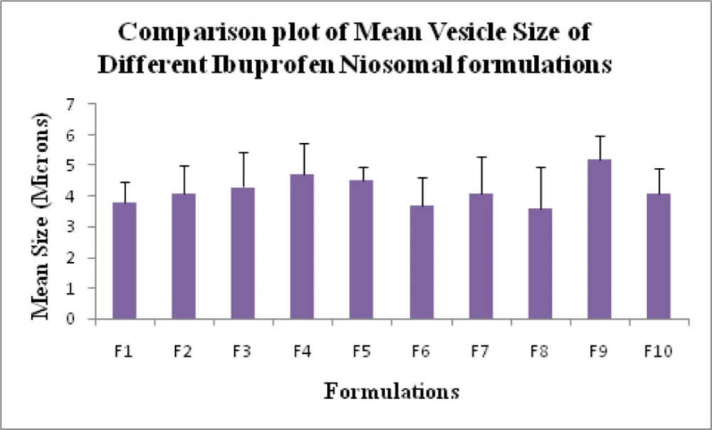
Mean vesicle size analysis of the different formulations of Ibuprofen niosomes were measured by an optical microscope. In this analysis, an optical combination of the 10 X eye-piece lens and 10X objective lens was used. The measurements of different formulations of microspheres were done in triplicate and volume mean diameter (Vd) and standard deviation were recorded (
42,
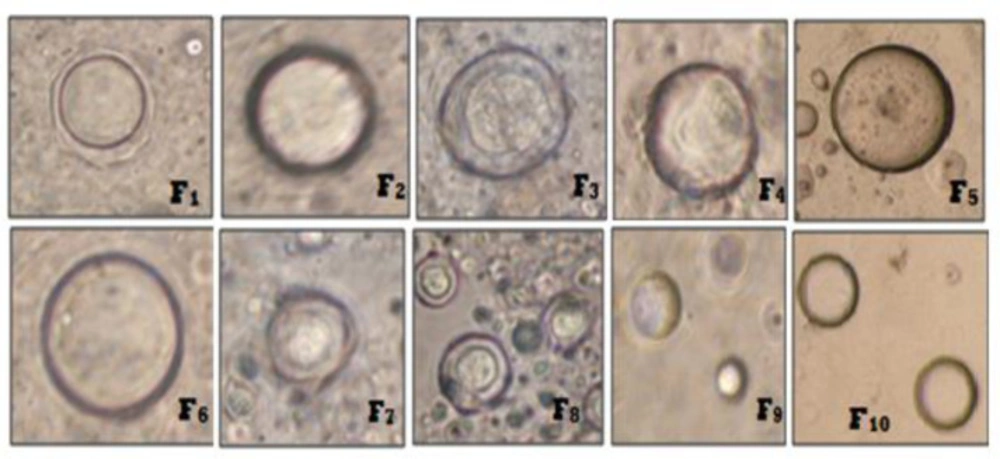
43). Besides, Phase contrast study was conducted to obtain topographical characteristics of vesicles, especially the shape and surface morphology. Different Ibuprofen loaded niosomal suspensions were deposited onto the separate glass slide and fixation was done by using a drop of glycerin. The glass slides were individually mounted on the phase contrast instrument and photographs were taken using phase contrast (Olympus Model BX 41, Japan).
Estimation of percent yield values
The total amounts of niosomes obtained were weighed by using a digital weighing balance (Citizen, Model CX 65, India). The percent yield was calculated by the following Equation (1):
Yield% = Amount of Niosomes (Drug + Cetosteryl alcohol + Surfactants) × 100
(Equation 1)
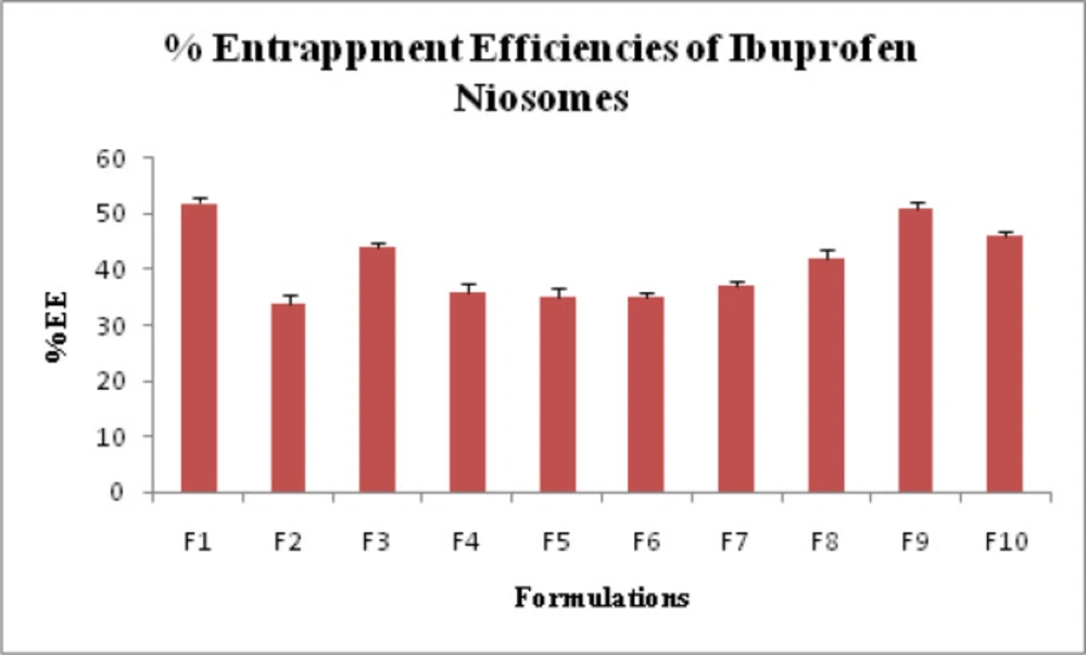
Estimation of drug entrapment efficiency (DEE)
The actual amount of Ibuprofen present in the different formulations of niosomes was estimated by ultra-centrifugation of niosomal suspension at 10,000 rpm for 1 h using at 4 °C. The amount of entrapped Ibuprofen was determined by lysis of the separated vesicles with Triton X-100. Thereafter, the filtered liquid was taken for the determination of Ibuprofen content spectrophotometrically by using a UV-VIS spectrophotometer (Shimadzu, model UV-1601 PC, Kyoto, Japan) at a wavelength of 221 nm against appropriate blank (44-46). The percent DEE was calculated by the following Equation 2:
DEE% = (Actual drug content/Theoretical drug content) × 100
(Equation 2)
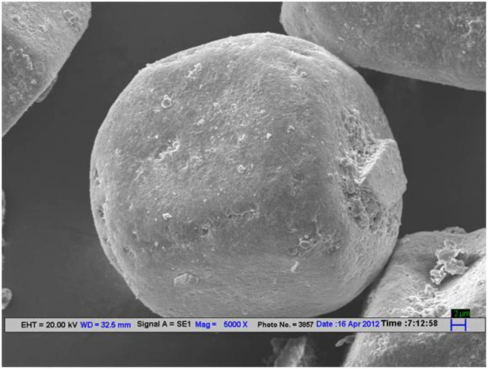
Scanning electron microscope (SEM) analysis
SEM images were taken for Tween 80 based Ibuprofen loaded niosomes (F
9). Scanning was performed using a scanning electron microscope (LEO 435VP model, Cambridge, UK). The working distance of 26 mm was maintained and the acceleration voltage used was 15 kV with the secondary electron image (SEI) as a detector (
47).
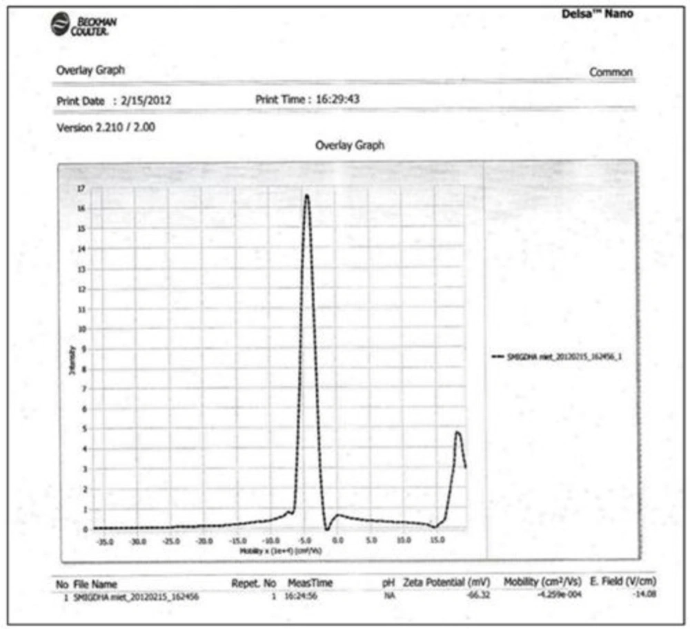
ZETA Potential study
The charges on Ibuprofen loaded vesicular surface were determined using Zetasizer DelsaTM Nano (Beckman Coulter, version 2.21) employing the phase analysis light scattering technique, which measures the particle electrophoretic mobility in a thermostated cell. The sample was analyzed 24 h after their preparation with an analysis time of 60 sec. The average zeta potential and charges were determined. The time-dependent correlation function on the scattered light intensity was measured at a scattering angle of 90°.
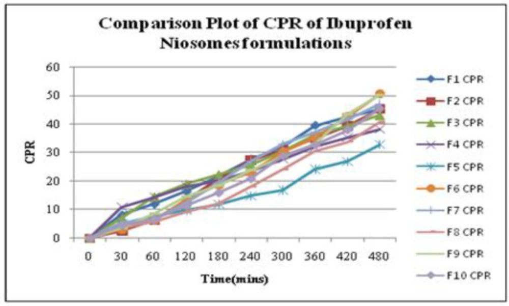
In-vitro drug release study
In-vitro release studies of Ibuprofen from niosomal formulations were determined by the “Membrane Diffusion” method. An amount equivalent of Ibuprofen was placed in a glass tube (diameter: 2.5 cm and length: 8 cm), covered with soaked osmosis cellulose membrane, that was placed in a beaker containing 50 mL of phosphate buffer (pH 5.8), which acted as receptor compartment. The temperature of the receptor medium was maintained at 37 ± 1 °C and agitated at 100 rpm speed using a magnetic stirrer. Aliquots of 2 mL samples were withdrawn periodically and sink conditions were maintained. The collected samples were analyzed at 221 nm in Double beam UV-VIS spectrophotometer using phosphate buffer (pH 5.8) as blank. The cumulative percent release (CPR) up to 8 hours was calculated for all niosomal formulations (
48,
49).
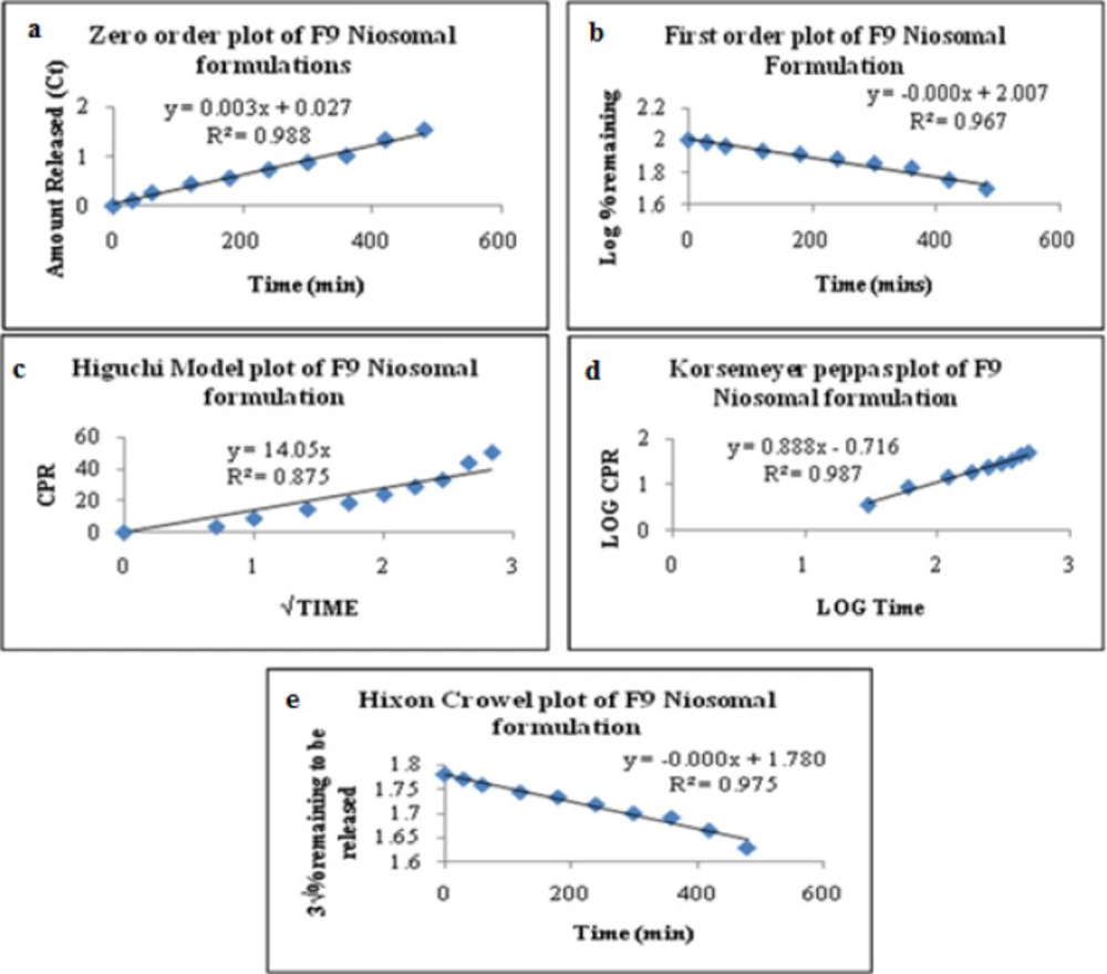
Release Kinetics of Ibuprofen Niosomes
To investigate the mode of drug release from Ibuprofen loaded niosomes, the release data were fitted with the following mathematical models (
50-
52).
Zero-order kinetics Equation 3:
Qt = k0.t
(Equation 3)
Where Qt is the amount of drug released at time t, k0 is the zero-order release rate constant, t is the time.
First-order kinetics Equation 4:
In Qt = In Q0 - k1.t
(Equation 4)
Where, Qt is the amount of drug released at time t, Q0 is the initial amount of drug in the solution, k1 is the first-order release rate constant.
Higuchi model kinetics Equation 5:
Qt = kH.t1/2
(Equation 5)
Where Qt is the amount of drug released at time t, kH is the Higuchi release rate constant.
Korsmeyer-Peppas model kinetics Equation 6:
Mt/M = KKP.tn
(Equation 6)
Where Mt is the fraction of drug released at time t, M∞ is the fraction of drug released at infinite time, KKP is the Korsmeyer-Peppas release rate constant, n is the release exponent.
Hixson-Crowell model kinetics Equation 7:
Q01/3 – Qt1/3 = KHC.t
(Equation 7)
Where Q0 is the initial amount of the drug in the dosage form, Qt is the remaining amount of drug in the dosage form at time t, KHC is the Hixson-Crowell release rate constant.
Formulation Process
Percent yield of mucilage from Balangu seeds
The percent yield of mucilage extracted from seeds of Lallemantia royaleana Benth. was found to be 8.67% w/w. Furthermore, the mucilage amount (%) used to develop niosomal gel was kept 1% and 1.5%.
Preparation of niosomal gel
The prepared niosomes were separated from aqueous medium by ultracentrifugation at 10,000 rpm at 40 °C and were gently added with a glass rod in the blank gel prepared by Carbopol-934 and
Lallemantia royaleana Benth. mucilage in different ratios. The composition of gel is given in
Table 2.
Evaluation of Niosomes Loaded Gel
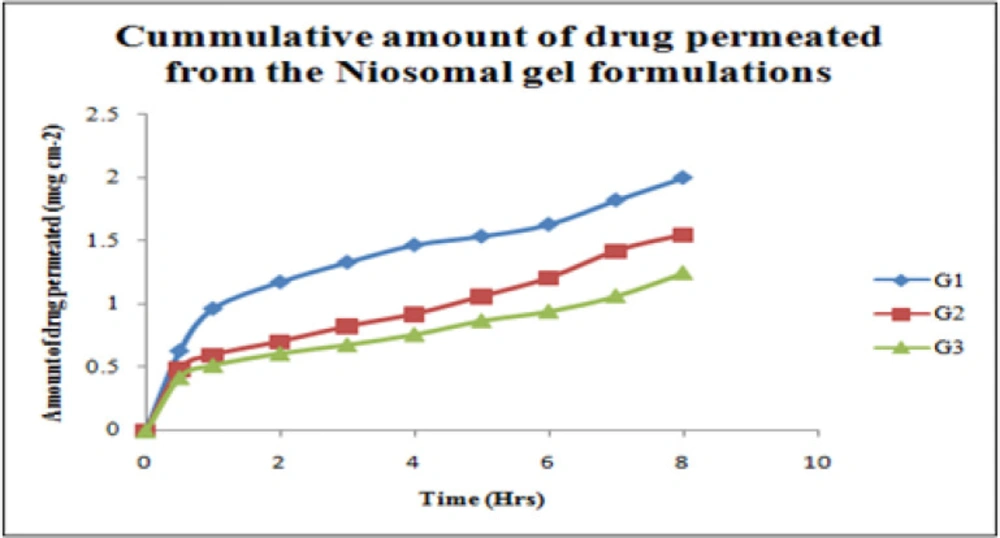
Ex-vivo diffusion studies
Franz Diffusion cell was used to performed
ex-vivo skin permeation studies for all Ibuprofen entrapped niosomal gel formulations (
53). The receptor compartment contained phosphate buffer (pH 5.8) which was constantly stirred at 100 rpm with a small magnetic stirrer and controlled temperature at 37 ± 2 °C throughout the experiment. The abdominal skin of male rats (weighing 200 g) was taken for this study. The shaved rat skin was mounted with the stratum corneum side facing upwards to the donor compartment, and the subcutaneous side was in contact with the receiver medium. The gel was placed into the donor compartment and covered with paraffin film. The sample aliquots from the receiver chamber were collected at 30 min 1, 2, 3, 4, 5, 6, 7, and 8 h respectively and analyzed by UV-VIS Spectroscopy at 221nm. The amount of drug permeated, drug flux, and the permeation coefficients were calculated for niosomes loaded gel formulations (
54,
55).
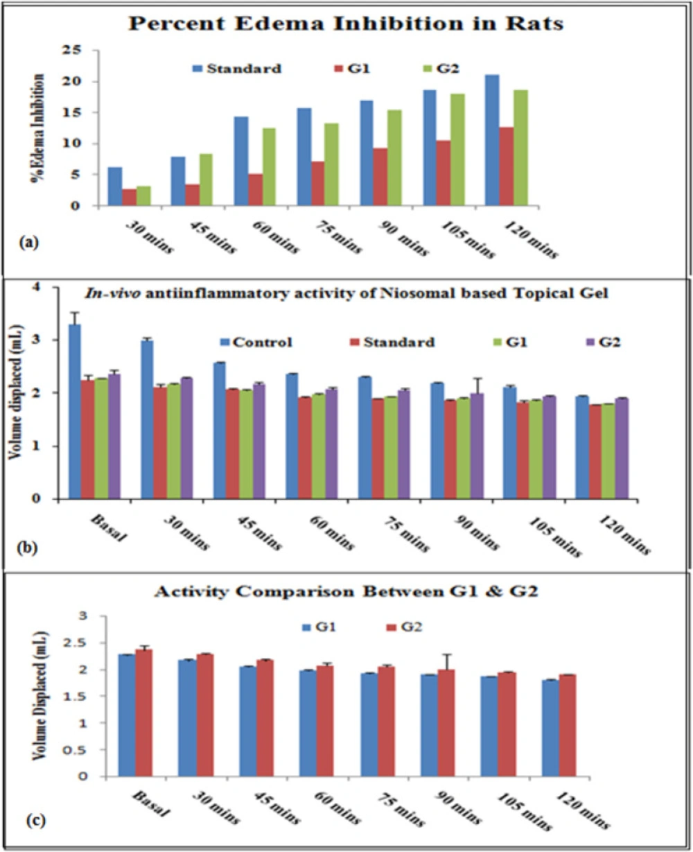
In-vivo anti-inflammatory study
The animal study has been conducted as per the Animals (Scientific Procedures) Act. All the animals used in research work were cared for by trained, accountable staff, and housed in proper facilities. Before the animal study, the animal approval was obtained by IAEC (CPCSEA No. 711/02/a/CPCSEA) Anti-inflammatory study was performed by “Carrageenan induced rat paw edema” model using 24 albino rats of either sex weighing (100-150 g) and divided into 4 groups [shown in
Table 3 (
56). In all groups, acute inflammation was induced by a sub-planter injection of 0.1 mL of freshly prepared 1% suspension of carrageenan in normal saline in the left hind paw of the rats. The paw edema volume was measured using “Plethysmometer” at every 15 min interval for 2 h after the injection of carrageenan. The average paw edema volume of all the groups were calculated and compared with that of control (
57).
The percent inhibition of edema was calculated by using the following Equation 8:
Edema inhibition% = 1 – VtVc × 100
(Equation 8)
Where, Vt = Mean edema volume of test, Vc = Mean edema volume of control.
Skin irritancy study
The skin irritation test was performed on the healthy albino rat (200 g) for the best formulation by applying niosomes loaded gel formulation on the shaved portion of rat skin. The test was performed primarily by examining the rat to notice any changes after the application of the formulation. Then photographic imaging of an exposed portion of rat skin was taken out before and after subsequent application for 72 h that is after the study period and these images were compared determining the difference with the images taken before applying the formulation (
58,
59).