Assessment of Cytotoxic and Apoptotic Effects of Salvia syriaca L. in Colorectal Adenocarcinoma Cell Line (Caco-2)

authors:

avatar Şevki Arslan , avatar Kübra Kocabıyık , avatar Doğukan Mutlu , avatar Gürkan Semiz , *

Department of Biology, Faculty of Arts and Sciences, Pamukkale University, Denizli, Turkey.

how to cite: Arslan Ş, Kocabıyık K, Mutlu D, Semiz G. Assessment of Cytotoxic and Apoptotic Effects of Salvia syriaca L. in Colorectal Adenocarcinoma Cell Line (Caco-2). Iran J Pharm Res. 2021;20(3):e124481. https://doi.org/10.22037/ijpr.2020.114254.14759.

Abstract

This work is aimed to elucidate cytotoxic and apoptotic effects of Salvia syriaca essential oil and its chemical composition by GC-MS. The human colon cancer cells (Caco-2) were treated with different essential oil concentrations for 24 h. Crystal violet test was used to determine cell viability at 630 nm by using an ELISA reader. Apoptotic processes were measured by Annexin V-FITC Apoptosis Assay Kit. Germacrene D (21.77%), trans-β-ocimene (14.66%), β-pinene (9.07%), α-cadinol (8.19%) and α-pinene (6.50%) were the main components of oil determined by GC-MS. Moreover, we observed that the cytotoxic effect was increased with an increasing dose of essential oil. The EC50 value was calculated as 63.5 µg/mL. An increase in the percentage of apoptotic cells was observed after treatment of Caco-2 cells with S. syriaca essential oil revealed by image-based cytometry. A nearly 6-fold increase was found in annexin-positive cells after treatment. In terms of mRNA levels, RT-PCR analysis indicated that, although Bax and Caspase-3 were increased, Bcl-2 was decreased after oil treatment. According to our results, S. syriaca essential oil has promising phytochemicals that might be useful in cancer treatment due to their relatively cytotoxic and apoptotic activities in Caco-2 cells.

Introduction

It is well established that most of the plants have been used widely as complementary and alternative therapy. In this regard, Turkey is a very rich country in terms of its flora, including around 12.000 plants species (1). The majority of the Turkish people use most of these plants for different purposes. Salvia species are one of the best-known plants used for medical purposes in Turkey as well as in the world. Salvia is considered one of the important genus in Lamiaceae with more than 1000 species (2). Most of the Salvia species are widely consumed in traditional medicine for the treatment of cold, stomach aches or sore throat, digestive problems and many other problems in Turkey (3-6). They have many different biological activities, including anti-tumoral, anti-carcinogenic, antioxidant, anti-diabetic, anti-inflammatory, anti-rheumatic, antimicrobial and hepatoprotective activities that are due to their high content of secondary metabolites, including flavonoids and terpenoids (7-16). In addition to their biological activities, some Salvia species are used in cosmetic and food industries (17). Therefore, Salvia species have paid attention due to their economic importance and their wide diverse biological activities. Among the Salvia species, Salvia officinalis L. (sage) is one of the most widely used and studied species (13, 14). In Turkey, there are 100 Salvia species, and 57 of them are endemic (18). That is, Anatolia shows remarkable richness for genus Salvia (19). One of the most common species is Salvia syriaca L. which is 30-60 (-80) cm, a rhizomatous and perennial herb in Turkey. Its stem is yellowish-green, erect and branched, eglandular-pubescent below and denser above (and rarely glandular). Leaves simple, (5) 6-13 (16) × (3) 4-8 (10) cm, ovate, chordate, rugose; petiole 3-6 cm. Calyx 5-10 mm, tubular, upper lip straight, tridentate. Corolla 8-12 mm, white, tube straight. The species is mainly spread in Inner Anatolia and North of the Middle East, recognized by the tidy habit, small white corolla and regularly ovate leaves (20, 21). Through its wide distribution, there is no deep information about the chemical composition and cytotoxic activity of S. syriaca essential oil. In this respect, the main object of the present study was to determine the cytotoxic and apoptotic effects of S. syriaca oil in Caco-2.

Experimental

Chemicals

The purchased chemicals were provided from Sigma-Aldrich Chemical Company (St Louis, Missouri, USA): fetal bovine serum (FBS), Dulbecco’s Modified Eagle’s medium (DMEM), bovine serum albumin (BSA). The Assay Kit (Annexin V-FITC Apoptosis, Biovision, CA, USA) was used to evaluate cell apoptosis. The remaining chemicals and solvents used in this study were purchased at the highest grade of purity.

Plant material

S. syriaca was collected from Burdur (Southwest of Turkey) during its flowering period (May-July) in 2018. It was taxonomically identified by Prof. Gürkan Semiz from herbarium specimens of the Biology Department, Pamukkale University. Voucher specimens were deposited in the Chemical Ecology Laboratory under code GSE2343. Small cut aerial parts of the plant were air-dried for one week at R.T.

Essential oil preparation

Air-dried plant materials (100 g) were subjected to hydro-distillation for 3 h using a Clevenger apparatus to obtain an oil. Anhydrous sodium sulfate solution was used to dry the obtained oil, and then the oil was kept in a glass vial at 4 °C until usage.

GC-MS analysis of the essential oil

About 15 μL of hexane diluted oil (1:100) was subjected to GC-MS analysis by using Agilent Technologies 7820A model GC system equipped with 5975 inert MSD. The samples were analyzed on a 30 m long HP5-MS capillary column (ID 0.25 mm, film thickness 0.25 mm, Hewlett Packard). The column temperature was programmed at 50 °C for 3 min then raised to 250 °C at a rate of 5 °C/min and kept constant at 250 °C for 5 min. The helium (flow rate 1.2 mL/min) as a carrier gas and SCAN technique was used. Wiley 7 MS and NIST02 libraries were used for the identification of the compounds based on comparing the mass spectral data and known compounds in the literature. The percentages were calculated from GC peaks with the normalization method.

Cell culture and cytotoxicity assay

Caco-2 cells provided from the European Collection of Animal Cell Culture (ECACC, UK) were used in this study. The cells were grown in a DMEM medium with 10% FBS, 1% of L-glutamine, 50 μg/mL streptomycin, and 50 IU/mL penicillin in a CO2 incubator. 5000 cells/well were sown to 96 well-plate. After 24 h, different concentrations of S. syriaca essential oil (1 µg/mL and 100 µg/m l9 were applied to cells. Dimethylsulfoxide (DMSO) was used for the preparation of oil solutions, and its concentration did not exceed 0.5%. For control cells, 0.5% DMSO concentration was also applied. After incubation of another 24 h, medium containing floating cells were taken out, and crystal violet solution was used to stain attached cells. The final color was measured at 630 nm. The absorbance values were used to calculate the EC50 concentration of the S. syriaca oil. This cytotoxicity experiment was repeated three times to determine the EC50 value.

Apoptosis

Cells were sown in six-well plates at a concentration of 2 × 10⁵ cells/wells and exposed to essential oils (EC50 doses). H2O2 was used as a positive control. After 24 h, cells were gathered and stained by using Annexin V-FITC Apoptosis Assay Kit (Biovision, CA, USA) as described in the product manual. For each treatment, 25 μL of cell suspension was loaded into one Arthur™ image-based cytometer slide and analyzed. For each treatment, 20 fields were analyzed, and around 7000 cells were counted. The percentage of apoptotic cells was calculated over the total cell population. This experimental set-up was performed two times, and the apoptotic cell percentage was represented as the mean of independent experimental sets ± SEM.

RNA isolation and determination of mRNA expression by RT-PCR

Caco-2 cells were treated with an EC50 dose of S. syriaca essential oil and incubated for 24 h. Then, cells were collected, and RNA was isolated by using InnuPREP RNA Mini Kit 2.0 (Analytic Jena, Germany) followed the instructions. Agarose gel electrophoresis was used to check the integrity of isolated RNA. The concentration of isolated RNAs was determined by measuring optical density (A260/A280 ratio). OneScript® Plus cDNA Synthesis Kit (ABM, USA) was used for cDNA synthesis. The reaction mixture consisted of 2 μg RNA, 0.5 μM oligo(dT)18 reverse transcription primer, dNTP, RT buffer, RNaseOFF Ribonuclease Inhibitor (20 Units), OneScript® Plus RTase (200 Units) and nuclease-free water. Quantitative real-time PCR assays for Bax, Bcl-2 and Caspase-3 were done by using Applied Biosystems™ StepOnePlus™ Real-Time PCR System (Thermo, USA). RT-PCR reactions were carried out in 10 µL volumes using ABM KiloGreen 2x qPCR MasterMix (ABM, USA). All samples were performed in duplicates. Nuclease-free water was used as a negative control. For the determination of fold changes in mRNA levels, the 2−ΔΔCt method was used as described previously (22). β-actin, a reliable housekeeping gene, was used as an internal control.

Statistical analysis

The results were given as means ± SD of at least three replicates. Minitab statistical software was used for statistical analyses. For comparisons of the groups, Student’s t-test was used. p < 0.05 was preferred for statistical significance.

Results and Discussion

The chemical composition of S. syriaca oil was given in Table 1. The obtained oil was pale yellow with a strong smell and a yield of 0.14%. A total of twenty-nine compounds have been identified, corresponding to 99.28% of the total composition of S. syriaca essential oil.

The main compounds were found as: germacrene-D (21.77 %), trans-β-ocimene (14.66%), β-pinene (9.70%), α-cadinol (8.19%), α-pinene (6.50%) and γ-cadinene (6.40%). In this study, we first time showed cytotoxic and apoptotic effects of S. syriaca essential oil on colon cancer cell line. In current literature, there have been few studies about the chemical composition of S. syriaca essential oils. Previously, Turkish S. syriaca oils were characterized by germacrene-D (33.83%) and bicyclogermacrene (12.47%) as the most important components (23). Even the major compound, namely germacrene-D, was found in both studies, the composition of the oils was totally different. Furthermore, twenty-two compounds were determined in the oil of S. syriaca obtained from Iran and germacrene-B (34.8%), germacrene-D (29.2%), α-ylangene (3.6%) and spathulenol (3.4%) were the main compounds (24). In a study from Jordan, the main compounds were found to be thymol, α-pinene and isobornyl acetate for S. syriaca (25). In another study from the West Azerbaijan Province (Iran), it was found that the main compounds of S. syriaca were 1,8-cineole (46.45%), camphor (27.58%) and bornyl acetate (4.66%) (26). It is well established that the chemical composition of plant essential oils was changed due to genetic differences, weather/soil conditions, time of harvest, the drying technique, etc. Due to these important factors, the chemical composition of S. syriaca obtained from this study differed from the other studies. In our study, the most abundant component was identified as germacrene-D (21.77%). The biological function of germacrene-D is not known completely in plants. It was thought that germacrene-D is responsible for the productions of other compounds in different organisms (27, 28) and also in plant-herbivore interactions as a deterrent (29, 30). The essential oils contain β-ocimene as a major compound (the second most abundant compound in our study) showed cytotoxic and anti-carcinogenic effects on different cell lines, including Caco-2 (31, 32).

In addition to the determination of chemical content of essential oil, the cytotoxic activity of S. syriaca oil was determined in the Caco-2 cell line. Caco-2 cells are important in testing cytotoxic activities of chemicals and experimental pharmacology studies. Moreover, it is well established that Salvia species are consumed by preparing tea using their leaves. Due to these reasons, Caco-2 cell lines were selected in this study. As seen in Figure 1, essential oil showed a cytotoxic effect on Caco-2 cells in a dose-dependent manner, and toxicity started at 5 μg/mL.

The EC50 value of the essential oil obtained from S. syriaca was found to be 63.5 µg/ml. The extracts prepared using different parts of S. syriaca and different solvents showed similar cytotoxic effects (33, 34).

Aqueous crude extract obtained from S. syriaca showed a cytotoxic effect, and EC50 value was found 94.85 µg/mL (35). Moreover, roots methanol extract of S. syriaca showed a cytotoxic effect on Caco-2 cells (34). As seen in these studies, EC50 values were different in each study due to their chemical content differences in extract or extraction methods. Similar to our results, essential oils obtained from other Salvia species showed cytotoxic activity in different cancer cells (35-37). However, in a comparative study, Firuzi et al. (2013) reported different extracts of S. syriaca had no or little effect on different human cancer cell lines among the other tested species (38). In order to understand the mechanisms of cytotoxic effect, apoptosis analysis was performed by applying EC50 dose to Caco-2 cells in this study. It is well established that one of the important strategies to treat cancer is the induction of apoptosis in tumor cells. Plant extracts or pure compounds obtained from extracts have been shown to induce apoptosis in different cancer cells. Therefore, screening of apoptosis in plant extracts is so important. As given in the method part, apoptosis was determined by Arthur image-based cytometer and the cell proportions were quantified as live, dead and apoptotic cells (Figures 2 and 3). H2O2, the apoptosis-inducing agent, was chosen as the positive control.

As shown in Figure 2, after 24 h of application with essential oil, 37% of the Caco-2 cells were apoptotic, 45% are viable, and 17% are dead. The ratio of apoptotic cell population in S. syriaca treatment was approximately 6-fold higher than the control. These results clearly showed that essential oil caused induction of apoptosis and have a cytotoxic effect. Also, in treated cells, a significant decrease was noticed in the percentage of live cells with the comparison to the control ones (Figures 2 and 3). Besides, RT-PCR results showed that S. syriaca essential oil increased the pro-apoptotic Bax (10.74-fold) and decreased anti-apoptotic Bcl-2 (2.45-fold) mRNA levels (Figure 4). Oil treatment increased Bax/Bcl-2 ratio as well. The essential oil also caused a 1.7-fold increase in Caspase-3 mRNA level (Figure 4). It is well known that the two main pathways of apoptosis are extrinsic and intrinsic pathways. Each pathway requires different caspase enzymes that cause activation of these pathways by Caspase-3. Based on the results obtained from our study, the intrinsic pathway of apoptosis was involved in S. syriaca-induced cell death.

Similar to our observation, other Salvia species also showed apoptotic effects in different cell lines (39-40).

Table 1

The chemical composition of the essential oil obtained from S. syriaca

NoCompounds*RI (%)NoCompoundsRI (%)
1tricyclene9191.0516myrtenal11890.30
2α-pinene9336.5017α-terpineol11980.46
3camphene9481.3318trans-carveol12170.05
4β-pinene9679.7019bornyl acetate12810.38
5myrcene9881.1820bicycloelemene13310.24
63-carene10140.2921α-cubenene13570.48
7α-terpinene10174.3222α-copaene13772.23
8d-limonene10331.8523β-elemene13883.27
9trans-β-ocimene104714.6624α-cedrene14161.41
10α-terpinolene10864.2825trans-β-caryophyllene14234.93
11linalool10991.1126germacrene-D147921.77
12trans-pinocarveol11440.2827γ-cadinene15246.40
13camphor11500.1328α-calacorene15360.40
14borneol11651.2529α-cadinol16508.19
15terpinene-4-ol11760.76Total99.28
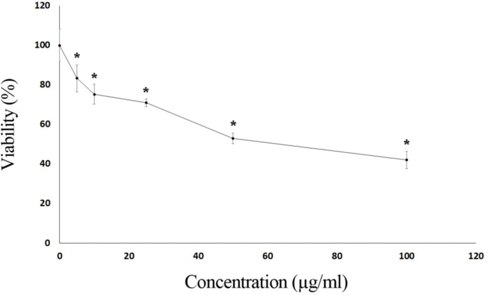
Cytotoxic effects of S. syriaca essential oil in CaCo-2 cells. Cells were exposed to different concentrations of essential oil for 24 h. Results are mean ± SD values for three independent experiments (*p < 0.05)
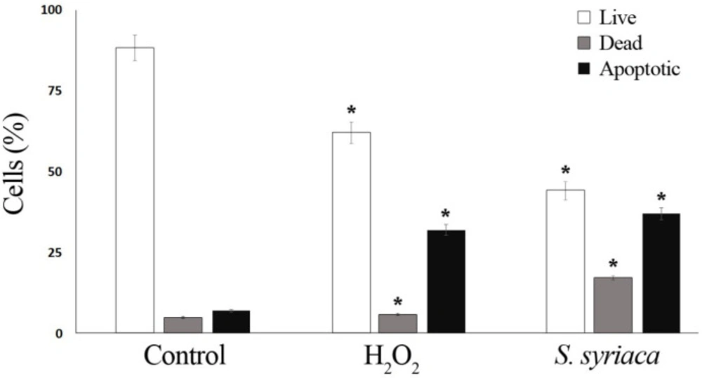
Apoptotic effects of S. syriaca essential oil by image-based cytometer. Caco-2 cells were assessed for apoptosis after a 24-hour incubation period with oil. H2O2 (25 mM) was applied as a positive control (*p < 0.05)
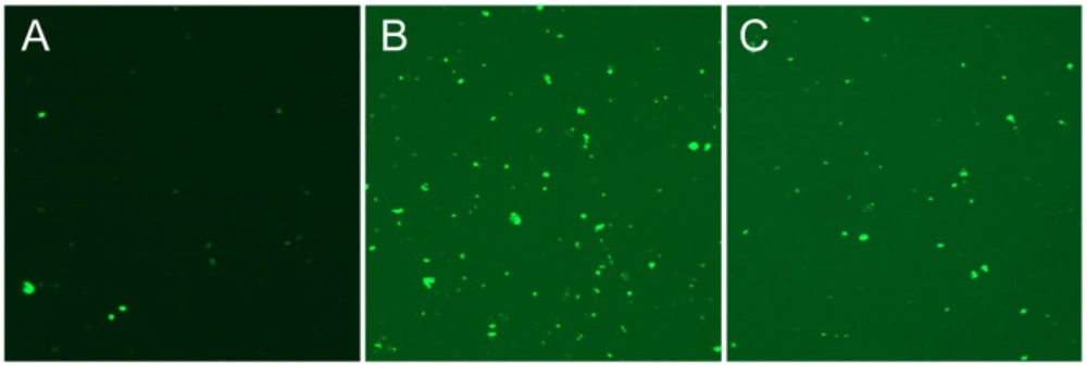
Annexin V staining of Caco2 cancer cells for 24 h with EC50 concentration of S. syriaca essential oil. (A) Control, (B) H2O2 treated and (C) S. syriaca treated cells
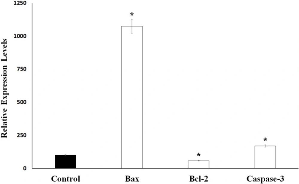
Bax, Bcl-2 and Caspase-3 mRNA levels in control and S. syriaca essential oil-treated Caco-2 cells. Individual gene expression levels were normalized by using β-actin. The value obtained from control cells was taken to be 100%, and the values obtained from the S. syriaca oil-treated cells were expressed as a percentage of control (*p < 0.05)

Conclusion

This is the first record of the cytotoxicity and apoptotic effects of S. syriaca in Caco-2 cells. The oil obtained from S. syriaca has promising phytochemicals that may be used in cancer treatment. Moreover, an activity-guided fractionation experiment should be done to test our hypothesis. Also, further works are necessary to find the molecule (s) responsible for cytotoxic and apoptotic effects of S. syriaca.

Acknowledgements

References

  • 1.

    M Güner A, Aslan S, Ekim T, Vural M, Babaç MT. Türkiye Bitkileri Listesi (Damarlı Bitkiler). Nezahat Gökyiğit Botanik Bahçesi ve Flora Araştırmaları Derneği Yayını. 1st ed. İstanbul; 2012. 15 p.

  • 2.

    Živković J, Ristić M, Kschonsek J, Westphal A, Mihailović M, Filipović V, Böhm V. Comparison of chemical profile and antioxidant capacity of seeds and oils from Salvia sclarea and Salvia officinalis. Chem. Biodiversity. 2017;14:e1700344.

  • 3.

    Altundag E, Ozturk M. Ethnomedicinal studies on the plant resources of east Anatolia, Turkey. Procedia Soc. Behav. Sci. 2011;19:756-77.

  • 4.

    Akbulut S, Bayramoglu MM. The trade and use of some medical and aromatic herbs in Turkey. Stud. Ethno. Med. 2013;7:67-77.

  • 5.

    Parsaeea H, Asilib J, Mousavic SH, Soofid H, Emamib SA, Tayarani-Najarane Z. Apoptosis induction of Salvia chorassanica root extract on human cervical cancer cell line. Iran. J. Pharm. Res. 2013;12:75-83. [PubMed ID: 24250574].

  • 6.

    Tayarani-Najaran Z, Asilib J, Aioubib E, Emami SA. Growth Inhibition and Apoptosis Induction of Salvia chloroleuca on MCF-7 Breast Cancer Cell Line. Iran. J. Pharm. Res. 2013;12:789-99. [PubMed ID: 24523759].

  • 7.

    Baričević D, Bartol T. The biological/pharmacological activity of the Salvia genus. In: Kintzios SE, editor. SAGE the genus Salvia. 1st ed. Amsterdam: Harwood Academic Publishers; 2000. p. 143-84.

  • 8.

    Abreu ME, Muller M, Alegre L, Munné-Bosch S. Phenolic diterpene and α-tocopherol contents in leaf extracts of 60 Salvia species. J. Sci. Food Agric. 2008;88:2648-53.

  • 9.

    Akkol EK, Göger F, Koşar M, Başer KHC. Phenolic composition and biological activities of Salvia halophila and Salvia virgata from Turkey. Food Chem. 2008;108:942-9. [PubMed ID: 26065756].

  • 10.

    Kivrak İ, Duru ME, Öztürk M, Mercan N, Harmandar M, Topçu G. Antioxidant, anticholinesterase and antimicrobial constituents from the essential oil and ethanol extract of Salvia potentillifolia. Food Chem. 2009;116:470-9.

  • 11.

    Alimpić A, Kotur N, Stanković B, Marin PD, Matevski V, Al Sheef NB, Duletić-Laušević S. The in-vitro antioxidative and cytotoxic effects of selected Salvia species water extracts. J. Appl. Bot. Food Qual. 2015;88:115-9.

  • 12.

    Alimpić A, Knežević A, Milutinović M, Stević T, Šavikin K, Stajić M, Marković S, Marin PD, Matevski V, Duletić-Laušević S. Biological activities and chemical composition of Salvia amplexicaulis Lam. extracts. Ind. Crop. Prod. 2017;105:1-9.

  • 13.

    Cutillas AB, Carrasco A, Martinez-Gutierrez R, Tomas V, Tudela J. Salvia officinalis L essential oils from Spain: determination of composition, antioxidant capacity, antienzymatic, and antimicrobial bioactivities. Chem. Biodiversity. 2017;14:e1700102.

  • 14.

    Ghorbani A, Esmaeilizadeh M. Pharmacological properties of Salvia officinalis and its components. J. Tradit. Complement. Med. 2017;7:433-40. [PubMed ID: 29034191].

  • 15.

    Jiang G, Liu J, Ren B, Zhang L, Owusu L, Liu L, Zhang J, Tang Y, Li W. Anti-tumor and chemosensitization effects of Cryptotanshinone extracted from Salvia miltiorrhiza Bge on ovarian cancer cells in-vitro. J. Ethnopharmacol. 2017;205:33-40. [PubMed ID: 28456578].

  • 16.

    Kolac UK, Ustuner MC, Tekin N, Ustuner D, Colak E, Entok E. The Anti-inflammatory and antioxidant effects of Salvia officinalis on lipopolysaccharide-induced inflammation in rats. J. Med. Food. 2017;20:1193-200. [PubMed ID: 29131698].

  • 17.

    Kaya A, Demirci B, Başer KHC. Glandular trichomes and essential oils of Salvia glutinosa L. S. Afr. J. Bot. 2003;69:422-7.

  • 18.

    Sen-Utsukarci B, Gurdal B, Bilgin M, Satana D, Demirci B, Tan N, Mat A. Biological activities of various extracts from Salvia cassia Sam ex Rech and chemical composition of its most active extract. Rec. Nat. Prod. 2019;13:24-36.

  • 19.

    Celep F, Dirmenci T, Güner Ö. Salvia hasankeyfense (Lamiaceae), a new species from Hasankeyf (Batman, South-eastern Turkey). Phytotaxa. 2015;227:289-94.

  • 20.

    Celep F. Revision of the genus Salvia L (Labiatae) in the Mediterranean and the Aegean geographic regions of Turkey [dissertation]. Ankara, Middle East Technical University. 2010:287.

  • 21.

    Davis PH. Flora of Turkey and the East Aegean Islands. Edinburgh: Edinburgh University Press; 1982. p. 7-29.

  • 22.

    Ozgun-Acar O, Celik-Turgut G, Gazioglu I, Kolak U, Ozbal S, Ergur BU, Arslan S, Sen A, Topcu G. Capparis ovata treatment suppresses inflammatory cytokine expression and ameliorates experimental allergic encephalomyelitis model of multiple sclerosis in C57BL/6 mice. J. Neuroimmunol. 2016;298:106-16. [PubMed ID: 27609283].

  • 23.

    Baser KHC, Demirçakmak B, Ermin N. Essential oil of Salvia syriaca L. J. Essent. Oil. Res. 1996;8:105-6.

  • 24.

    Sefidkon F, Mirza M. Chemical composition of the essential oils of two Salvia species from Iran, Salvia virgata Jacq and Salvia syriaca L. Flavour Fragr. J. 1999;14:45-6.

  • 25.

    Flamini G, Cioni PL, Morelli I, Bader A. Essential oils of the aerial parts of three Salvia species from Jordan: Salvia lanigera S spinosa and S syriaca. Food Chem. 2007;100:732-5.

  • 26.

    Forouzin F, Jamei R, Heidari R. Comparison of essential oil components and antioxidant activity between Salvia syriaca and Salvia aristata in their natural habitats in west Azerbaijan province, Iran. J. Pharm. Pharmacol. 2015;3:400-4.

  • 27.

    Bülow N, König WA. The role of germacrene D as a precursor in sesquiterpene biosynthesis: Investigation of acid catalyzed, photochemically and thermally induced rearrangements. Phytochem. 2000;55:141-68.

  • 28.

    Telascrea M, de Araújo CC, Marques MOM, Facanali R, de Moraes PLR, Cavalheiro AJ. Essential oil leaves of Cryptocarya mandioccana Meisner (Lauraceae): Composition and intraspecific chemical variability. Biochem. Syst. Ecol. 2007;35:222-32.

  • 29.

    Bruce TJA, Birkett MA, Blande J, Hooper AM, Martin JL, Khambay B, Prosser I, Smart LE, Wadhams LJ. Response of economically important aphids to components of Hemizygia petiolata essential oil. Pest Manag. Sci. 2005;61:1115-21. [PubMed ID: 16059962].

  • 30.

    Kiran SR, Devi PS. Evaluation of mosquitocidal activity of essential oil and sesquiterpenes from leaves of Chloroxylon swietenia DC. Parasitol. Res. 2007;101:413-8. [PubMed ID: 17520288].

  • 31.

    Mahmoud GI. Biological effects, antioxidant and anticancer activities of marigold and basil essential oils. J. Med. Plants Res. 2013;7:561-72.

  • 32.

    Misra LN, Vyry Wouatsa NA, Kumar S, Venkatesh Kumar R, Tchoumbougnang F. Antibacterial, cytotoxic activities and chemical composition of fruits of two Cameroonian Zanthoxylum species. J. Ethnopharmacol. 2013;148:74-80. [PubMed ID: 23567034].

  • 33.

    Kasabri V, Afifi FU, Abu-Dahab R, Mhaidat N, Bustanji YK, Abaza IF, Mashallah S. In vitro modulation of metabolic syndrome enzymes and proliferation of obesity related-colorectal cancer cell line panel by Salvia species from Jordan. Rev. Roum. Chim. 2014;59:693-705.

  • 34.

    Abdallah Q, AlDeeb I, Bader A, Hamam F, Saleh K, Abdulmajid A. Anti-angiogenic activity of middle east medicinal plants of the Lamiaceae family. Mol. Med. Rep. 2018;18:2441-8. [PubMed ID: 29901194].

  • 35.

    Al-Kalaldeh JZ, Abu-Dahab R, Afifi FU. Volatile oil composition and antiproliferative activity of Laurus nobilis, Origanum syriacum, Origanum vulgare, and Salvia triloba against human breast adenocarcinoma cells. Nutr. Res. 2010;30:271-8. [PubMed ID: 20534330].

  • 36.

    Erdogan E, Everest A, De Martino L, Mancini E, Festa M, De Feo V. Chemical composition and in-vitro cytotoxic activity of the essential oils of Stachys rupestris and Salvia heldreichiana, two endemic plants of Turkey. Nat. Pro. Comm. 2013;8:1637-40.

  • 37.

    Khosravi Dehaghi N, Ostad SN, Maafi N, Pedram S, Ajani Y, Hadjiakhoondi A, Khanavi M. Cytotoxic activity of the essential oil of Salvia verticillata L. Res. J. Pharmacogn. 2014;1:27-33.

  • 38.

    Firuzi O, Miri R, Asadollahi M, Eslami S, Jassbi AR. Cytotoxic, antioxidant and antimicrobial activities and phenolic contents of eleven Salvia species from Iran. Iran. J. Pharm. Res. 2013;12:801-10. [PubMed ID: 24523760].

  • 39.

    Russo A, Carmen F, Rigano D, Cardile V, Arnold NA, Senatore F. Comparative phytochemical profile and antiproliferative activity on human melanoma cells of essential oils of three lebanese Salvia species. Ind. Crops Prod. 2016;83:492-9.

  • 40.

    Russo A, Formisano C, Rigano D, Senatore F, Delfine S, Cardile V, Rosselli S, Bruno M. Chemical composition and anticancer activity of essential oils of Mediterranean sage (Salvia officinalis ) grown in different environmental conditions. Food Chem. Toxicol. 2013;55:42-7. [PubMed ID: 23291326].