Materials
Scopolamine, D-galactose, pentylenetetrazole, pentobarbital, and flumazenil were purchased from Sigma-Aldrich (Steinheim, Germany). Diazepam, celecoxib, and naloxone were kindly donated by Darou Pakhsh Pharmaceutical Factory (Tehran, Iran). Dimethyl sulfoxide (DMSO) was obtained from Merck (Darmstadt, Germany). Dorema ammoniacum gum with the voucher specimen no. 8061 was obtained from the Department of Pharmacognosy at Shahid Beheshti University of Medical Sciences (Tehran, Iran). A 500 mL round-bottomed flask connected to a Clevenger apparatus was used for the essential oil extraction by water distillation. The essential oil in the volume of 1.3 mL was obtained from 100 grams of DAG. The density of the essential oil was estimated to be 0.384 g/mL, using the gravimetric method at room temperature. The essential oil was stored in the refrigerator until the day of the experiment.
Animals and drug administration
All experimental procedures were executed in accordance with the institutional procedures of the Animal Experimentation Committee of Shahid Beheshti University of Medical Sciences, Tehran, Iran (Code number: IR.SBMU.PHNM.1395.411). Male C57BL/6J mice (weight, 22-25 g) were used in the D-galactose-induced accelerated aging model and the rest of the experiments were conducted on male NMRI mice (weight, 18-22 g). The mice were kept in the animal house under the same ambient conditions (22 ± 2 ºC, 50% ± 10% humidity) and a 12-h diurnal light cycle with free access to food and water. The animals were randomly divided into groups of ten, and each mouse was used only once. The mice were intraperitoneally treated with the DAG-EO (diluted in sesame oil) in the volume of 5 mL/kg. Scopolamine, D-galactose, pentylenetetrazole, pentobarbital, celecoxib, and naloxone were dissolved in normal saline (0.9%, w/v) and administered in the volume of 10 mL/kg by intraperitoneal (i.p.) route. Diazepam and flumazenil were dissolved in DMSO. The injection volume of diazepam and flumazenil was decreased to a constant volume of 5 mL/kg body weight (i.p.) to reduce the toxicity of DMSO. In all experiments, the animals in the vehicle group received sesame oil.
Biological activity
D-galactose- and scopolamine-induced memory impairment
The mice were subcutaneously treated with D-galactose at a dose of 100 mg/kg once daily for 49 consecutive days to generate D-galactose-induced aging mice (
13). Sham group received normal saline instead of D-galactose. Scopolamine-induced memory impairment was induced by the injection of a single dose of scopolamine (1 mg/kg). Memory-related behavioral responses were measured in the step-through passive avoidance and Y-maze tasks to evaluate the effect of DAG-EO on D-galactose- and scopolamine-induced memory impairment models.
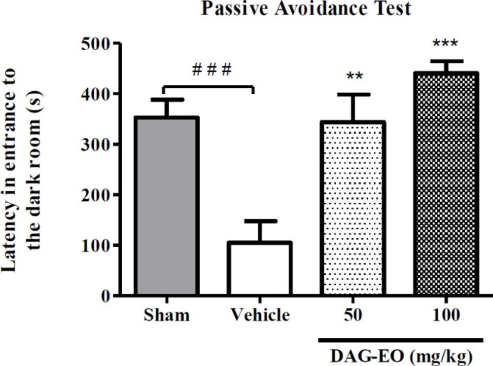
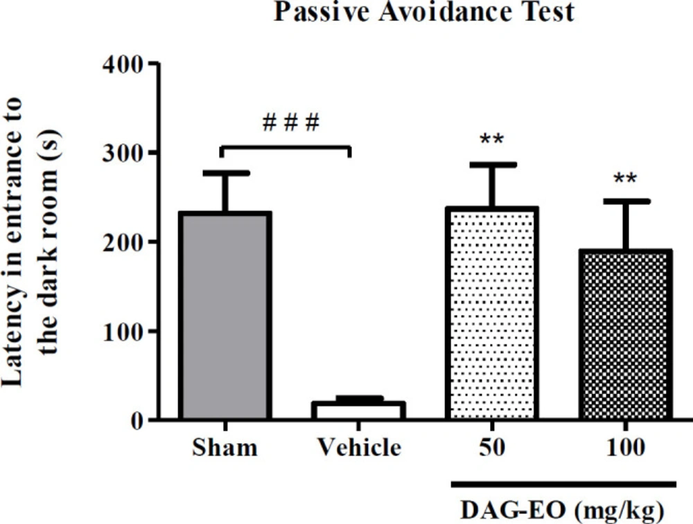
Step-through passive avoidance test
DAG-EO (50 and 100 mg/kg) were administered once daily for 7 consecutive days within the last 7 days injection of D-galactose. The step-through passive avoidance test was performed as previously described (
14). To summarize, one hour after the last administration of the essential oil, each mouse was placed in the bright compartment of a light-dark apparatus and received a constant current electrical stimulation (50 Hz, 0.2 mA, 2 s) when they entered the dark compartment. On the next day, the mice were again placed into the illuminated part of the apparatus, and latency in the entrance to the dark compartment was recorded. In the scopolamine-induced memory impairment test, the mice were treated with different doses of the essential oil (50 and 100 mg/kg). Following 15 min, they received scopolamine (1 mg/kg) and 15 min later, the step-through passive avoidance test was performed as described above.
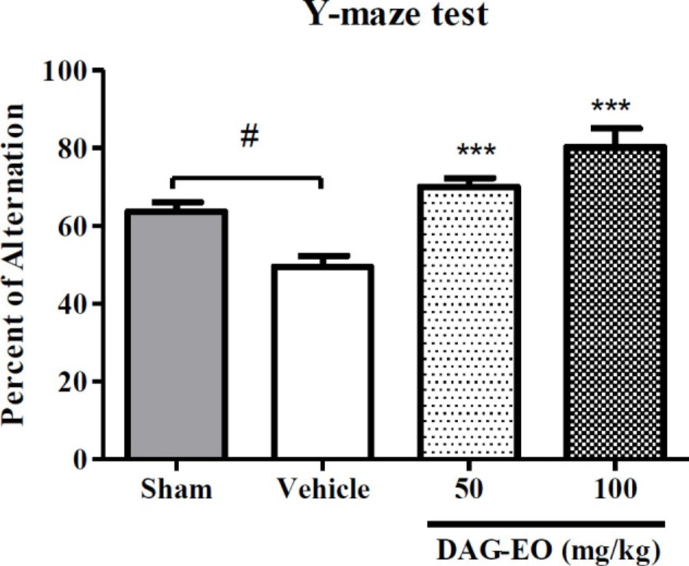
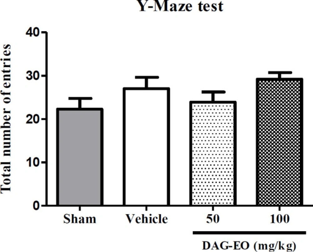
Y-maze test
Y-maze apparatus made of three opaque plastic arms (A, B, and C) at a 120° angle from each other and a central zone was used according to the method described by Huh
et al. (
15). One hour after the last administration of DAG-EO in D-galactose-induced memory impaired group and 15 min after the injection of scopolamine in the scopolamine-induced memory impaired mice, each mouse was placed in the central zone of the apparatus and allowed to freely explore the three arms. The number of arm entries was recorded over 10 min, and the percentage of spontaneous alternation was calculated by the following Equation:
Spontaneous alternation% = [(Number of alternations)/(Total arms entries-2)] × 100
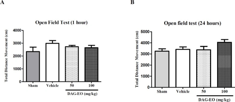
Open field test
The open field test was performed to evaluate the motor activity of the mice following the administration of DAG-EO (
16). A cubic chamber (40 × 40 cm) made of clear Plexiglas wall (40 cm high) was used in this experiment. Locomotion of each mouse was recorded for 10 min in the open field arena, using a digital camera placed above the chamber. The test was performed one hour after the last administration of DAG-EO on day 49 and 24 h later on day 50. An automated tracking system (Ethovision XT software, Noldus, The Netherlands) was used in order to analyze the recorded videos, and total distance movement in cm was considered as the total locomotor activity of the animals.
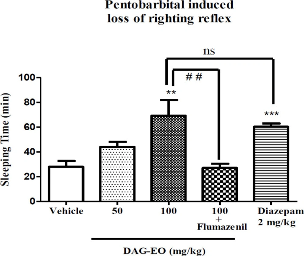
Pentobarbital-induced sleep test
This test was employed to evaluate the hypnotic effect of the essential oil (
17). The mice at different groups received DAG-EO (50 and 100 mg/kg), diazepam (2 mg/kg) or vehicle. Pentobarbital at the dose of 60 mg/kg was used for sleep induction 30 min following each treatment, and sleep duration (the duration between loss and recovery of righting reflex) was recorded.
Pentylenetetrazole-induced seizures test
The anticonvulsant activity of the essential oil was evaluated in PTZ and MES induced seizure tests. PTZ test was conducted according to what explained by Mohammadi-Khanaposhtani
et al. (
18). To summarize, the mice were treated with DAG-EO, diazepam as the positive control, and vehicle. The ability of each treatment in the protection of mice against the lethal dose of PTZ (100 mg/kg) was evaluated by recording the number of death following 30 min. Flumazenil (10 mg/kg) and naloxone (1 mg/kg) were used to find out the possible mechanism of action(s).
Maximal electroshock induced seizures test
The ability of DAG-EO to prevent MES induced seizures was evaluated in this test. Thirty minutes after the administration of DAG-EO, diazepam or vehicle, an electrical stimulation (10 Hz, 37.2 mA, and 0.3 s) was applied by the apparatus (Borj Sanat, Iran) through the ear electrodes, and the number of mice protected from hind limb tonic extension (HLTE) was recorded following each treatment (
19). Flumazenil (10 mg/kg) and naloxone (1 mg/kg) were used to find out the possible mechanism of action(s).
Acetic acid-induced writhing test
This is a test to evaluate the analgesic activity of an agent. In this test, any writhing (stretch, tension to one side, an extension of hind legs and finally contraction of the abdomen) was considered as a positive response (
20). Writhing was induced by intraperitoneal injection of acetic acid (1% v/v) in the volume of 0.1 mL 30 min after administration of DAG-EO or celecoxib as a positive control at different doses and the writhing episodes were recorded for 20 min. The following equation was used to evaluate the percentage of inhibition against abdominal writhing. In this equation, Nc and Nt refer to the number of constrictions or writhes in the control group and treated group, respectively.
Protection% = (Nc-Nt)/Nc × 100
Acute toxicity
Non-fatal dose and median lethal dose
Following acute administration of DAG-EO at different doses, the mice were observed for 3 days. The maximum non-fatal dose (the dose that had not induced any mortality) and median lethal dose (LD50) were estimated based on the number of dead mice during the experiment. During 49 days of the experiment, the mice were observed and any change in the weight of animals was recorded.
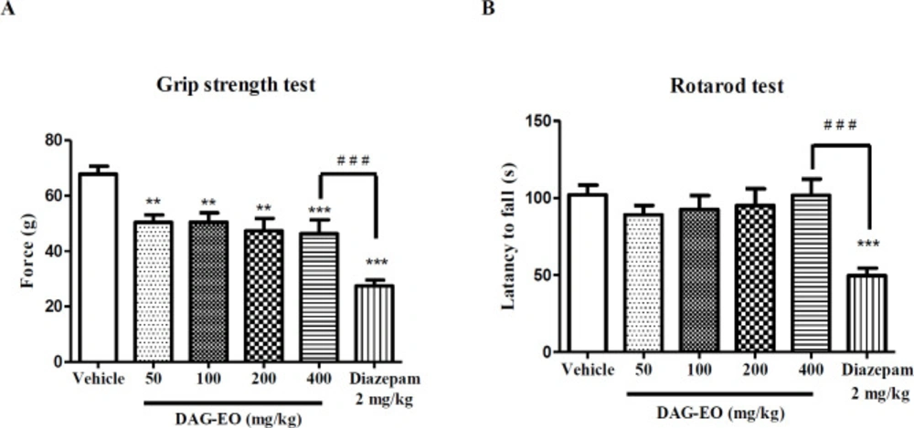
Grip strength test
Neuromuscular function and the maximal force developed by mice could be assessed in the forelimb grip strength test (
21). This test was performed with a grip strength meter (GS 5000, Borj Sanat Co.). Thirty minutes after the administration of DAG-EO (50 and 100 mg/kg), diazepam (2 mg/kg) or vehicle, the mice were placed on the tension pad of the apparatus, and the maximum force (gram) was recorded automatically by the apparatus. Each mouse was tested three times and the averaged value was reported as the final grip strength (
22).
Rotarod test
Motor impairing properties of DAG-EO was assessed in the rotarod test. The mice were trained daily for 3 consecutive days on the rotarod apparatus (rod diameter: 3 cm) rotating at a constant speed of 6 rpm. During each training session, the animals were placed on a rotating rod for 3 min with an unlimited number of trials, and only the mice that were able to remain on the rotating rod for 1 min were chosen for the test day. On the test day, the mice were intraperitoneally pretreated with vehicle, DAG-EO, and diazepam (2 mg/kg). After 30 min, the test was conducted and the latency to fall during 120 s was recorded (
23).
Statistical analysis
In the seizure tests, ED50s of the essential oil as the mean value with a 95% confidence interval was calculated using the Probit-Regression method (SPSS software, Chicago, IL; version 17.0). In the acetic acid-induced writhing test, ID50s values were calculated using nonlinear regression analysis. In the rest of the experiments, the results were compared using One-way Analysis of Variance (ANOVA), and the differences among the mean values were tested with the Tukey post-test in Graph Pad Prism software (San Diego, CA; version 5.0), when appropriate. The data was presented as the mean ± standard error of the mean (SEM). In all tests, p < 0.05 was considered a statistically significant difference.