Recently, there is much attention dedicated to develop and characterize of materials at nanoscale for various and new applications. In this study, SPION and Gemcitabine were simultaneously encapsulated in PLGA ± PEG to form a multifunctional drug delivery system. Optimum size and encapsulation efficiency were considered with respect to the radiosensitization, hyperthermia, and diagnostic applications as well as the preparation parameters were investigated systematically. Additional to the above issues, physicochemical characteristics of the optimized nanoparticles were studied. Then SPION-PLGA and PLGA-Gem nanoparticles were prepared with the same optimized parameters and the toxicity of these nanoparticles was compared with Gemcitabine alone in human breast cancer cell line (MCF-7).
Optimization of preparation parameters of polymeric nanoparticles
In this study, for optimization of preparation parameters of nanoparticles, various applications such as hyperthermia (with ultrasound or RF waves), sonoporation, radiosensitization in radiotherapy and contrast enhancement of ultrasound and MR images were considered.
For final selection of the optimal preparation parameter of (SPION- PLGA-Gem) ± PEG nanoparticles, size and encapsulated efficiency (EE) were considered and the examined variables were the volume percentage of sucrose (as cryoprotectant); and weight fractions of Gemcitabine and SPION to the polymer. Moreover, the presence and absence of PEG was examined in order to improve the physicochemical properties of nanoparticles for in-vivo studies.
For selection of volume fractions, sonication time, washing and other parameters, the previous studies and experiments were taken into account.
Then, for better comparison of various formulations, weight fractions for (PLGA-Gem) ± PEG, (SPION-PLGA) ± PEG and PLGA ± PEG nanoparticles were selected similar to the (SPION-PLGA-Gem) ± PEG nanoparticles.
Optimization of formulation with sucrose
In order to evaluate the effect of sucrose in nanoparticles size, 0%, 1% and 3% sucrose per 20 mg of PLGA ± PEG were examined. As it is evident from the data in
Table 1. 3% sucrose has considerable effect on the optimization of nanoparticle size. Additionally, there has no considerable deference in nanoparticle size between the 1% and 3% sucrose. Therefore, for evaluation of the other parameters, 1% sucrose per 20 mg of polymer was used to increase the weight fraction of nanoparticles in the final product.
Optimization of formulation with concentration of SPION
To optimize the formulation with SPION, different concentrations of SPION (2, 4 and 10 mg per 1 mL chloroform, equivalent with 0.08, 0.16 and 0.4 of SPION/PLGA) were used. From the data presented in
Table 2. it can be seen that although the size of the nanoparticles at SPION concentration of 4 mg/mL is higher than 2 mg /mL, but both sizes of nanoparticles are in the desirable range (about 200 nm), and thus the concentration of 2 mg/mL was neglected. Additionally, since the loading content and encapsulation efficiency of SPION and Gem in concentration of 10 mg/mL is better than 4 mg/mL, the optimum concentration is 10 mg/mL
Optimization of formulation with weight ratio of Gem/polymer
For this purpose, two fractions of 0.04 and 0.08 were examined. Nanoparticles that were characterized in
Table 3. were prepared at fixed conditions of 1% sucrose, 10 mg/mL of SPION/chloroform. As it can be seen in
Table 3. weight fractions of 0.04 and 0.08 had no considerable effect on nanoparticle size and encapsulation efficiency, therefore to improve the loading content of Gemcitabine, the fraction of 0.04 was selected.
Addition of PEG to formulation
To evaluate the effect of PEG, in all the experiments, 20% of PLGA was replaced with PEG. According to the results that were listed in
Table 2 and
Table 3. The existence of PEG has no considerable effect on the size and encapsulation efficiency of nanoparticles.
Optimized parameters for preparation of SPION-PLGA-Gem nanoparticles
With respect to the previous results, for the SPION-PLGA-Gem formulation, the selected parameters included 25 mg of PLGA ± PEG in 1 mL dichloromethane, 1 mg Gem per 200 mL of deionized water (equivalent to Gem/polymer of 0.04), 10 mg SPION per 1 mL of chloroform (equivalent to SPION/polymer of 0.4) and 1% sucrose per 20 mg of PLGA ± PEG.As it was mentioned before, nanoparticles made from PEG-PLGA exhibit in-vivo long-circulation properties (20, 21). Therefore, for examination of in-vitro toxicity of the prepared nanoparticles, PEG was not used.
| PDI(Mean) | (nm)*(Mean ± SD) | Sucrose(%) | PEG(mg) | SPION)mg/mL) | PLGA(mg) | |
|---|
| 0.23 | 89.0±1044.0 | 0 | 5 | 10 | 20 | 0.08 |
| 0.17 | 178.5±9.6 | 1 | 5 | 10 | 20 | 0.08 |
| 0.15 | 8.3±170.2 | 3 | 5 | 10 | 20 | 0.08 |
| 0.15 | 66.6±678.5 | 0 | 0 | 10 | 25 | 0.08 |
| 0.09 | 173.4±0.5 | 1 | 0 | 10 | 25 | 0.08 |
| 0.05 | 3.1±171.2 | 3 | 0 | 10 | 25 | 0.08 |
| PDI*Mean | *Mean±SD | %EE (Gem) Mean±SD | %EE(SPION)Ŧ Mean±SD | SPION)mg/mL) | | PLGA | PEG | Sucrose(%) |
|---|
| 0.17 | 178.5±9.6 | 17.1±0.4 | 0.6±40.2 | 10 | 0.08 | 20 | 5 | 1 |
| 0.09 | 173.4±0.5 | 13.8±0.3 | 3.4±44.9 | 10 | 0.08 | 25 | 0 | 1 |
| 0.15 | 160.0±4.1 | 15.2±1.3 | 3.2±45.1 | 4 | 0.08 | 20 | 5 | 1 |
| 0.10 | 163.2±1.2 | 11.6±0.9 | 5.1±47.3 | 4 | 0.08 | 25 | 0 | 1 |
| 0.15 | 5.2±165.3 | 15.2±1.6 | 3.1±45.1 | 2 | 0.08 | 20 | 5 | 1 |
| 0.10 | 3.4±161.1 | 11.6±2.3 | 5.1±47.3 | 2 | 0.08 | 25 | 0 | 1 |
Obtained by UV-Vis spectrophotometer.
Obtained by atomic absorption spectrophotometer
| PDI*Mean | *Mean±SD | %EE (Gem) Mean±SD | %EE(SPION)Ŧ Mean±SD | SPION)mg/mL) | | PLGA | PEG | Sucrose(%) |
|---|
| 0.08 | 8.0±174.0 | 16.1±2.0 | 47.7±2.6 | 10 | 0.04 | 20 | 5 | 1 |
| 0.17 | 178.5±9.6 | 17.1±0.4 | 0.6±40.1 | 10 | 0.08 | 20 | 0 | 1 |
| 0.08 | 10.0±180.0 | 16.0±1.0 | 48.2±2.1 | 10 | 0.04 | 25 | 5 | 1 |
| 0.09 | 173.4±0.5 | 13.8±0.3 | 3.4±44.9 | 10 | 0.08 | 25 | 0 | 1 |
Obtained by UV-Vis Spectrophotometer.
Obtained by atomic absorption spectrophotometer
| Formulation | Zave(nm)**(mean±SD) | Size (nm)(TEM) | PDI*(mean±SD) | Zeta potential (mV)(mean±SD) |
|---|
| SPION | 2.2±20.1 | 7.0±0.5 | 0.20±0.05 | +20.2±2.0 |
| PLGA | 190.6±8.6 | - | 0.03±0.01 | -15.3±0.5 |
| SPION- PLGA | 170.3±4.6 | - | 0.16±0.04 | -12.1±1.0 |
| PLGA-Gem | 175.2±8.3 | - | 0.05±0.02 | -14.2±1.1 |
| SPION-PLGA-Gem | 180.2±10.3 | 180.0±20.0 | 0.08±0.02 | -13.3±1.0 |
Poly Disperse Index (PDI), Zave and zeta potential (mV) were obtained by DLS.
| Formulation | This study(mean ± SD) | Other studies* | References |
|---|
| SPION (TEM) | 7.0±0.5 | 4-20 | (37-41) |
| PLGA-SPION | 170.0±4.6 | 293-110 | (38, 39, 42) |
| PLGA-Drug | - | 50-190 | (18, 43) |
| PLGA-Gem | 175.0±8.3 | 132-206 | (1, 3, 36, 37) |
| SPION-PLGA-Drug | 180.0±10.0 | 200-300 | (40, 44) |
Other studies with approximately similar synthesis and preparation methods
| Formulation | Encapsulation efficiency (%)
| Loading content(%)
|
|---|
| Gem( UV-Vis) | SPION(AAS) | Gem | SPION |
|---|
| SPION-PLGA | - | 50.1±1.1 | - | 28.6 |
| PLGA-Gem | 13.2±1.3 | - | 3. 8 | - |
| SPION-PLGA-Gem | 16.1±2.2 | 48.2±2.1 | 2.8 | 27.8 |
| Formulation | Survival (%) | Enhancement ratio | P-value* |
|---|
| PLGA-SPION | 93.1±1.2 | - | - |
| Gem | 50.0±0.0 | 1.00 | - |
| PLGA-Gem | 32.7±1.1 | 1.53 | 0.015 |
| SPION-PLGA-Gem | 26.2±2.3 | 1.89 | 0.006 |
P-values are calculated compared to Gemcitabine group
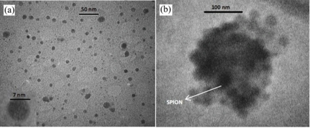
TEM image of SPIO nanoparticles (a), TEM image of SPION-PLGA-Gem nanoparticles (b).The samples were deposited onto a copper grid (300 meshes). The acceleration voltage was set to 100 kV for SPION and 80 kV for SPION-PLGA-Gem nanoparticles
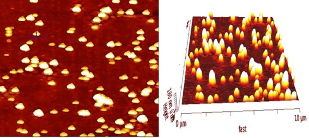
AFM image of SPION-PLGA-Gem nanoparticles. Lyophilized nanoparticles (100 µg) were dispersed in ultrapure water with bath sonicator (0.1 mg/ml) and were dispensed onto the glass slide of microscope
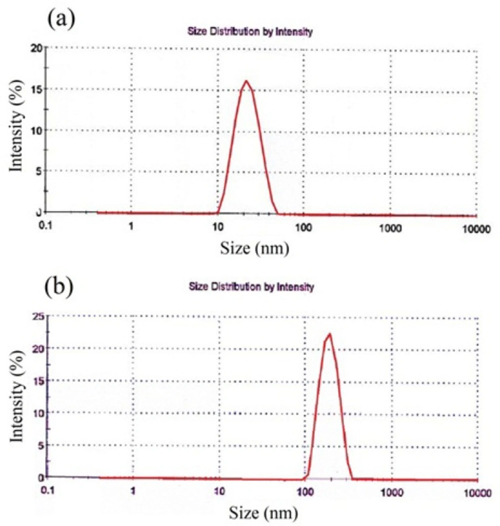
Size distribution of nanoparticles obtained by AFM, SPIONs (a), SPION-PLGA-Gem nanoparticles (b
In-vitro cumulative release of Gemcitabine from SPION-PLGA-Gem nanoparticles at the specified intervals (2, 4, 8, 12 hours; 1, 2, 4 and 7 days) in PBS (7.4 pH). The data were obtained by UV-Vis spectrophotometry
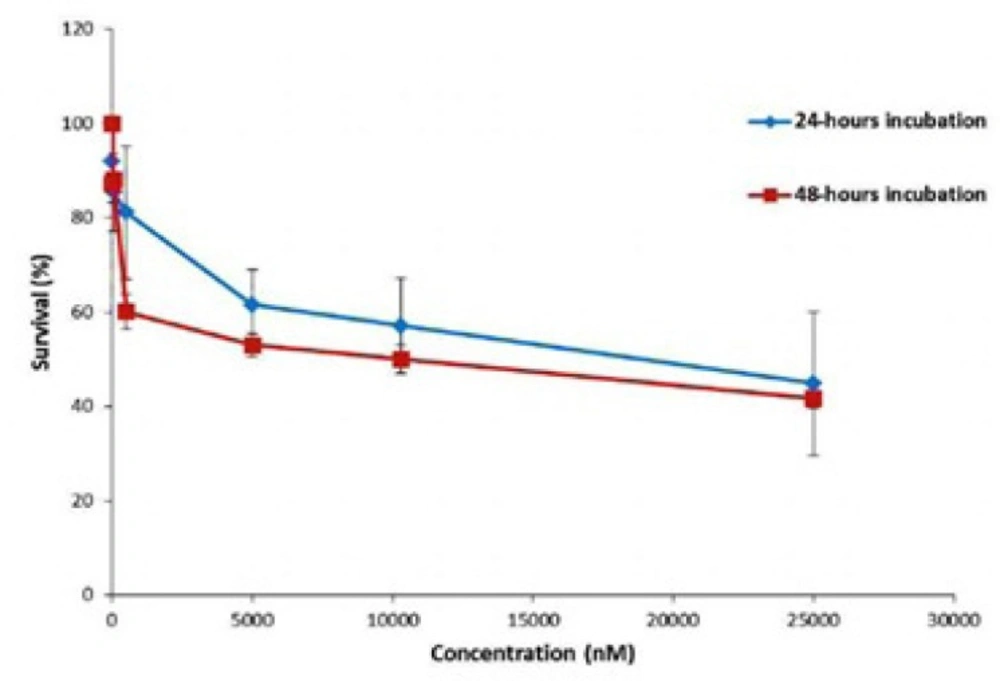
Survival (%) of MCF-7 cells treated by Gemcitabine after 24 and 48-hours incubation. The serial concentrations of Gemcitabine are 0.01-25 µM
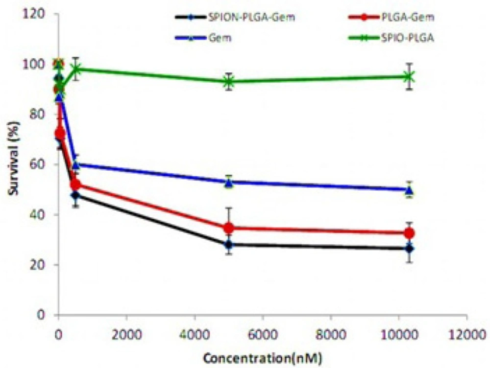
Survival (%) of MCF-7 cells of various treatment groups after 48-hours incubation. The concentrations of formulations are equivalent the concentration of 0.01 µM to 10.3 µM of Gemcitabine
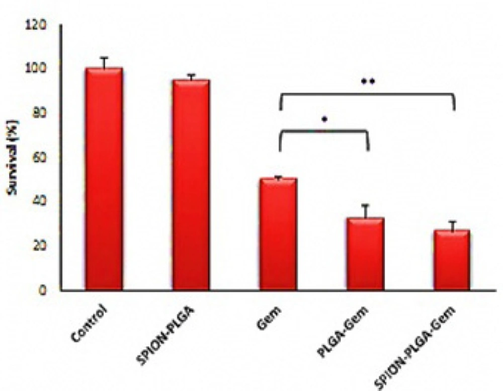
Survival (%) of MCF-7 cells of various treatment groups after 48-hours incubation. The concentration of formulations is equivalent to IC50 of Gemcitabine (10.3 µM). *p≤0.05 compared to Gemcitabine group, **p≤0.01 compared to Gemcitabine group
Physicochemical properties of nanoparticles
Morphology and size distribution
Morphology of prepared nanoparticles obtained by TEM and AFM in
Figure 1 and
2. illustrates that SPIONs prepared by chemical coprecipitation method are spherical with uniform distribution and SPIO-PLGA-Gem nanoparticles prepared by a double emulsion method are also have nearly spherical shape and is uniformly distributed. Entrapped SPIONs in these nanoparticles are obvious as dark spherical points.
Size, size distribution and zeta potential
Hydrodynamic diameter, Poly Disparity Index (PDI), zeta potential and size distribution profile of nanoparticles were obtained by dynamic light scattering (DLS) method. According to
Table 4. the average hydrodynamic diameter of these nanoparticles is 20.1 ± 2.2 nm with PDI of 0.20 ± 0.05. The size of SPIONs that was obtained by TEM is 7.0 ± 0.5 nm. Zeta potential of these nanoparticles is 20.2 ± 2.0 mV. These nanoparticles have narrow size distribution (
Figure 3 (a)).
As listed in
Table 4. the average size of SPION-PLGA-Gem nanoparticles that was obtained by TEM is 180.0 ± 20.0 nm. The average hydrodynamic diameter of these nanoparticles obtained by DLS is 180.2 ± 10.3 nm with PDI of 0.08 ± 0.02. Zeta potential of these nanoparticles is -13.3 ± 1.0 mV. SPION-PLGA-Gem nanoparticles disperses easily in water and be collected by permanent magnet. As it illustrated in
Figure 3 (b). the size distribution of SPION-PLGA-Gem nanoparticles is Gaussian, narrow, and symmetrical.From the data listed in
Table 4. the average Hydrodynamic diameters of polymeric nanoparticles with minor differences were less than 200 nm. The size of the PLGA nanoparticles (without drug and SPION), is slightly more than all other provided nanoparticles (190 ± 8.6). This indicates that the existence of SPION in the formulation, on the one hand, increases PDI and on the other hand, may not have a considerable impact on the size and make slight adjustments to it. This may be due to the role of the SPION nanoparticles in stability and prevention of aggregation of polymeric nanoparticles.
In
Table 5. the sizes of prepared nanoparticles are compared with the sizes of the same nanoparticles provided in the other studies. As it can be seen, the sizes of these nanoparticles are in an appropriate range, compared with the other studies using similar preparation methods.
Drug loading and entrapment efficiency
As can be seen from
Table 6. the SPION encapsulation efficiency, using AAS method, is about 50%. Of course this amount is affected by the loading content of SPIONs in the polymeric nanoparticles. SPION loading content in provided polymeric nanoparticles is about 28%. Gemcitabine loading capacity in polymeric nanoparticles is between 2.8% for the SPION-PLGA-Gem formulation and 3.8% for the PLGA-Gem formulation.
Efficiency of Gemcitabine encapsulation is 16.1 ± 2.2 (%) and 13.2 ± 1.3 (%) in these formulations, respectively.
Higher performance of SPION encapsulation (4 folds) in SPION-PLGA-Gem formulation compared to Gemcitabine may be effective for radiosensitivity and imaging purposes. The cause of the issue is that in these applications, concentrations of Gemcitabine with minimal toxicity are used, while considering the lower toxicity of SPION compared to Gemcitabine, there is no special restrictions on the use of SPION compared with Gemcitabine. Using higher density of SPION leads to the effectiveness of diagnosis and treatment.
Therefore, this amount of the difference in Encapsulation Efficiency between SPION and Gemcitabine appears to be favorable for application purposes.
In-vitro release of Gemcitabine
Due to chemical instability and poor cell harvest, Gemcitabine has a very short half-life in plasma after intravenous injection (
25). Therefore, it should be used with high doses leading to high systemic toxicity (
1,
3). To solve this problem, in the past decades, nanotechnology has an improvement, and has emerged as a basis for the treatment of a wide range of different tumors. Nanotechnology leads to a prolongation of the release and increasing the entrance of drug into the cell (
1). Nano platforms increase anti-tumor effects with negligible toxicity and causes controlled transfer and accumulation in tumor area and protection of drug molecules against biodegradability and plasma clearance (
2).
In this study, PLGA nanoplatforms were used. The advantages of this polymer could be biodegradability, biocompatibility, decreasing their systemic side effects, facilitating intratumoral encapsulation, rapid clearance from the biological system and high efficiency of drugs transmission and transportation (
17,
18,
26). Therefore, they have been used in many micro-formulations and nanoparticles (
27-
30). For example, fluorouracil (
31), doxorubicin (
7), docetaxel (
32,
33), paclitaxel (
34,
35) and cisplatin (
20) could be mentioned.
Following the physicochemical characterization of NPs,
in-vitro Gemcitabine release profile from SPION-PLGA-Gem NPs has been conducted in a PBS solution at pH 7.4. As illustrated in
Figure 4. it can be seen that in the first 24 h, release rate grows with a slight slope and reaches more than 60% after 48 h. After this time the growth of the curve is very slight and slow. Therefore, the time of 50% release (
T1/2) of about 18 h implies the drug release from the controlled drug delivery system is controllable and sustains in the natural conditions (pH 7.4). Therefore, it is expected that the provided nanoplatforms are suitable for controlled transfer, accumulation in tumor area and protecting of drug molecules from biodegradability and plasma clearance.
The release curve shows that in 48 h, more than 60 percent of the drug is released from the formulation and then the trend of release growth will be slow.
Toxicity of Gemcitabine hydrochloride and nanoparticles
In this study with the aim of in-
vivo applications, nanoparticles of SPION-PLGA-PEG-Gem were also prepared and the results showed that addition of PEG to PLGA has no considerable effect on physical properties of nanoparticles (
Table 4), but for
in-vitro toxicity experiments, nanoparticles without PEG were used.
The viability (%) of the cells under treatment by Gemcitabine (with concentration between 0.01 µM to 25 µM) was studied using 24 h and 48 h MTT assays. As it can be seen in
Figure 5. the repeatability of the toxicity data in 48 h incubation is better than the 24 h incubation. Moreover, According to the slope of the release curve in the first 24 h and slowing the release curve growth after 48 h (
Figure 4), it can be concluded that to assess the toxicity of the nanoparticles, incubation time of 48 h is more appropriate and therefore considered for evaluation of toxicity of nanoparticles.
After determining the range of treatment concentration of Gemcitabine, the MCF7 cells were treated by different formulations with the same procedure, in the range of concentrations equivalent to the concentrations (0.01 μM to 10.3 μM) of Gemcitabine (released in 48 h).
Figure 6 illustrates the survival of MCF-7 cells under various treatment groups by nanomolar equivalent concentrations of Gemcitabine with 48-hours incubation using MTT assay. This figure obviously shows the effectiveness of the treatment with SPION-PLGA-Gem compared to Gemcitabine and the other nanoparticles, especially at higher concentrations.
The range of inhibitory concentration of 10% (IC10) for Gemcitabine is about 10 nM. The toxicity of the other formulations containing 10 nM Gemcitabine is relatively equivalent to the toxicity of 10 nM Gemcitabine. SPION-PLGA nanoparticles with very higher concentrations (equivalent to 25 μM of Gemcitabine in other formulations) have no considerable toxicity (less than 10% toxicity). This suggests that the use of SPION in different formulations has a great advantage for therapeutic applications (hyperthermia and radiosensitization) or diagnostic applications (as a contrast agent in MR and ultrasound imaging). According to
Figure 6. treatment with polymeric nanoparticles considerably had better result than Gemcitabine. The survivals of cell treated by concentration of formulations that were equivalent to IC50 of Gemcitabine were listed in
Table 7. The survivals were 26.5% and 32.7% for SPIO-PLGA-Gem and PLGA-Gem respectively (that were equivalent to DER of 1.89 and 1.53).As illustrated in
Figure 7. statistical differences between these groups are significant (
p-values of 0.006 and 0.015 for SPIO-PLGA-Gem and PLGA-Gem groups compared with Gemcitabine group). It means the good treatment efficiency of formulations for MCF7 cells.In this study nanoparticles of SPION-PLGA-PEG-Gem were also prepared and the results showed that addition of PEG to PLGA had no considerable effect on physical properties of nanoparticles (
Zave = 174.0 ± 8.0 nm, PDI = 0.08 ± 0.03 and zeta potential = 15 ± 1.6 mV) but for
in-vitro experiments, nanoparticles without PEG were used.