
Introduction
Experimental
Results
| Groups | Dosage | ALT(Ul/lit) | AST(Ul/lit) | ALP(Ul/lit) |
|---|---|---|---|---|
| E0 | normal saline 0.9% | 56.71±28.62 | 79.14±22.61 | 279.57±194.74 |
| E1 | 10mg/kg FA 37% | 100±5.09a | 240.57±47.46a | 523.86±124.08a |
| E2 | 10mg/kg FA+5mg ME | 49.43±14.08b | 115±81.50c | 171.57±85.64b |
| E3 | 10mg/kg FA+10mg ME | 58.86±13.33c | 105.14±29.96c | 147.71±30.45 b |
| E4 | 10mg/kg FA+15mg ME | 63±10.49c | 187.60±87.23 | 204.80±102.60 b |
| E5 | 10mg/kg FA+20mg ME | 61.60±34.13c | 209.40±152.06 | 154.80±25.37 b |
| E6 | 10mg/kg FA+50mg ME | 50.60±10.47c | 143.20±25.47 | 114.40±11.95 b |
| E7 | 10mg/kg FA+100mg ME | 43.60±12.78b | 132.80±41.40 | 131.40±46.53 b |
: p= 0.002, in compared with control
: p < 0.001, in compared with FA group
: p < 0.005. in compared with FA group
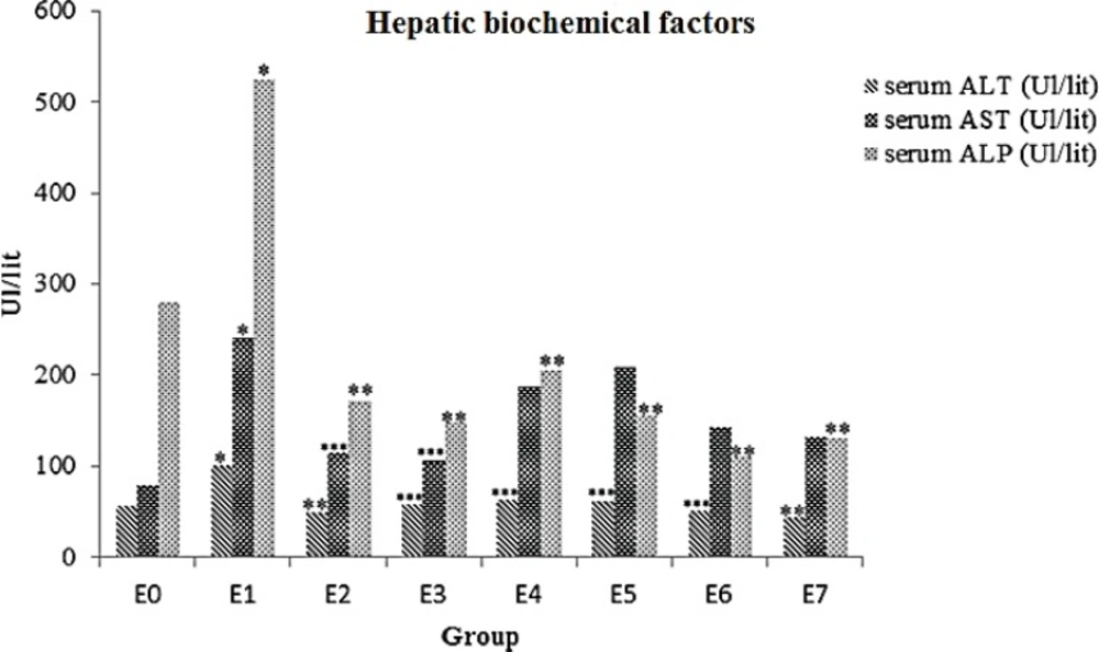
Effects of ME on serum levels of ALT, AST and ALP in FA- intoxicated mice. Data are expressed as the mean ± SD, n = 7. * p= 0.002, compared to the control group, ** p < 0.001 and *** p < 0.005 compared to the FA group. E0 group: normal saline; E1 group: 10mg/kg FA; E2 group: 5 mg ME + FA; E3 group: 10 mg ME + FA; E4 group: 15 mg ME + FA; E5 group: 20 mg ME + FA; E6 group: 50 mg ME + FA; E7 group: 100 mg ME + FA
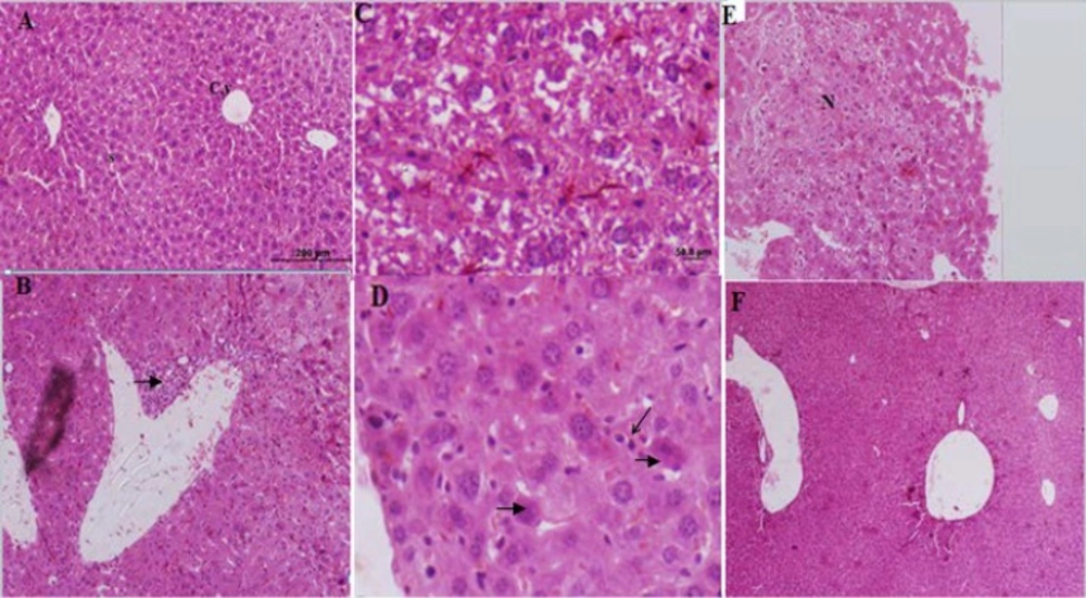
Histological figure from a mice liver (A): showed a normal hepatocyte in control group. X100 (B, C, D and E): liver sections in FA treated groups (Group E1) visible infiltration of inflammatory cells, hepatocytes degenerations, increased kupffer cells, hypereosinophilic cytoplasm and necrosis. (F): Improved liver changes in E2 group
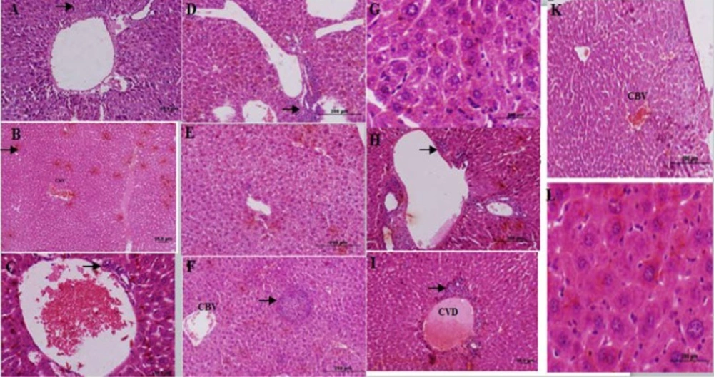
Histological figure from a mice liver (A): lymphocyte infiltration observed in E3 group. (B): congested blood vessel (CBV) and lymphocyte infiltration in E4 group. (C): hepatocyte view of E5 group central with venous distension, and mild lymphocyte infiltration (D-G): E6 group hepatocytes view with lymphocytes accumulation (D), vacuole formation (E), lymphocytes infiltration (F) and kupffer cells accumulation (G). (H-L): liver sections in FA treated group (E7 group) lymphocytes accumulation (H), central venous distension (I), congested blood vessel (K) and kupffer cells accumulation (L).
