Tadalafil is a potent and selective cyclic guanosine monophosphate specific type (V) phosphodiesterase inhibitor used for the treatment of erectile dysfunction, benign prostatic hyperplasia and pulmonary arterial hypertension (
1-
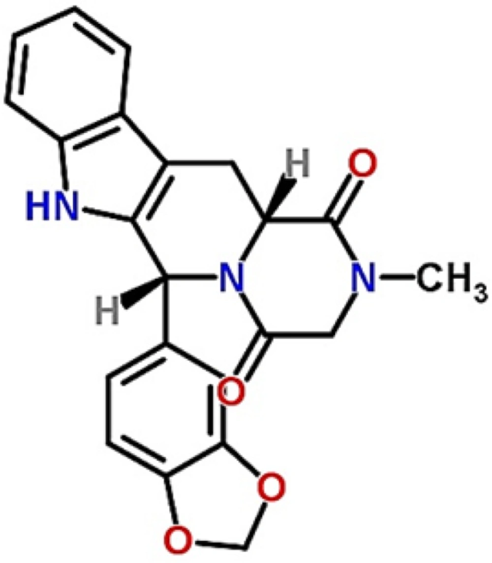
3). The chemical structure of tadalafil is shown in
Figure 1. Longer duration of action (approximately 36 h) and minimized potential for vision abnormalities are the most important advantageous of tadalafil in comparison with other drugs in this pharmacological category (
4). Since it belongs to class II of classification system, poor aqueous solubility and wettability is an important issue and in spite of good permeability, its bioavailability is limited by solubility and dissolution rate, leads to variability in blood concentrations and irreproducibility in clinical responses (
5). Different groups have utilized various formulation strategies for improving the solubility and dissolution rate of tadalafil that the most reported ones include preparation of solid dispersion by using water soluble polymers and copolymers (such as poly(vinyl pyrrolidone) k 30, poly(ethylene glycol) 6000, poloxamer 188 and 407 (
6), formation of inclusion complex with ß-cyclodextrins and microporous silica (
7,
8), the use of amorphization methods (such as vitrification, cryogenic grinding, ball milling, spray drying and freeze drying) (
9). However, to the best of our knowledge a few studies have been conducted in the field of nanoparticles preparation by antisolvent precipitation-ultrasonication method.
Drug particle size reduction has emerged as an effective and versatile option for surmounting solubility issue (
10,
11). According to Noyes-Whitney equation, dissolution rate increases when the surface area increases by reducing the size to nanometer (
12).
Nanosuspension or nanocrystal suspension is a pure particulate system that is composed of submicron (average particle size in the range of 200-600 nm) colloidal dispersion with a surfactant as the stabilizer (
13). As the first time, in 2000, nanosuspensions have been commercialized in the pharmaceutical market. Increased saturation solubility, increased adhesiveness to surfaces/cell membranes and increased dissolution rate are special properties of nanosuspensions
(
14). Techniques used to produce drug nanosuspensions can be classified into two major groups: top-down and bottom-up technologies (
15). One of the most bottom-up promising techniques is nanoprecipitation that is cost-effective, rapid to perform and suited for scaling up (
16,
17).
In this method, nanoparticles could be formed in different ways such as pH controlled precipitation, antisolvent precipitation with or without surfactant and sonoprecipitation (
18). Sonoprecipitation, a combination of antisolvent precipitation and ultrasoication, is an effective method of controlling the nucleation and crystallization process (
19).
Ultrasound amplifies the mass transfer when it propagates through a liquid medium, and initiates an important phenomenon known as cavitation. Furthermore, it increases micro-mixing, reduces particle growth and agglomeration, so it is possible to obtain particles with uniform size distribution (
20). In a study, Xia
et al. (
21) succeed to prepare nitrendipine nanosuspensions with diameter of about 209 ± 9 nm with an enhanced dissolution rate and bioavailability. Also, the same results were found in the preparation of carvedilol (
22), itraconazole (
23), clarithromycin, cefixime, glipizide nanosuspensions by other groups. In this technique, in addition of the type of antisolvent and stabilizers, process parameters such as the precipitation temperature, the power input and the time length of ultrasonication play an important role (
21).
Formulation parameters such as drug concentration, stabilizer concentration and antisolvent to solvent ratio also need to be optimized because of their important influence on supersaturation degree and nucleation rate (
24).
The aim of this study was to prepare tadalafil nanosuspension by sonoprecipitation technique and optimize process parameters in order to enhance solubility and dissolution rate of the drug.
Materials
The raw tadalafil was purchased from Dr. Reddy’s Pharmaceutical Co., Ltd., India. Tween80, sodium hydroxide (NaOH), monobasic potassium phosphate (KH2PO4), methanol, ethanol, dimethyl sulfoxide (DMSO), acetone and high-performance liquid chromatography (HPLC) grade acetonitril were obtained from Merck, Germany. Deionized water was prepared with Millipore water purification system, Germany.
Methods
Preparation of tadalafil Nanocrystals by Sonoprecipitation
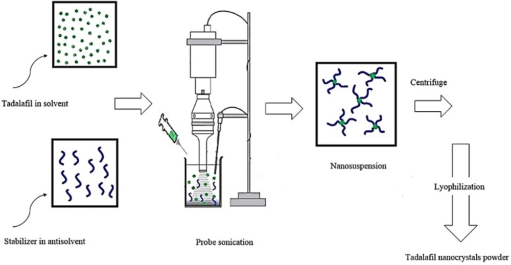
Tadalafil nanosuspensions were prepared through sonoprecipitation technique. The experimental process for the preparation of tadalafil nanosuspension is illustrated in
Figure 2. In a typical procedure, combination of two organic solvents, acetone: DMSO (88:12 v/v), was selected as water-miscible solvent and deionized water was used as antisolvent. Tadalafil coarse powder was completely dissolved in the organic phase and then was filtered through a syringe filter, PTFE membrane with pore size of 0.45 µm (Simplepure, USA) to remove the possible particulate impurity. A range of surfactants and polymers (such as hydroxypropyl methylcellulose in grades of 6, 15 and 50 cp, poly (ethylene glycol) 400 and 6000, polyoxyl 40 stearate, poloxamer 188 and 407, poly (vinyl pyrrolidone) k30, poly (vinyl alcohol), sodium carboxymethyl cellulose, Tween 80 and sodium lauryl sulfate) were screened as stabilizer and finally Tween 80 was selected in accordance with the size criteria (data not shown). The antisolvent phase was prepared by dispersing a stabilizer in distilled water that was cooled to 5±1 °C in an ice-water bath and treated with an ultrasonic probe (Hielscher UP400S, 400W, 24 kHz, Germany) at power input of 280 W and a cycle of 0.5 per second. In the next step, precipitation initiated by drop-wise adding of drug solution phase within 5 min. As the nanosuspension emerged, size and polydispersity index (PDI) were evaluated. Then, the obtained nanosuspensions were concentrated by centrifugation at 16000rpm for 50 min using an ultracentrifuge (3-30K, Sigma, Germany) and washed three times with deionized water. Furthermore, the content of drug was determined in supernatant by HPLC analysis for calculation of yield. Finally, the obtained nanoparticle residue was frozen at -80°C for 24 h and subsequently lyophilized for 48 h at a temperature of 58 °C under vacuum by using freeze dryer (Alpha 1-2 LDplus, Christ, Germany). The dried powder was then collected and stored in air tight container for further use.
Experimental design
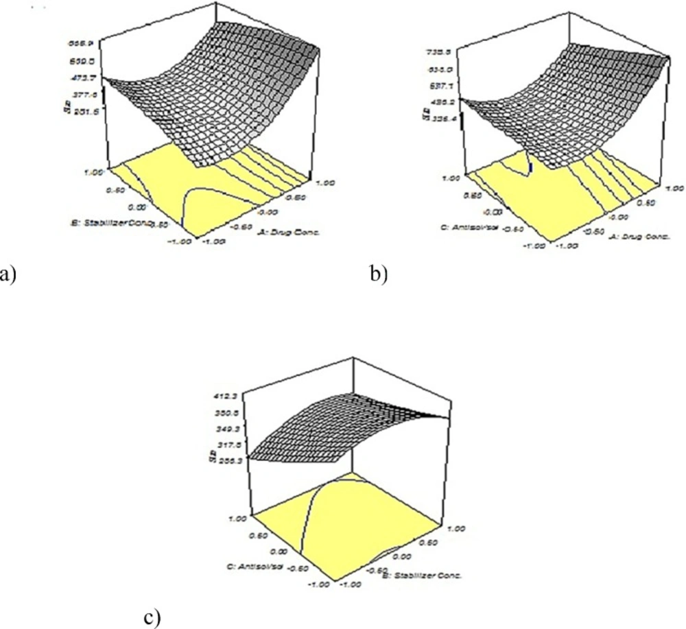
Response surface methodology based on central composite design was employed for evaluation of the formulation factors
i.e., drug concentration (A), stabilizer concentration (B), antisolvent to solvent volume ratio (C) and mean particle size (y) was assessed as the response. The details of design are shown in
Table 1. Based on the results of preliminary experiments, the experimental range of each variable was chosen. The experiments were designed using Design-Expert
® software (ver. 7.0.0, stat-ease
®, USA). To reduce systematic errors, the experiments were completely randomized.
By Analysis of collected data for responses, the relationship linking the main factors and their interactions to the responses were determined and presented as a general form in the following Equation (1).
A quadratic model Equation (2) was fitted to the response using the Design-Expert® software.
Where y represents the predicted response, A, B and C represents the independent variable and ß represents the coefficient. The three dimension (3D) response surface graphs were plotted using origin 7.0 software according to the equation.
Physicochemical Characterization of tadalafil nanocrystals
Particle size and zeta potential analysis
The mean particle size (z-average), polydispersity index (PDI) and zeta potential were determined by photon correlation spectroscopy (PCS) using Zeta-sizer (Nano-ZS, Malvern Instruments, UK). The real refractive index and the imaginary refractive index were set at 1.76 and 0.01, respectively. The z-average and PDI values were obtained by averaging of three measurements. Before the measurement, a small aliquot of nanosuspensions was diluted with 5 mL of deionized water to have a suitable scattering intensity and then sonicated to create a homogenous suspension.
Yield of nanoprecipitation process
Nanosuspensions were centrifuged (3-30K, Sigma, Germany) at 16000 rpm for 50 min. The concentration of dissolved tadalafil in the supernatant was determined using a reverse phase high performance liquid chromatography (RP-HPLC) (Smart line 1000 pump, Smart line Diode array 2800 UV detector, Knauer, Germany) as was reported by Cheng and
et a.l (
26). EZ CHROM software was used to record and evaluate the data collected during and following chromatographic analysis. The chromatographic separation was accomplished using a Perfectsil target C
18 column (150
4.6 mm, 5µm) (ODS-3, MZ, Germany) protected by a guard column (10×4.6mm) which was packed with the same C
18 material. Acetonitrile and 20 mm phosphate buffer (pH 7.0) in a ratio of 40:60 was used as the mobile phase. The column was maintained at 25 °C and equilibrated for 60 min with the analytical mobile phase before injection. The injection volume was 20 µL, and the mobile phase was pumped isocratically at a flow rate of 1.0 mL/min. The eluent was analyzed at 283 nm and the retention time of the drug was 9.45 min. The yield of the process was calculated using the Equation 3.
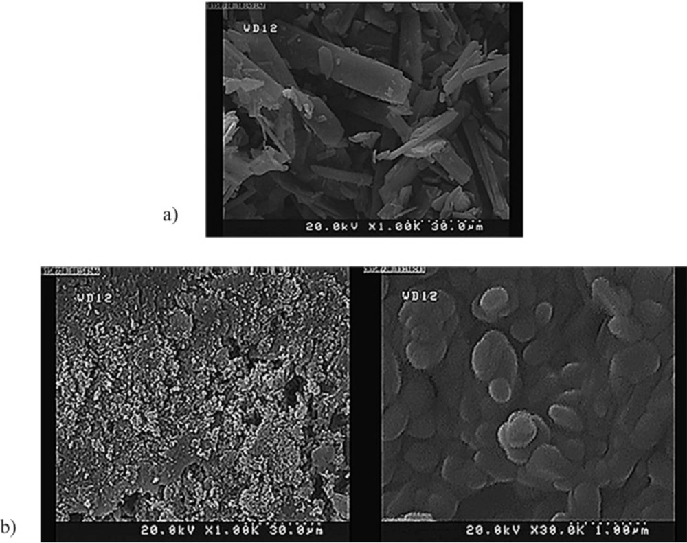
Particle morphology
The morphology of tadalafil and the tadalafil nanocrystals was evaluated through field emission scanning electron microscope (S-4160, Hitachi, Japan). Prior to analysis, the samples were diluted with ultra-purified water to obtain a suitable concentration. Then, the samples were spread on a sample holder, dried under vacuum and eventually coated with gold.
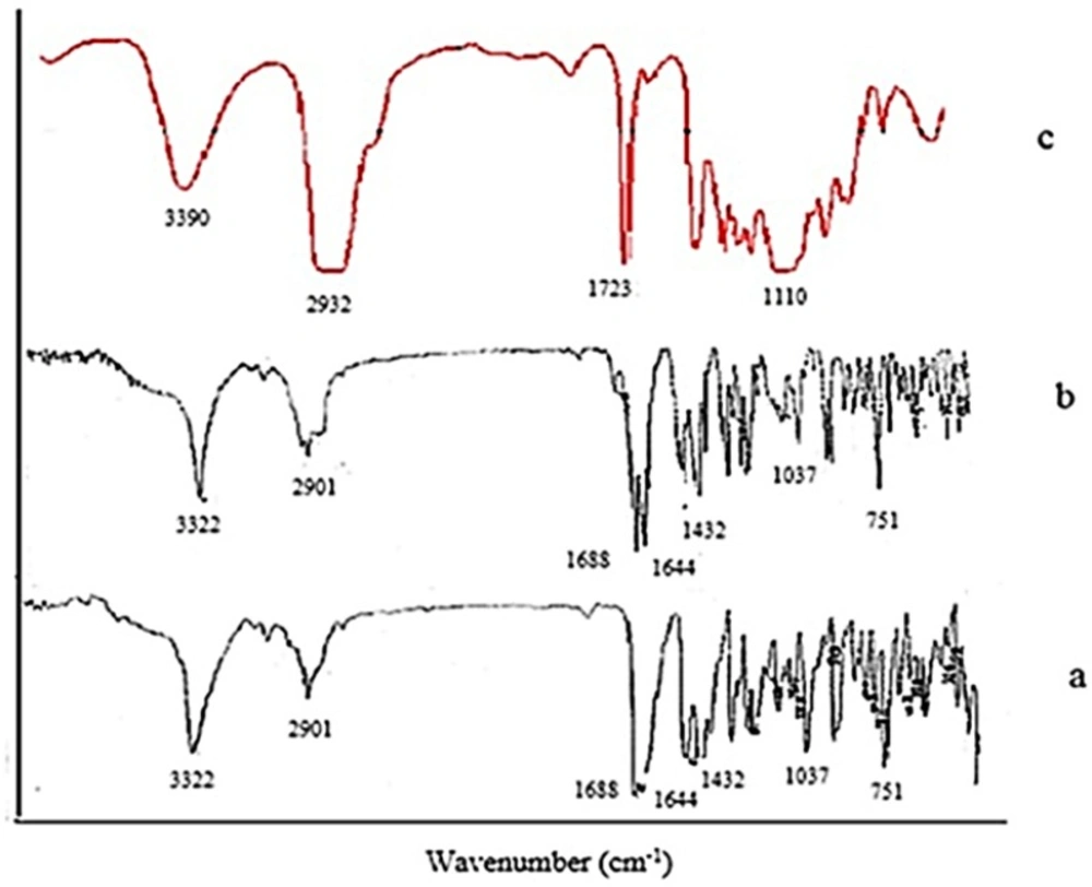
Fourier transformed infrared spectroscopy (FTIR)
FTIR technique was applied to determine the molecular structures of raw tadalafil and the interaction between stabilizer and drug nanocrystals. FTIR spectra were recorded by FTIR spectrometer (Nicolet Magna-IRTM, USA) within the spectral region of 400 and 4000cm−1 at a resolution of 2 cm−1. The IR spectra were obtained in a KBr disc.
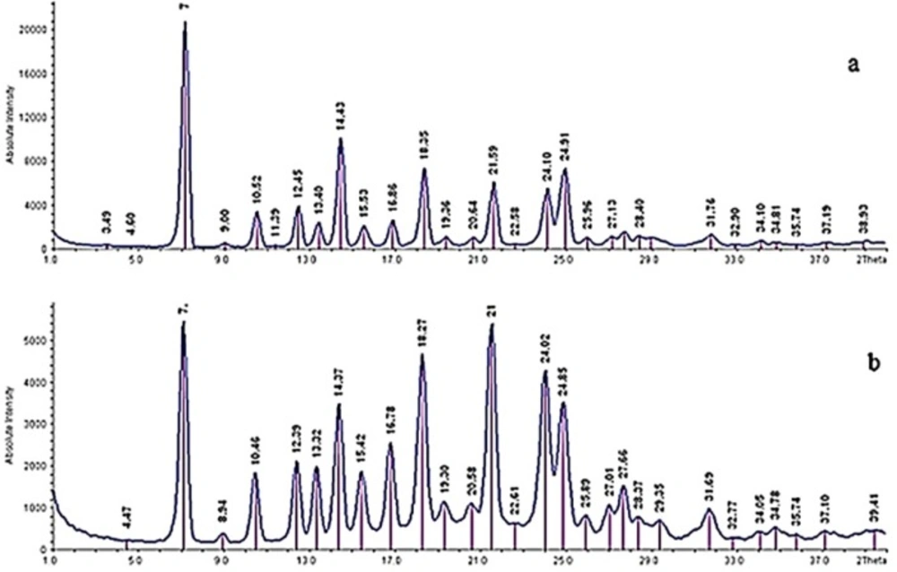
X-ray powder diffraction (XRPD)
The crystal forms of the samples were detected using a powder X-ray diffractometer (IPDS II, STOE, Germany). The current and voltage using Cu Kαl radiation were 30 mA and 40 kV, respectively. The obtained data were typically collected from 1 to 40 with a step size of 0.06 at a rate of 1/s. the output is given as intensity versus 2θ.
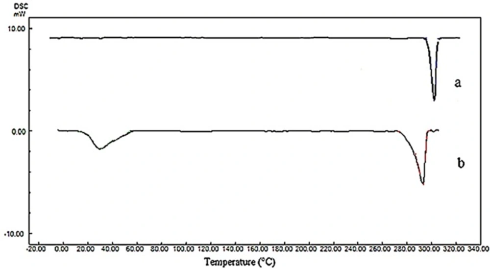
Differential scanning calorimetry (DSC)
The thermal analysis was performed using differential scanning calorimeter (DSC-60, Shimadzu, Japan). Approximately, 3 mg of each samples was placed in an aluminum pan. The heating and cooling scans were performed from -10 °C to 320 °C at the heating and cooling rates of 10 °C/min in a dry N2 atmosphere. An empty aluminum pan was used as a reference. The melting temperature and enthalpy were calculated from the DSC thermograms.
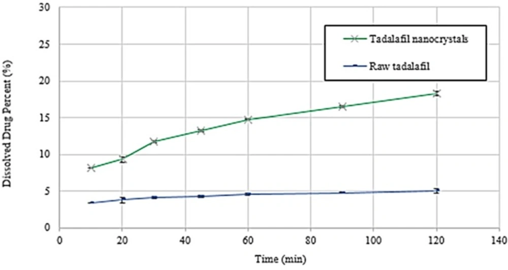
Dissolution rate study
The dissolution rate experiments of raw tadalafil and tadalafil nanocrystals were carried out according to the USP 32 apparatus II (paddle) method. Phosphate buffer (pH 7.4) was used as the dissolution medium. The stirring speed and the bath temperature were 100 rpm and
37.0 ± 0.5 °C, respectively. Unprocessed tadalafil (2 mg) and tadalafil nanocrystals (containing 2 mg tadalafil) were added to 900 mL of dissolution medium. Then, aliquots equivalent to 5 mL were withdrawn after 10, 20, 30, 45, 60, 90 and 120 min and immediately replaced with the same volume of phosphate buffer. All of samples were passed through a 0.45 µm syringe filter and injected into the HPLC system for analysis of drug concentration. Furthermore, the obtained dissolution profile data of the raw tadalafil
and tadalafil nanocrystals were evaluated and compared using the dissolution efficiency (DE%) in 30 and 90 min. The measurements were repeated three times. This concept was proposed by Khan and Rhodes (
27) and is defined as follows:
Where y is the percent of drug dissolved at any time t, y100 denotes 100% dissolution, and the integral represents the area under dissolution curve between time zero and t.
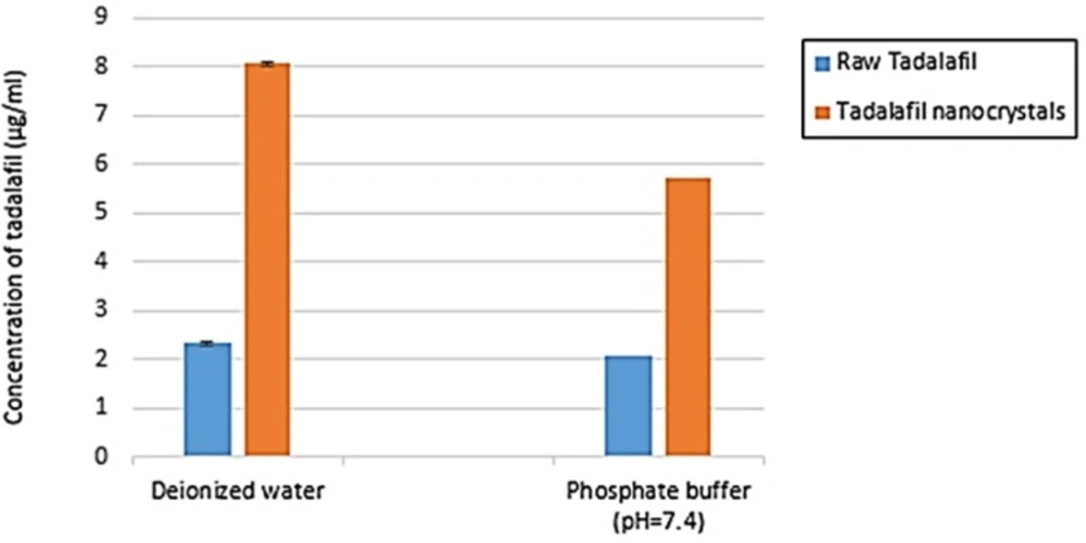
Saturation solubility
The saturation solubility of raw tadalafil and tadalafil nanocrystals were evaluated in two mediums: deionized water and phosphate buffer (pH=7.4) at 25 °C. Excess amounts of lyophilized powder was dispersed in 10 mL of medium and placed on a stirrer for 48 h to ensure that the solubility equilibrium had been reached. The samples were centrifuged and the resulting supernatant was filtered and then analyzed by HPLC. The measurements were repeated three times.
Statistical analysis
The reported data represented the mean value ± standard deviation (SD). Significance of difference was evaluated using Student t-test and one-way ANOVA at the probability level of 0.05 using SPSS 19 for Windows (SPSS, Chicago, IL, USA) and Design-Expert® software (ver. 7.0.0, stat-ease®, USA).