Effects of Silver Sulphadiazine on Production of Extracellular Proteins by Strains of Staphylococcus Aureus Isolated from Burns Wound

authors:

avatar Vahideh Javid Khojasteh a , * , avatar Souad Alfakhri b , avatar Howard Anthony Foster c

Department of Microbiology,Faculty of Medicine, Guilan University of Medical Sciences, Rasht, Iran .
Biomedical Sciences Research Institute, School of Environment and Life Sciences, University of Salford, The Crescent, Salford M5 4WT, UK .
Biomedical Sciences Research Institute, School of Environment and Life Sciences, University of Salford, The Crescent, Salford M5 4WT, UK.

how to cite: Javid Khojasteh V, Alfakhri S, Foster H A. Effects of Silver Sulphadiazine on Production of Extracellular Proteins by Strains of Staphylococcus Aureus Isolated from Burns Wound. Iran J Pharm Res. 2016;15(2):e125172. https://doi.org/10.22037/ijpr.2016.1835.

Abstract

Previous studies had shown that sub-inhibitory concentrations of silver sulphadiazine (AgSD) stimulated the production of Toxic Shock Syndrome Toxin-1 in certain strains (responder strains) of Staphylococcus aureus and that protease production was also affected. No changes were detected in other strains (non-responders).
Extracellular proteins from eleven responder and non-responder strains grown with and without AgSD were separated by SDS PAGE.
There were three classes of response, responder strains that showed enhancement of synthesis of certain proteins, non-responder strains that showed no change and responder strains that showed a general decrease in exoprotein production in the early stages of growth.
The results showed that the effects of AgSD were complex and that S. aureus strains were heterogenous with respect to their response to sub-inhibitory concentrations of AgSD.

Introduction

Staphylococcus aureus is a major common cause of skin and soft tissue infections, although the majority of serious infections occur in the health care setting and in hospitals (1). Treatment of severe S. aureus infections can be puzzling and mortality rate remains between 20-25% (2). Over the past 40 years, methicillin resistant S. aureus had become widespread in worldwide hospitals (3). Increasing incidence of antibiotic resistance poses a serious risk to human health (4). The invasion of host tissues by S. aureus αβγδβ5). Approximately 70 different extra-cellular proteins were detected in strain N315 using a proteomics approach (6) but some expected proteins were not detected e.g. nuclease. Over 100 extracellular proteins were detected by Royan et al., (7). Many of these exoproteins have been shown to play a direct role in the pathogenicity of the organism or to enhance virulence (5-8).

Silver sulphadiazine (AgSD) is very effective for prevention and treatment of infection in burn wounds which is widely used (9-11). As it is likely that at least some areas of treated wounds will be exposed to low concentrations we have determined the effects of sub-inhibitory concentrations on toxin synthesis and in previous reports we showed that the production of Toxic Shock Syndrome Toxin-1 and exoproteases were affected by sub-inhibitory concentrations of silver sulphadiazine (12, 13). Silver sulphadiazine (AgSD) increased levels of TSST-1 in strains T4 and RN4282 (termed responder strains) but no changes in TSST-1 were seen in strain T1 (termed a non-responder strain). AgSD also repressed metalloprotease production in strain T4 and stimulated thiol-protease so that the overall protease activity remained the same. There were no effects on proteolytic activities in strain T1. In order to further define the effects of AgSD in this study we investigated the effects of AgSD on extracellular protein production in a number of responder and non-responder strains isolated from burn wounds.

Experimental

Bacterial strains and culture conditions

Eleven strains of S. aureus were used for this study (Table 1). T1 (FRI1189S) was originally isolated from a case of menstrual toxic shock syndrome (MTSS) and supplied by M. S. Bergdoll; T4 was from a burned patient with confirmed non-menstrual toxic shock syndrome (NMTSS, Bury and Rochdale Healthcare Trust, UK), RN4282 was obtained from T.J. Foster, University of Dublin, Ireland and 8325-4 from R. Novick, New York University of Medicine, USA. All other strains were isolated from burn wounds (12). S. aureus RN4282, T4, B6, B23, B45 and B54 were responders, T1, B3 and B35 were non-responders, 8325-4 had not been tested and B10 did not produce TSST-1 and was included as a control. All strains were confirmed as S. aureus by performing Gram stain, catalase, oxidase and Staphaurex (Murex Biotech Limited, Kent, UK) tests.

Table 1

Strains of Staphylococcus aureus used in this study and their characteristics

StrainGender/AgeIsolationSite of BurnToxin SapiResponder Status
T1unknownMTSSTSST-1SAPI2NR
T4unknownNMTSSTSST-1SAPI2R
B3M/3yRight armTSST-1SAPI2R
B6M/4yHandTSST-1SAPI2NR
B10F/10mChestEnt CNT
B23M/unknownChestTSST-1/ Ent A SAPI2R
B35M/11ySkinTSST-1SAPI2NR
B45M/11yAbdomenTSST-1SAPI1R
B54F/unknownunknownTSST-1SAPI1R
8325-4RN4282Laboratory strainLaboratory strainTSST-1TSST-1 SAPI1NTR

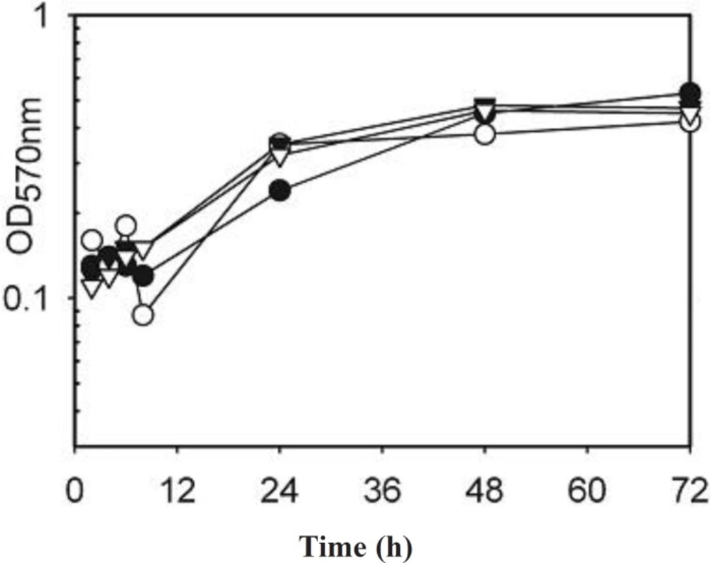
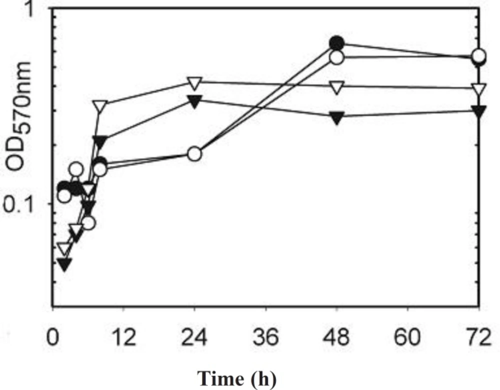
Kinetics of exoprotein production in sub-inhibitory concentrations of silver sulphadiazine

Overnight broth cultures of S. aureusμ-1 of AgSD and incubated at 37 °C as before. Flasks were removed at 2, 4, 8, 24, 48 and 72 h, turbidities were determined using a Unicam SP600 spectrophotometer at 550 nm using distilled water as a blank. Cultures with optical densities above 0.7 were diluted 1:10 with distilled water. The remainder was centrifuged for 20 min at 1500 x g and 4 °C and the supernatant fluids removed and stored at –20 °C.

Preparation of protein

Ice-cold propanol-2 (100 mL) was added to 100 mL of culture supernatant fluid in a 250 mL centrifuge tube, kept on ice for 30 min, and centrifuged at 10,000 rpm for 15 min. The pellet was collected and washed with 50% (v/v) of propanol-2 in electrophoresis buffer and centrifuged again at 10,000 rpm for 15min.The pellet was collected and dissolved in 10 mL of electrophoresis buffer (EB; 10 mM Tris-HCl, 1mM EDTA, both Sigma-Aldrich, UK, pH 8). The sample was placed in a dialysis tube (Medical International Ltd, UK) and dialysed against EB for at least 2 h at 5 ºC. The preparations were removed and stored at 0 ºC until used for electrophoresis.
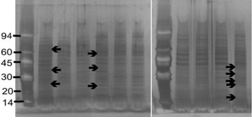
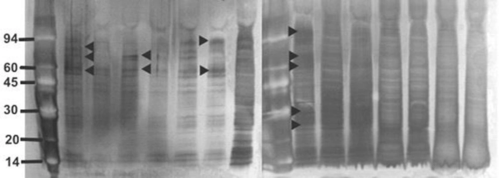
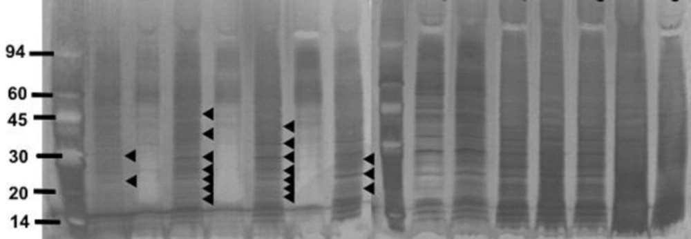
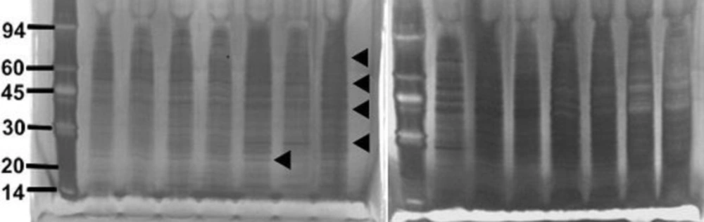
Electrophoresis

Sample (250 μL) was mixed with 250 μL of 5 % (w/v) SDS and 10 % (v/v) β-mercaptoethanol in electrophoresis buffer solution (Pharmacia Biotech, UK) in a 1.5 mL Eppendorf tube and boiled for 5 min. SDS-PAGE (Sodium Dodecyl Sulphate-Polyacrylamide Gel Electrophoresis) was performed using Phastgel 10-15 gradient polyacrylamide gels (Pharmacia) with a continuous 10-15 % gradient and 2 % cross-linking. Protein preparations (3 µL) were applied and electrophoresis performed at 250V (10.0 mA) for 60 Vh. Protein markers (α-lactalbumin 14,000; trypsin inhibitor, 20,000; carbonic anhydrase, 30,000; ovalbumin, 43,000; bovine serum albumin, 67,000; phosphorylase b, 94,000: Amersham Biosciences, UK) were included on each gel for calibration.

Detection of proteins

The proteins were detected by staining with silver stain (Amersham Biosciences, UK). All staining solutions were prepared freshly before use as direction provided with the kit. The gels were washed x 3 with distilled water for two min on a rotary shaker. Dry gel solution (35 mL; Invitrogen Ltd) was added and shaken for 15 min. The gel was placed between two cellophane sheets (Invitrogen Ltd) and left overnight to dry.

The gels were photographed using an Alpha Imager™2200 system (Alpha Innotech Corporation, UK) and the Rƒ values and the molecular weight of each protein band was automatically calculated using the Alpha Imager™2200 and Alpha Ease™ software using the protein markers to calibrate each gel.

Estimation of protein concentration

Protein concentration was measured by the Bradford method (14) using Bradford Reagent and Bovine serum albumin as standard (both from Sigma Aldrich, UK).

Results

The strains of S. aureus were cultured for 24 h with and without a sub-inhibitory concentration of AgSD and exoproteins isolated from the culture supernatants. The total amounts of exoprotein varied from 8.7 to 13.3 mg 100 mL-l. Total protein remained the same with AgSD in the non-responder strains but increased by 6-15% with AgSD in responder strains. In strain B10, which does not produce TSST-1 and cannot be classified as a responder/non-responder, total exoprotein increased by 5 % in the presence of AgSD which proteins were fractionated by SDS PAGE. There was no clearly detectable increase in overall protein production in responder strains compared to non-responder strains. There were three patterns of exoproteins produced when cultures were grown with and without AgSD. All three strains previously identified as non-responders showed few detectable changes in the proteins (T1, B3, B35; Figure 1). Three strains previously identified as responder strains (T4, B54 and B23), showed a visible increase in the relative amounts of some proteins (arrowed). Some responder strains showed a reduction with AgSD (RN4282, B54) and two responder strains showed no change (B6, B45). Strain 8325-4 had not previously been classified and B10 did not produce TSST-1, both showed a reduction with AgSD (Figure 1). Most strains produced 20+ detectable bands but some bands were unique to particular strains.

As the differences may have been due to changes in growth rate between the strains and between different strains the electrophoresis was repeated for strains incubated with and without AgSD for 2 to 72 h and the growth curves determined (Figure 2). There was generally little effect of AgSD on growth but there were some differences in growth rate between strains (Figure 2). Strains B10, T1 and B45 had similar growth curves and had entered stationary phase by 48h, strain B45 was the only strain to show a decline after 48 h. Strains RN4282, B3, B23, B54 and 8325-4 had similar growth curves, grew more gently and were still in exponential phase by 72 h. Strains B6, B35 and T4 showed growth decelerating after 24h and were in stationary phase by 48 h (Figure 2). The kinetics of exoprotein production in three representative strains is shown in Figures 3. In the responder strains e.g. T4, there was inhibition of exoprotein synthesis by AgSD up to 4 h, especially of proteins of 60-90 kD (Figure 3). Stimulation of proteins of 60 and 94 kD occurred after 6h and there was a general stimulation of protein by AgSD after 8 h, especially proteins of 22, 30, 60, 70 and 97 kD. There were few differences with and without AgSD at later times (Figure 3a). In contrast, in the responder strain B6, production of exoproteins was reduced in the presence of AgSD up to 8 h, particularly in the range of 15-45 kD. Amounts had recovered by 24h and there were few further differences up to 72 h (Figure 3b). In non-responder strain T1, there were few differences during the growth cycle except for reduced amounts of a band at 22 kD at 6h and bands at 40, 50 and 70 kD after 8 h (Figure 3c).

It was possible to tentatively identify some of the proteins on the basis of their molecular weight by comparison with published data (12, 13).

Figure 1. SDS PAGE of extracellular protease from Staphylococcus aureus strains culture incubated with and without silver sulphadiazine for 24h.

Lanes1 and 8 molecular weight markers (kD); 2, T4 control; 3, T4+AgSD; 4, T1 control; 5, T1+AgSD; 6, 8325-4 control; 7, 8325-4+AgSD; 9, B35 control; 10, B35+AgSD; 11, B3 control; 12, B3+AgSD; 13, B54 control; 14, B54+AgSD
lanes 1 and 8 molecular weight markers (kD); 2, RN4282 control; 3, RN4282+AgSD; 4, B10 control; 5, B10+AgSD; 6, B6 control; 7, B6+AgSD; 9, B45 control; 10, B45+ AgSD; 11 B23 control; 12, B23+AgSD.

Figure 2. Growth curves of Staphylococcus aureus strains incubated with and without AgSD for 2 to 72 hours.

T4 control Ḝ● , T4+AgSD ○ , T1 control ▼ ,T1+AgSDΔ.
RN4282 control ● , RN4282+AgSD ○, B23 control ▼ , B23+AgSD Δ.

Figure 3. SDS-PAGE of extracellular proteins produced by Staphylococcus aureus Strains at different times 2, 4, 6, 8, 24, 48, 72 hours of incubation in the presence and absence of silver sulphadiazine (25 μg mL-1AgSD).

SDS-PAGE of extracellular proteins produced by Strains T4, at different times in the presence and absence of silver sulphadiazine
SDS-PAGE of extracellular proteins produced by Strain B6, at different times in the presence and absence of silver sulphadiazine
SDS-PAGE of extracellular proteins produced by Strain T1, at different times in the presence and absence of silver sulphadiazine

Discussion

The effects of sub-inhibitory concentrations of silver sulphadiazine on exoprotein synthesis were complex. There were three classes of response, responder strains that showed enhancement of synthesis of certain proteins, non-responder strains that showed no change and responder strains that showed a general decrease in exoprotein production in the early stages of growth.

The extracellular proteins produced by S. aureus strains were dissimilar in different growth phases. Some of the exoproteins were produced throughout growth, some were only produced in exponential phase, some were produced only in late exponential phase and some were only produced in stationary phase in both responder and non-responder strains. A number of factors have been shown to affect exoprotein synthesis. Expression of hla, tst and spa was strongly repressed in the presence of sodium chloride (1 M) or sucrose (20 mM), expression of tst requires O2 and CO2 and is affected by concentration of Mg2+ (15-19).

The synthesis of many exoproteins is regulated by agr and sar, so that is highest in stationary phase which was reached by most stains at 48 h in the present study. The cell density was approximately 10 to 20 times lower than in shaker (aerated) cultures (unpublished observations) but cell densities are still higher than the1.2 x 108 cfu mL-1 needed for induction of agr by 24-48h depending on the strain.

The control of exoprotein gene expression is a complex interplay between different regulatory systems (20-28). Sar regulates expression of agr mainly during the transition between exponential and stationary phases of growth and also has direct effects on expression of some exoproteins independent of agr (29-32).

It has also been reported that strains that produce TSST-1 produce lower amounts of extracellular proteins (32-34). No mechanism has been proposed for this. In the present study the TSST-1 negative strain B10 did not produce significantly more exoprotein than the TSST-1 producing strains. The reduction in exoprotein production in TSST-1 positive strains may not be a general phenomenon or may not have been expressed under the growth conditions used here.

The increases seen in this study were modest compared to the 8-32 fold increases in TSST-1 and changes in proteases seen in our previous study (12, 13). One difference is that static cultures were used in present study, whereas shaken cultures (aeration) were used in previous studies. The production of TSST-1 has been shown to be affected by aeration and CO2 levels (18, 19)and specific growth rate (33) and there is clearly an interaction with the stimulation of TSST-1 synthesis by AgSD and aeration which is not simply due to the higher cell densities formed by aerated cultures.

Overall the effects of sub-inhibitory concentrations of AgSD were complex with three classes of responses recorded in the strains tested. This may reflect differences in susceptibility to AgSD and, although MIC’s were similar, this could be tested by investigating the effects of different concentrations of AgSD. The results support the work of Kaur et al. (34) who showed that 70 μg/mL of Ag+ decreased synthesis of haemolysin, deoxyribonuclease and protein A but increased coagulase production in that the effect of Ag+ are dependent on exoprotein.

Some of the proteins were tentatively identified on the basis of their apparent molecular weight when compared to published data but there were a number of proteins that could not be identified. The pattern of exoproteins will depend on whether a particular strain carries the gene for a particular protein and may be complicated by activation of exoenzymes by cleavage by proteases e.g. lipase and serine protease (35) and by modification of surface proteins such as FnbA and protein A by proteoytic cleavage (27) the cleavage products of which will be present in the medium. The proteins that could not be identified from results published in previous studies may reflect the use of different strains and culture followed by MALDI/TOF (Matrix Assisted Laser Desorption Ionization/ Time of Flight) mass spectroscopy should allow identification of the proteins.

Acknowledgements

References

  • 1.

    System NNIS. National Nosocomial Infections Surveillance (NNIS) System report, data summary from January 1992 to June 2002. AJIC. 2002;30:458.

  • 2.

    Cosgrove SE, Sakoulas G, Perencevich EN, Schwaber MJ, Karchmer AW, Carmeli Y. Comparison of mortality associated with methicillin-resistant and methicillin-susceptible Staphylococcus aureus bacteremia: a meta-analysis. Clin inf dis. 2003;36:53-59.

  • 3.

    Diekema D, Pfaller MA, Schmitz FJ. Survey of infections due to Staphylococcus species: frequency of occurrence and antimicrobial susceptibility of isolates collected in the United States, Canada, Latin America, Europe and the Western Pacific region for the SENTRY Antimicrobial Surveillance Program, 1997–1999. Clin inf dis. 2001;32:S114-S132.

  • 4.

    Schito G. The importance of the development of antibiotic resistance in Staphylococcus aureus. Clin micro and infe. 2006;12:3-8.

  • 5.

    Fischetti VA, Novick RP, Portnoy DP, Rood JI. Gram-positive pathogens. ASfM. 2006;2:379-385.

  • 6.

    Ziebandt AK, Becher D, Ohlsen K, Hacker J, Hecker M, Engelmann S. The influence of agr and σB in growth phase dependent regulation of virulence factors in Staphylococcus aureus. Proteo of micro patho. 2007;4:225-247.

  • 7.

    Royan S, Sharp L, Nair SP, Crean S, Henderson B, Poole S, Scott GL, Evans AW. Identification of the secreted macromolecular immunogens of Staphylococcus aureus by analysis of serum. FEMS Immunology & medical microbiology. 2000;29:315-321. [PubMed ID: 11118913].

  • 8.

    Jarraud S, Mougle C, Thioulouse J, Lina G, Meugnier H, Forey F, Nesme X, Etienne J, Vandenesch F. Relationships between Staphylococcus aureus genetic background, virulence factors, agr groups (alleles), and human disease. Infe and immu. 2002;70:631-641.

  • 9.

    Fox CL, Modak SM. Mechanism of silver sulfadiazine action on burn wound infections. Antimicrobial agents and chemo. 1974;5:582-588.

  • 10.

    Fox CL, Rappole B, Stanford W. Control of pseudomonas infection in burns by silver sulfadiazine. Surg gyneco & obst. 1969;128:1021-1026.

  • 11.

    Hamilton-Miller JS, Shah, Smith C. Silver sulphadiazine: a comprehensive in vitro reassessment. Chemo. 1993;39:405-409.

  • 12.

    Edwards-Jones V, Foster H. The effect of topical antimicrobial agents on the production of toxic shock syndrome toxin-1. JMM. 1994;41:408-413. [PubMed ID: 7966218].

  • 13.

    Edwards-Jones V, Foster H. Effects of silver sulphadiazine on the production of exoproteins by Staphylococcus aureus. JMM. 2002;51:50-55. [PubMed ID: 11800472].

  • 14.

    Bradford MM. A rapid and sensitive method for the quantitation of microgram quantities of protein utilizing the principle of protein-dye binding. Anal biochem. 1976;72:248-254. [PubMed ID: 942051].

  • 15.

    Yarwood JM, Schlievert PM. Oxygen and carbon dioxide regulation of toxic shock syndrome toxin 1 production by Staphylococcus aureus MN8. J clin micro. 2000;38:1797-1803.

  • 16.

    Sarafian S, Morse S. Environmental factors affecting toxic shock syndrome toxin-1 (TSST-1) synthesis. JMM. 1987;24:75-81. [PubMed ID: 3612745].

  • 17.

    Reeves MW. Effect of trace metals on the synthesis of toxic shock syndrome toxin 1. Rev of infe dis. 1989;11:145-150.

  • 18.

    Taylor D, Holland K. Production of toxic shock syndrome toxin 1 by Staphylococcus aureus under aerobic and anaerobic conditions and the effect of magnesium ion limitation. Rev of infe dis. 1989;11:151-156.

  • 19.

    Ross RA, Onderdonk AB. Production of toxic shock syndrome toxin 1 by Staphylococcus aureus requires both oxygen and carbon dioxide. Infe and immu. 2000;68:5205-5209.

  • 20.

    Recsei P, Kreisworth B, O'Reilly M, Schlievert P, Gruss A, Novick RP. Regulation of exoprotein gene expression in Staphylococcus aureus by agr. MGG. 1986;202:58-61. [PubMed ID: 3007938].

  • 21.

    Chan PF, Foster SJ. The role of environmental factors in the regulation of virulence-determinant expression in Staphylococcus aureus 8325-4. Microbio. 1998;144:2469-2479.

  • 22.

    Peng H, Novick RP, Kreiswirth B, Kornblum J, Schlievert P. Cloning characterization, and sequencing of an accessory gene regulator (agr) in Staphylococcus aureus. J of bact. 1988;170:4365-4372. [PubMed ID: 2457579].

  • 23.

    Novick R, Projan S, Kornblum J, Ross H, Kreiswirth B, Moghazeh S. Theagr P2 operon: An autocatalytic sensory transduction system in Staphylococcus aureus. MGG. 1995;248:446-458. [PubMed ID: 7565609].

  • 24.

    Janzon L, Arvidson S. The role of the delta-lysin gene (hld) in the regulation of virulence genes by the accessory gene regulator (agr) in Staphylococcus aureus. The EMBO journal. 1990;9:1391. [PubMed ID: 2328718].

  • 25.

    Ji GY, Beavis RC, Novick RP. Cell density control of staphylococcal virulence mediated by an octapeptide pheromone. Proceedings of the National Academy of Sciences. 1995;92:12055-12059.

  • 26.

    Balaban N, Novick RP. Autocrine regulation of toxin synthesis by Staphylococcus aureus. Proceedings of the National Academy of Sciences. 1995;92:1619-1623.

  • 27.

    Karlsson A, Saravia-Otten P, Tegmark K, Morfeldt E, Arvidson S. Decreased amounts of cell wall-associated protein A and fibronectin-binding proteins in Staphylococcus aureus sarA mutants due to up-regulation of extracellular proteases. Infe and immu. 2001;69:4742-4748.

  • 28.

    Novick RP. Autoinduction and signal transduction in the regulation of Staphylococcal virulence. Molec micro. 2003;48:1429-1449.

  • 29.

    Karlsson A, Arvidson S. Variation in extracellular protease production among clinical isolates of Staphylococcus aureus due to different levels of expression of the protease repressor SarA. Infe and immu. 2002;70:4239-4246.

  • 30.

    Garvis S, Mei JM, Ruiz-Albert J, Holden DW. Staphylococcus aureus SvrA: a gene required for virulence and expression of the agr locus. Microbio. 2002;148:3235-3243.

  • 31.

    Said-Salim B, Dunman PM, McAleese FM, Macapagal D, Murphy E, McNamara PJ, Arvidson S, Foster TJ, Projan SJ, Kreiswirth BN. Global regulation of Staphylococcus aureus genes by Rot. J of bact. 2003;185:610-619. [PubMed ID: 12511508].

  • 32.

    Vojtov N, Ross HF, Novick RP. Global repression of exotoxin synthesis by Staphylococcal superantigens. Proceedings of the National Academy of Sciences. 2002;99:10102-10107.

  • 33.

    Wright JD, Holland KT. The effect of cell density and specific growth rate on accessory gene regulator and toxic shock syndrome toxin‐1 gene expression in Staphylococcus aureus. FEMS microbiology letters. 2003;218:377-383. [PubMed ID: 12586420].

  • 34.

    Kaur P, Saxena M, Sareen M, Vadehra DV. Effect of silver ion resistance in Staphylococcus aureus on production of extracellular enzymes & toxins. The IJMR. 1987;86:716-721.

  • 35.

    Rollof J, Normark S. In vivo processing of Staphylococcus aureus lipase. J of bact. 1992;174:1844-1847. [PubMed ID: 1548232].