Celecoxib Treatment Alters p53 and MDM2 Expression via COX-2 Crosstalk in A549 Cells

authors:

avatar Mehdi Gharghabi a , avatar Farhang Rezaei a , avatar Fereshteh Mir mohammadrezaei b , avatar Mohammad Hossein Ghahremani a , *

Department of Toxicology-Pharmacology, School of Pharmacy, Tehran University of Medical Sciences, Tehran, Iran.
Department of Biology, Faculty of Science, University of Mazandaran, Mazandaran, Babolsar, Iran.

how to cite: Gharghabi M, Rezaei F, Mir mohammadrezaei F, Ghahremani M H. Celecoxib Treatment Alters p53 and MDM2 Expression via COX-2 Crosstalk in A549 Cells. Iran J Pharm Res. 2016;15(2):e125182. https://doi.org/10.22037/ijpr.2016.1849.

Abstract

Cyclooxygenase-2 (COX-2) has a pivotal role in the pathogenesis of the lung cancer. It is known that COX-2 negatively regulates the activity of a number of tumor suppressors, including p53. Consequently, inhibition of COX-2 signaling is anticipated to be a promising approach to stabilize p53 functionality. In this regard, we investigated the effect of COX-2 signaling blockade on p53 and COX-2expression in A549 cells. Cell viability was assessed using MTT and protein expression was measured using Western Blot assay. Results revealed that Celecoxib dose-dependently induced growth inhibition within 24 h. However, prolonged exposure to the drug up to 48 h led to increase cell viability compared to the corresponding control. Western blot analysis demonstrated that Celecoxib could augment p53 expression within 24 h, independently of COX-2 inhibition. In contrast, Celecoxib treatment not only returned p53 to the control level, but also strikingly induced COX-2 expression within 48 h. Of further relevance, Celecoxib exposure could significantly result in MDM2 elevation at 48 h. These findings represent p53 as a molecular target being interconnected with COX-2 signaling axis upon Celecoxib treatment. Moreover, our data point toward the possibility that Celecoxib treatment may not be a proper therapeutic strategy in lung cancer cells owing to its potential role in the activation of oncogenes, including COX-2 and MDM2 which seemingly confers a chemoresistance circumstance to the cell. Consequently, these results underscore intensive preclinical assessment prior to applying COX-2 inhibitors in the treatment of lung tumors.

Introduction

Lung cancer is a leading cause of cancer death worldwide, in which amplification of oncogenes contributes to inflammation and tumorogenesis (1, 2). A vast array of mediators orchestrates inflammation signaling pathways destined to invasion, metastasis, and angiogenesis in lung cancer (-). Meanwhile, COX-2 is a key modulatory molecule engaged in the pathogenesis and development of the lung neoplasia (-). Consistently, growing evidences has emphasized COX-2 blockers as a promising therapeutic approach in a variety of malignancies, in particular lung tumors (-). Accumulating investigations have substantiated that Celecoxib exerts its effect partly through COX-2-independent mechanisms, including inhibition of Akt signaling (-). Akt is a crucial signaling molecule that delivers mitogenic and anti-apoptotic signals (17, 18). It is known that Akt promotes p53 degradation via MDM2 (19). p53 is a well-established tumor suppressor protein and key regulator of apoptotic responses, while MDM2 is a major modulator of p53 activity via binding to and thereby promoting its degradation (20). Thus, the consequent Akt blockade can affect p53. On the other hand, several lines of evidence have addressed functional crosstalk between p53 and COX-2 signaling pathway (11, 12 and -). Given these molecular interconnections stated above, we sought to investigate the effect of COX-2 signaling blockade on p53 and COX-2 expression pattern in A549 cells.

Experimental

Reagents

RPMI 1640 media, FBS (Fetal Bovine Serum), trypsin, penicillin, and streptomycin were purchased from Biosera (Austria). MTT [3-(4, 5-dimethylthiazol-2-yl)-2, 5-diphenyltetrazolium bromide] was purchased from Sigma (USA). Celecoxib was from Tehran University of Medical Sciences, Faculty of Pharmacy (Iran). Western blot detection kit and polyvinyl difluoride (PVDF) membrane were provided from Roche (Germany). Anti-FHIT antibody was from Abcam (USA). p53 and COX-2 antibodies were provided from cell signaling (U.S.A).MDM2 antibody was from Novus (USA). β-actin antibody was from Santa Cruz Biotech (USA). The rest of the reagents, including chemicals were from Merck (Germany).

Cell culture

Parental A549 cells were obtained from EACC. Cells were cultured in RPMI-1640 media with 10 % fetal bovine serum, penicillin (100 IU/mL), and Streptomycin (100 mg/mL) at 37 °C in a 5 % CO2 humid atmosphere.

Drug treatment

Cells were exposed to different concentrations of Celecoxib (10-100µM). Subsequently, treated cells were harvested within 24 and 48 h. Dimethyl sulfoxide(DMSO) (equal volume to added drug) was used as the vehicle control in all the experiments.

Cell viability assay

The effect of Celecoxib on cell viability was assessed using MTT method. Briefly, cells were seeded at 10,000 cells per well in 96-well plate and were treated with Celecoxib for 24 and 48 h. Subsequently, MTT solution was added at final concentration of 5 mg/mL and cells were incubated (37 °C, 5% CO2) for additional 4 h. The formazan crystals were dissolved using 60μL of DMSO and the absorbance was measured using micro plate reader (Anthos 2020, Austria) at 570 nm against690 nm. The cell viability was finally calculated as percentage of control group.

Western Blotting

After incubation period, cells were harvested into Laemli lysis buffer (Tris62.5mM, pH 6.8, dithiothreitol 50mM, sodium dodecyl sulphate (SDS) 2 %, Glycerol 10 % and Bromophenol Blue 0.25 % (w/v). Equal amount of protein samples were subjected to 12 % SDS–PAGE; and transferred to PVDF membranes. Membranes were then gently blocked with 1 % casein for 4h at room temperature and incubated with anti-p53 (1:1000), anti-COX-2 (1:1000), and anti-MDM2 (1/1000) antibodies overnight at 4 °C. Anti-β-actin antibody (1:2000) was used as internal control. The blots were then exposed to HRP-conjugated secondary antibody (1:10000) for 1 h at room temperature. Protein bands were revealed using chemiluminescence kit on X-ray film. The protein densitometry analysis was carried out using Image J software (USA) and expressed as Mean±SE of three independent experiments.

Statistical analysis

All of the experiments were conducted at least three times independently. Results were presented as Mean±SE. Multiple comparisons were carried out using One-way Analysis of Variance (ANOVA) followed by a Tukey–Kramer post hoc analysis. P values less than 0.05 were regarded as statistically significant.

Results

Celecoxib-induced growth inhibition

We examined the effect of different doses of Celecoxib (10-100µM) on A549 cell proliferation. The lower stimuli (10, 25µM) did not provoke significant growth inhibition at both time points compared to the corresponding control (data not shown). Hence, we investigated the effect of higher doses of Celecoxib on cell proliferation. As plotted in Figure 1, Celecoxib exerted marked growth inhibition upon 50,100µM within 24 h, whereas prolonged exposure interestingly increased cell viability compared to the corresponding 24 h (P values<0.05 and 0.001, respectively). These results imply that Celecoxib biphasically affects cell proliferation in a dose-and time-dependent manner.

Celecoxib-induced growth inhibition in A549 cells. Cell viability assay was performed in parental cells. Cells were seeded in 96-well plates and exposed to Celecoxib for 24h and 48h. Proliferation assay was carried out using MTT method. Results were calculated as percent control and presented as Mean±SE of three separate experiments (*p<0.05, **p<0.01, and ***p<0.001 compared to control

Celecoxib treatment enhances 53 expression

COX-2 is known to be an oncogene being interconnected with a number of tumor suppressors, such as p53. It has been appeared that COX-2 negatively regulates p53 expression. Consequently, we aimed to examine whether COX-2 signaling blockade can affect on p53 expression. In this respect, we evaluated p53 expression upon Celecoxib treatment in A549 cells within 24, 48 h. As depicted in Figure 2C, Celecoxib dose-dependently augmented p53 expression within 24 h (1.5-2-fold of control, P values <0.05, 0.01, respectively)which was returned to the control level at 48 h.These results express that Celecoxib modulates p53 expression in a dose- and time-dependent manner in wild-type A549 cells.

The effect of Celecoxib on p53 (B), COX-2 (C), and MDM2 (D) expression in wild-type A549 cells. Cells were treated for 24 and 48 h and harvested within the indicated time points. The protein expression profile was analyzed by western blotting. ß-actin was used as internal control. The protein expression was calculated as ratio to actin and presented as Mean±SE of three independent experiments (*p<0.05, **p<0.01, and ***p<0.001 compared to the corresponding control

COX-2 expression alterations in response to Celecoxib treatment

In our quest to understand the effect of Celecoxib on COX-2 signaling pathway, we exposed cells to Celecoxib and COX-2 expression alterations were assessed within 24 and 48 h. As demonstrated in Figure 2C, Celecoxib negligibly attenuated COX-2 level within 24 h in parental cells, whereas incubation within 48 h elevated COX-2 expression compared to the corresponding control (1.5-fold, P value<0.05). Such findings indicate that COX-2 signaling inhibition strictly depends on the timing of the stimuli which implies that Celecoxib treatment may not be a proper approach within longer incubation periods.

MDM2 is markedly induced following Celecoxib treatment

MDM2 is considered as the major protein modulating the expression of p53.Considering the effect of Celecoxib on parental cells at 48 h, we attempted to examine whether Celecoxib affects MDM2 expression at this time point. As depicted in Figure 2D, Celecoxib remarkably led to elevate MDM2 expression level within 48 h (5-fold of control, P value<0.01), implying COX-2 expression correlates with MDM2 signaling modulation in A549 cells.

Discussion

COX-2 signaling pathway has been addressed as an indispensable mediator in the pathogenesis of a variety of malignancies, in particular lung cancer (6, 7). Consistent with this notion, it has been appeared that COX-2 pathway blockade can be a desirable approach in the treatment of a range of cancer models, including the lung neoplasia (27). Previous experiments have delineated functional crosstalk between COX-2 and a subset of molecular targets, such as p53 (-). Considering these statements, the role of COX-2 inhibitors as a possible candidate to induce cell death through apoptotic machinery has been investigated in numerous reports thus far (23, 28).

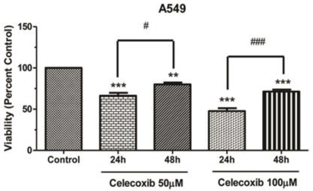
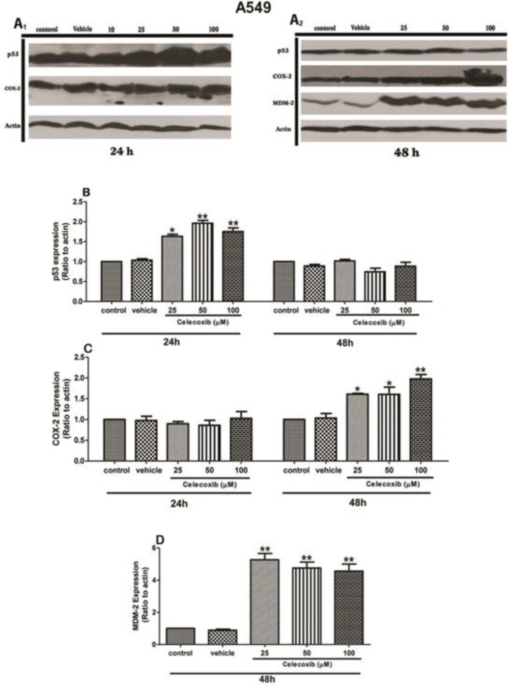
In this respect, we aimed to examine the effect of Celecoxib on p53 expression and persued p53 signaling interaction with COX-2 and MDM2 pathways upon Celecoxib treatment in A549 cells. The cell viability assay has shown that Celecoxib caused a dose-dependent growth inhibition in wild-type A549 cells within 24 h. However, prolonged exposure to the drug conversely led to increase cell viability percent, suggesting that Celecoxib treatment biphasically modulates cell growth depending on the dose and timing of the treatment. In accordance with this, our western blot results have also demonstrated that Celecoxib enhanced p53 expression within 24h in parental cells in a dose-dependent manner (1.5-2-fold of control, P values <0.05, 0.01, respectively). It has been appeared that Akt is a self-evident repressor of the p53 expression through MDM2 and consistently, several studies have reported that Celecoxib can inhibit Akt signaling pathway hence, the resultant p53 up-regulation might be ascribed to disruption of Akt signaling induced by Celecoxib(-, 29). Although Celecoxib could increase p53 level within 24h, COX-2 expression was barely affected at this time point, indicating that Celecoxib augmented p53 expression, independently of COX-2 inhibition (Figures 2B, C). This result suggests p53 as a COX-2-independent molecular target. Considering this finding, we next sought to determine whether inhibition of COX-2 pathway affects prolonged p53 signaling. Paradoxically, Celecoxib exposure lowered p53 expression within 48h, so that p53 returned to the control level at this time point compared to the corresponding 24 h (Figure 2B). Coincidentally, Celecoxib strikingly could result in COX-2 induction at 48 h (Figure 2C).The resultant COX-2 elevation upon Celecoxib treatment can be attributed to p53 that can induce COX-2 expression through Ras/Raf/MAPK cascade according to the prior experiments(24). Accompanying this, MDM2 expression was markedly augmented at 48h, indicating a direct correlation between these signaling pathways (Figure 2D). Thus, it seems that p53 triggers a negative regulatory feedback which finely tunes the cellular homeostasis by manipulating the expression of both MDM2 and COX-2.From the clinical standpoint, such findings can point toward the possibility that Celecoxib treatment may not be a proper therapeutic strategy in lung cancer cells owing to its potential role in the activation of counter-regulatory pathways, resulting in the induction of crucial oncogenes, such as COX-2 and MDM2. In supporting this issue, a recent investigation has also substantiated that Celecoxib promotes cell invasion and confers cells resistant to chemotherapy through activation of MEK-ERK signaling cascade (30). Consistently, it has been appeared that ERK promotes tumorogenesis via MDM2 signaling axis (31).Conceivably, blockage of MDM2 and/or other contributing mediators induced by Celecoxib might be an effective approach to hamper Celecoxib-induced cancer promotion, although elucidating such assumptions demands further investigations. Taken together, such a scenario underscores intensive preclinical assessment before applying COX-2 inhibitors in the treatment of lung tumors.

Acknowledgements

References

  • 1.

    Choi JS, Zheng LT, Ha E, Lim YJ, Kim YH, Wang YP, Lim Y. Comparative genomic hybridization array analysis and real-time PCR reveals genomic copy number alteration for lung adenocarcinomas. Lung. 2006;184:355-62. [PubMed ID: 17086460].

  • 2.

    Choi YW, Choi JS, Zheng LT, Lim YJ, Yoon HK, Kim YH, Wang YP, Lim Y. Comparative genomic hybridization array analysis and real time PCR reveals genomic alterations in squamous cell carcinomas of the lung. Lung Cancer. 2007;55:43-51. [PubMed ID: 17109992].

  • 3.

    Peebles KA, Lee JM, Mao JT, Hazra S, Reckamp KL, Krysan K, Dohadwala M, Heinrich EL, Walser TC, Cui X, Baratelli FE, Garon E, Sharma S, Dubinett SM. Inflammation and lung carcinogenesis: applying findings in prevention and treatment. Expert Rev. Anticancer Ther. 2007;7:1405-21. [PubMed ID: 17944566].

  • 4.

    Lee JM, Yanagawa J, Peebles KA, Sharma S, Mao JT, Dubinett SM. Inflammation in lung carcinogenesis: new targets for lung cancer chemoprevention and treatment. Crit. Rev. Oncol. Hematol. 2008;66:208-17. [PubMed ID: 18304833].

  • 5.

    Sethi G, Shanmugam MK, Ramachandran L, Kumar AP, Tergaonkar V. Multifaceted link between cancer and inflammation. Biosci. Rep. 2012;32:1-15. [PubMed ID: 21981137].

  • 6.

    Hosomi Y, Yokose T, Hirose Y, Nakajima R, Nagai K, Nishiwaki Y, Ochiai A. Increased cyclooxygenase 2 (COX-2) expression occurs frequently in precursor lesions of human adenocarcinoma of the lung. Lung Cancer. 2000;30:73-81. [PubMed ID: 11086200].

  • 7.

    Soslow RA, Dannenberg AJ, Rush D, Woerner BM, Khan KN, Masferrer J, Koki AT. COX-2 is expressed in human pulmonary, colonic, and mammary tumors. Cancer. 2000;89:2637-45. [PubMed ID: 11135226].

  • 8.

    Wolff H, Saukkonen K, Anttila S, Karjalainen A, Vainio H, Ristimaki A. Expression of cyclooxygenase-2 in human lung carcinoma. Cancer Res. 1998;58:4997-5001. [PubMed ID: 9823297].

  • 9.

    Khuri FR, Wu H, Lee JJ, Kemp BL, Lotan R, Lippman SM, Feng L, Hong WK, Xu XC. Cyclooxygenase-2 overexpression is a marker of poor prognosis in stage I non-small cell lung cancer. Clin. Cancer Res. 2001;7:861-7. [PubMed ID: 11309334].

  • 10.

    Altorki NK, Keresztes RS, Port JL, Libby DM, Korst RJ, Flieder DB, Ferrara CA, Yankelevitz DF, Subbaramaiah K, Pasmantier MW, Dannenberg AJ. Celecoxib, a selective cyclo-oxygenase-2 inhibitor, enhances the response to preoperative paclitaxel and carboplatin in early-stage non-small-cell lung cancer. J. Clin. Oncol. 2003;21:2645-50. [PubMed ID: 12860939].

  • 11.

    Hida T, Kozaki K, Muramatsu H, Masuda A, Shimizu S, Mitsudomi T, Sugiura T, Ogawa M, Takahashi T. Cyclooxygenase-2 inhibitor induces apoptosis and enhances cytotoxicity of various anticancer agents in non-small cell lung cancer cell lines. Clin. Cancer Res. 2000;6:2006-11. [PubMed ID: 10815926].

  • 12.

    Kang KB, Zhu C, Yong SK, Gao Q, Wong MC. Enhanced sensitivity of celecoxib in human glioblastoma cells: Induction of DNA damage leading to p53-dependent G1 cell cycle arrest and autophagy. Mol. Cancer. 2009;8:66. [PubMed ID: 19706164].

  • 13.

    Liu W, Chen Y, Wang W, Keng P, Finkelstein J, Hu D, Liang L, Guo M, Fenton B, Okunieff P, Ding I. Combination of radiation and celebrex (celecoxib) reduce mammary and lung tumor growth. Am. J. Clin. Oncol. 2003;26:S103-9. [PubMed ID: 12902866].

  • 14.

    Hsu AL, Ching TT, Wang DS, Song X, Rangnekar VM, Chen CS. The cyclooxygenase-2 inhibitor celecoxib induces apoptosis by blocking Akt activation in human prostate cancer cells independently of Bcl-2. J. Biol. Chem. 2000;275:11397-403. [PubMed ID: 10753955].

  • 15.

    Leng J, Han C, Demetris AJ, Michalopoulos GK, Wu T. Cyclooxygenase-2 promotes hepatocellular carcinoma cell growth through Akt activation: evidence for Akt inhibition in celecoxib-induced apoptosis. Hepatology. 2003;38:756-68. [PubMed ID: 12939602].

  • 16.

    Wu T, Leng J, Han C, Demetris AJ. The cyclooxygenase-2 inhibitor celecoxib blocks phosphorylation of Akt and induces apoptosis in human cholangiocarcinoma cells. Mol. Cancer Ther. 2004;3:299-307. [PubMed ID: 15026550].

  • 17.

    Coffer PJ, Jin J, Woodgett JR. Protein kinase B (c-Akt): a multifunctional mediator of phosphatidylinositol 3-kinase activation. Biochem. J. 1998;335(Pt 1):1-13. [PubMed ID: 9742206].

  • 18.

    Khwaja A. Akt is more than just a Bad kinase. Nature. 1999;401:33-4. [PubMed ID: 10485701].

  • 19.

    Ogawara Y, Kishishita S, Obata T, Isazawa Y, Suzuki T, Tanaka K, Masuyama N, Gotoh Y. Akt enhances Mdm2-mediated ubiquitination and degradation of p53. J. Biol. Chem. 2002;277:21843-50. [PubMed ID: 11923280].

  • 20.

    Momand J, Zambetti GP, Olson DC, George D, Levine AJ. The mdm-2 oncogene product forms a complex with the p53 protein and inhibits p53-mediated transactivation. Cell. 1992;69:1237-45. [PubMed ID: 1535557].

  • 21.

    Shigemasa K, Tian X, Gu L, Shiroyama Y, Nagai N, Ohama K. Expression of cyclooxygenase-2 and its relationship to p53 accumulation in ovarian adenocarcinomas. Int. J. Oncol. 2003;22:99-105. [PubMed ID: 12469191].

  • 22.

    Subbaramaiah K, Altorki N, Chung WJ, Mestre JR, Sampat A, Dannenberg AJ. Inhibition of cyclooxygenase-2 gene expression by p53. J. Biol. Chem. 1999;274:10911-5. [PubMed ID: 10196169].

  • 23.

    Swamy MV, Herzog CR, Rao CV. Inhibition of COX-2 in colon cancer cell lines by celecoxib increases the nuclear localization of active p53. Cancer Res. 2003;63:5239-42. [PubMed ID: 14500353].

  • 24.

    Han JA, Kim JI, Ongusaha PP, Hwang DH, Ballou LR, Mahale A, Aaronson SA, Lee SW. P53-mediated induction of Cox-2 counteracts p53- or genotoxic stress-induced apoptosis. EMBO J. 2002;21:5635-44. [PubMed ID: 12411481].

  • 25.

    Choi EM, Heo JI, Oh JY, Kim YM, Ha KS, Kim JI, Han JA. COX-2 regulates p53 activity and inhibits DNA damage-induced apoptosis. Biochem. Biophys. Res. Commun. 2005;328:1107-12. [PubMed ID: 15707991].

  • 26.

    Corcoran CA, He Q, Huang Y, Sheikh MS. Cyclooxygenase-2 interacts with p53 and interferes with p53-dependent transcription and apoptosis. Oncogene. 2005;24:1634-40. [PubMed ID: 15608668].

  • 27.

    Pold M, Krysan K, Pold A, Dohadwala M, Heuze-Vourc'h N, Mao JT, Riedl KL, Sharma S, Dubinett SM. Cyclooxygenase-2 modulates the insulin-like growth factor axis in non-small-cell lung cancer. Cancer Res. 2004;64:6549-55. [PubMed ID: 15374967].

  • 28.

    Grosch S, Tegeder I, Niederberger E, Brautigam L, Geisslinger G. COX-2 independent induction of cell cycle arrest and apoptosis in colon cancer cells by the selective COX-2 inhibitor celecoxib. FASEB J. 2001;15:2742-4. [PubMed ID: 11606477].

  • 29.

    Liu CH, Bao HG, Ge YL, Wang SK, Shen Y, Xu L. Celecoxib inhibits insulin-like growth factor 1 induced growth and invasion in non-small cell lung cancer. Oncol. Lett. 2013;5:1943-7. [PubMed ID: 23833672].

  • 30.

    Wang ZL, Fan ZQ, Jiang HD, Qu JM. Selective Cox-2 inhibitor celecoxib induces epithelial-mesenchymal transition in human lung cancer cells via activating MEK-ERK signaling. Carcinogenesis. 2013;34:638-46. [PubMed ID: 23172668].

  • 31.

    Yang JY, Zong CS, Xia W, Yamaguchi H, Ding Q, Xie X, Lang JY, Lai CC, Chang CJ, Huang WC, Huang H, Kuo HP, Lee DF, Li LY, Lien HC, Cheng X, Chang KJ, Hsiao CD, Tsai FJ, Tsai CH, Sahin AA, Muller WJ, Mills GB, Yu D, Hortobagyi GN, Hung MC. ERK promotes tumorigenesis by inhibiting FOXO3a via MDM2-mediated degradation. Nat. Cell Biol. 2008;10:138-48. [PubMed ID: 18204439].