General
Rink amide MBHA (4-methylbenzhydrylamine) resin and all of the Fmoc-protected amino acids were purchased from Nova Biochem. Boc-HYNIC reagent was synthesized as previously reported method by Abrams
et al. (
27). Solvents, coupling reagents, pyridine, and other reagents were purchased from Fluka, and used without further purification. All aqueous solutions were made using double distilled ultrapure water and filtered by 22 µ filters before biological uses. The cell lines were cultured in Dulbecco’s Modified Eagle’s Medium (DMEM) and Roswell Park Memorial Institute (RPMI 1640) mediums supplemented with 10% (v/v) fetal bovine serum (FBS), 2mM L-Gln, 100 units/mL penicillinG, 100 µg/mL streptomycin from Gibco at 37 °C, 95% humidity, 5% CO
2 in a sterile incubator. Sodium pertechnetate (Na
99mTcO
4) was obtained from commercially available
99Mo/
99mTc generator (AEOI, Tehran, Iran).A JASCO 880-PU intelligent pump reverse phase high performance liquid chromatography (RP-HPLC) with a multiwave length UV detector and a flow-through Raytest-Gabi γ-detector was used for HPLC analysis. A CC 250×4.6mmNucleosil 120-5 C18 column from Teknokroma was used for analytical HPLC, and a VP 250×30 mm Nucleosil 100-5 C18 column was used for semi-preparative HPLC purification. The mobile phase was 0.1% (v/v)TFA/H
2O (Solvent A) and ACN (Solvent B)with the following gradients. Method I for analytical HPLC: 0-5 min 5% B (95% A), 5-20 min 5-100% B, 20-25 min 100% B, 25-30 min 100-5% B, 30-35 min 5% B; flow = 1 mL/min; λ = 280 nm. Method II for semi-preparative HPLC: 0-2 min 20% B (80% A), 2-17 min 20-50% B, 17-19 min 50-100% B), 19-21 min 100% B), 21-25 min 100-20% B, 25-27 min 20% B; flow = 15 ml/min, λ = 280 nm. Mass spectrum was recorded on an Agilent 1100/Bruker Daltonic (Ion trap) VL instrument (LC-MS) using electro-sprayionization (ESI) mode. Quantitative gamma counting was performed on a well-type NaI γ-counter EG&G/ORTEC Model 4001M. Male albino mice and male athymic nude mice were obtained from animal house of research laboratories (AEOI, Tehran, Iran). U373MG cells were obtained from National Cell bank of Iran (NCBI, Pasteur Institute of Iran).
Synthesis
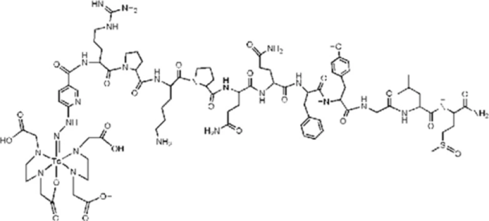
HYNIC-Tyr8-Met(O)11-SP was synthesized according to the standard Fmoc solid phase peptide synthesis chemistry on Rink Amide MBHAresin with loading capacity of 0.69 mmol/g. Briefly, the treatment of diisopropylcarbodiimide (DIC) and N-hydroxybenzotriazole (HOBT)activated carboxyl groups of the Fmoc amino acids to react with the N-terminal amino groups of growing peptide on Rink Amide MBHA resinfor stepwise amino acid addition. The carbodiimide/HOBT coupling strategy was used to minimize the racemization of chiral amino acids and to increase the yield of reactions as the strategy needs equimolar application of amino acids and coupling reagents theoretically. Coupling of each amino acid was performed in the presence of 3 mol excess of Fmocamino acid, 3 mol excess of HOBT, 3 mol excess of DIC and 5 mol excess of diisopropylethylamine (DIPEA) in dimethylformamide (DMF), even though the first Fmoc amino acid was coupled to amino groups of the resin in the presence of 5 mol excess of reagents to achieve the maximum loading on resin active sites. The Kaiser test was used to assess the fullness of coupling reactions and the Fmoc groups were removed by adding 20% piperidine in DMF. Finally, coupling of Boc-HYNIC to peptide was performed in the presence of 1.2 mol excess of Boc-HYNIC, 2.5 mol excess of 2-(7-aza-1H-benzotriazole-1-yl)-1,1,3,3-tetramethyluronium hexafluorophosphate (HATU), and 5 mol excess of DIPEA in DMF. The cleavage step from the resin and the final deprotection of all remained protecting groups was done in a standard cocktail containing trifluoroaceticacid (TFA), triisopropylsilane (TIPS), thioanisole, and water (92.5:2.5:2.5:2.5).The crude peptide was precipitated into cold petroleum ether/diisopropyl ether (50:50). Then, the precipitate was dissolved in water/methanol (50:50) and purified by semi-preparative RP-HPLC (method II).The purified product was lyophilized in vacuum and characterized by analytical HPLC and LC-MS.
Radiolabeling with 99mTc
Labeling of HYNIC-peptide was performed as previously described (
28-
31) with
99mTc in the presence of ethylenediamine-N,N'-diacetic acid (EDDA) and tris(hydroxymethyl)methylglycine (Tricine) as coligands using an exchange labeling approach with both present together. In brief, the labeling process was done by adding 10.75 μL of the stock solution (20 μg of peptide), 15 mg of Tricine and 5 mg of EDDA as coligands in 0.5 mL of water. Then, 40 μg SnCl
2 (10 μL of freshly prepared 4 mg/mL SnCl
2in nitrogen-purged 0.1 M HCl) was added to this solution. Instantly, 30-35 mCi (1110-1295MBq) of freshly eluted sodium pertechnetate (Na
99mTcO
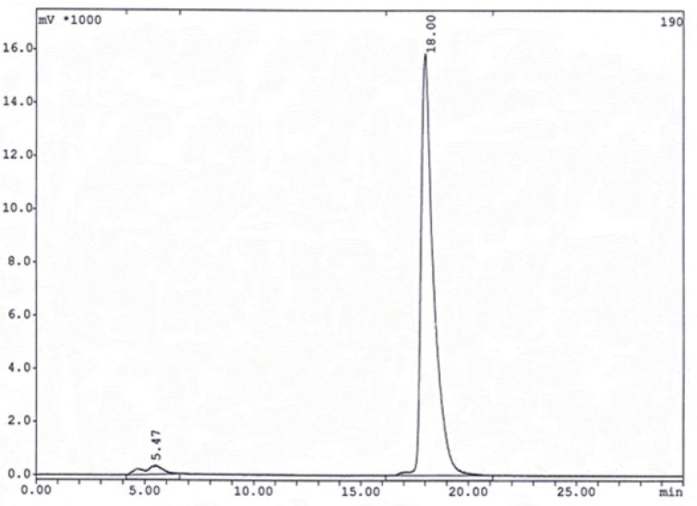
4) in 0.5 mL normal saline was added to the solution and incubated for 15 min at 95 °C in a shielded container. After cooling down to room temperature, the labeling yield was checked on RP-HPLC.
In-vitro evaluation of radiolabeled peptides
Stability in aqueous solution and human serum
The stability of radiolabeled peptide in aqueous solution was evaluated by incubation of the reaction mixture at room temperature (25 °C)up to 24 h.Stability in human serum at 37 °C was tested in parallel after adding 100 μL of reaction mixture to 1ml of fresh human serum. The incubation mixtures were sampled at 1, 4, and 24 h time points. Serum samples (100-200 μL) were treated with ethanol (200-400 μL) and centrifuged (4000 g, 5 min, 4 °C) to precipitate the serum proteins. 20-100 μL aliquots from the supernatant were separated to assess the degradation of 99mTc labeled peptide by RP-HPLC (method I). 100 μL aliquots from the 25 °C incubated mixture were passed through a 22 μ filter to eliminate possible 99mTc colloids and then, 5-20 μL of filtrate were analyzed by RP-HPLC as well.
Protein binding
For protein binding estimation, freshly labeled peptide solution was filtered through a 22 μ filter and a 100 μL aliquot of filtrate was incubated in 1ml fresh human serum for 2 h. After ethanol treatment and centrifugation (4000 g, 5 min, 4 °C), all the supernatant was completely removed. A slight wash with 1x phosphate-buffered saline (PBS) was done on the surface of the sediment and the eluate was added to the removed supernatant. The total activity of the supernatant and the activity of the sediment were measured by well-type NaI γ-counter to determine the percentage of radio peptide bound or transferred to albumin and other serum proteins.
Log p-values
In a 2 mL microtube, 0.5 mL of the99mTc labeled peptides in PBS was mixed with 0.5 mL of octanol. The tube was vigorously vortexed over a period of 10 min and centrifuged at 4000g for 5 min. Three aliquots of 100 μL were sampled from eachlayer and counted in theγ counter. The averaged activities from the aqueous and the octanol layers were used to calculate the log p-values. The octanol-to-water partition coefficient (Po/w) of the radiolabeled peptides was calculated by dividing the counts of the octanol phase by that of the aqueous phase.
In-vitro cell line studies
All the main cell studies were performed on U373MG human Glioblastoma cell lines. Cells were cultured in DMEM was supplemented with L-glutamine, antibiotics (penicillin, streptomycine, gentamycine) and10% FBS. They were grown in culture until a sufficient number of cells were available. At proper times, cells were detached by trypsin-EDTA (0.25% and 0.02%) from the culture flask and diluted with fresh medium to start a new culture or the cell line tests.
Internalization and surface binding studies in U373MG cell lines
Internalization of the
99mTclabeled HYNIC-Tyr
8-Met(O)
11-SP was studied as previously described method(
32,
33) with some modifications. In brief, newly detached U373MG cells were seeded in six-well plates 12 h before the experiment. Their culture medium was replaced by 1.2 mL of previously warmed fresh medium and cells were allowed to adjust to the medium for 1 h at 37 °C, 5% CO
2. In order to determine nonspecific internalization, one line of wells per plate was incubated with 10000 fold excess of unlabeled SP (25 nmol peptide in 150 μL PBS per well) at 37°C/5% CO
2 for 15 min to block NK
1Rs. Then about 150kBq (2.5 pmol peptide) in 150 μL diluted solution of freshly radiolabeled peptide was added to each of the wells and were incubated (in triplicates) for 0.5, 1, 2 and 4 h at 37 °C/5% CO
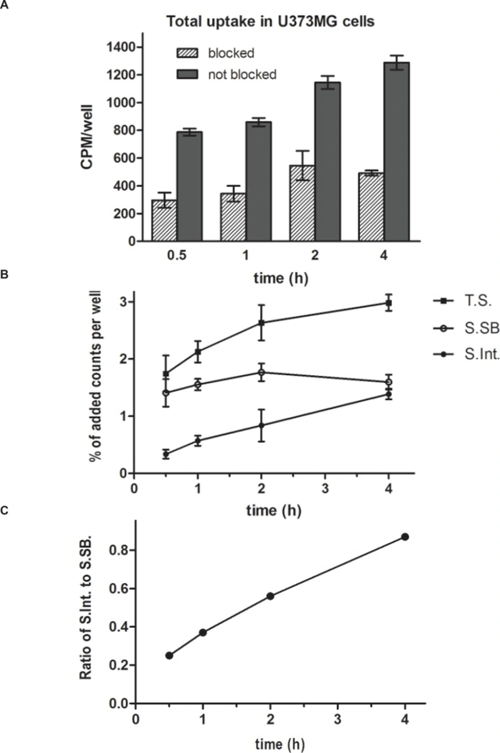
2, respectively. As the final volume of blocked wells was 1.5 mL, the content volume of unblocked wells was adjusted to 1.5 mL by adding 150 μL PBS per well. At each time point, radioactive media were aspirated and wells were washed twice with cold PBS (1x, pH 7.2) to remove free radio peptides. After that, cells were treated with 1 mLGly buffer (0.2M, pH adjusted to 2.8 with 1 M HCl) at room temperature for 5 min (twice) to remove cell-surface bound radio peptide. Finally, the cells were detached from the plates by three steps incubation with 1N NaOH at room temperature for 5 min to determine the internalized radio peptide. All consequent fractions were measured with a NaI γ-counter and results were expressed as percentage of total activity per well (free + cell-surface bound + internalized). Nonspecific accumulations were subtracted from totals to determine the specific accumulations.
Saturation binding experiment (Kd and Bmax)
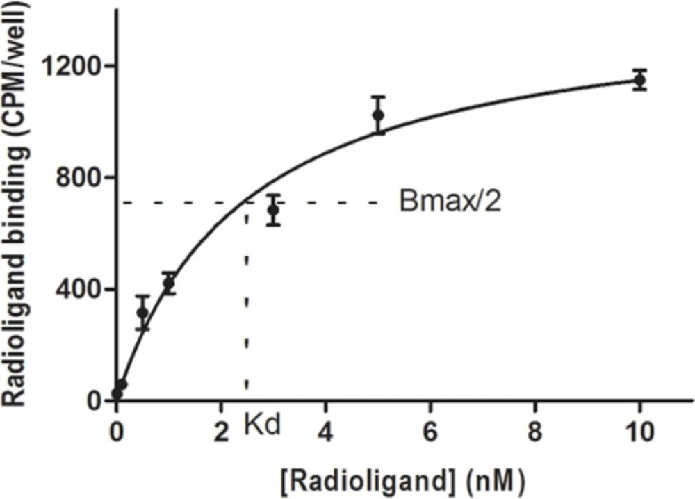
For saturation binding experiment, U373MG cells were incubated with increasing concentrations of 99mTc labeled HYNIC-Tyr8-Met(O)11-SP in the presence and absence of excess unlabeled SP to determine nonspecific (NSB) and total binding (T), respectively. In brief, newly detached U373MG cells were suspended in fresh RPMI medium and aliquots of 800000 cell/1mL were prepared in 5 mL test tubes. In a series of tubes, various concentrations of radiolabeled peptide (0.01-10 nM) in triplicate were the only added but in the other series the radiolabeled peptide was added as well with a fixed amount of excess unlabeled SP. The tubes were incubated for 90 min with a slight shaking every 5 min and were centrifuged at 200 g. Then the supernatants were removed and the surface of cell pellets were washed with cold PBS 1x slightly. The radioactivity of the supernatants was counted to determine the free radiopeptide amounts. The radioactivity of the pellets was counted to measure the NSB and T. The affinity for the radio ligand (Kd) and the maximal number of receptor binding sites (Bmax) were calculated from specific binding (SB) curve using the nonlinear regression analysis of Graphpad Prism 5th (SB = T - NSB).
In-vivo evaluation of radiolabeled peptide
Male albino mice in a weight range of18-24 g were used for normal mice treatments and malenude mice (6 to 8 weeks old) were used for in-vivo tumor treatments. The mice were housed at controlled room temperature (25 ºC) in a 12 h light/12 h dark schedule. They were kept in standard cages with free access to food and water except during the experiments. The nude mice were grown and kept in isolated sterile condition. All animal experiments were carried out in compliance with our institutional ethical guidelines.
Biodistribution in normal mice
Freshly labeled 99mTc-HYNIC-Tyr8-Met(O)11-SP (purity>98%) was diluted to 3.3 nmol/mL with normal saline and a dose of 800 µCi/ 330 pmol in 100 µL was injected to each mouse via the tail vein. Also agroup of three animals (blocked group) were given150 µg of SP in 50 µL salineco-injected with the radiopeptide to determine the non-specific biodistribution of the radiopeptide by blocking the receptor-positive organs. After 1, 4, and 24 h post injection the mice in groups of three animals were sacrificed and organs of interest were excised. All organs and tissues were weighed, and the radioactivity was determined by gamma counter. Results were expressed as percentage of injected dose per gram (% ID/g) of tissue.
Biodistribution in tumor bearing nude mice
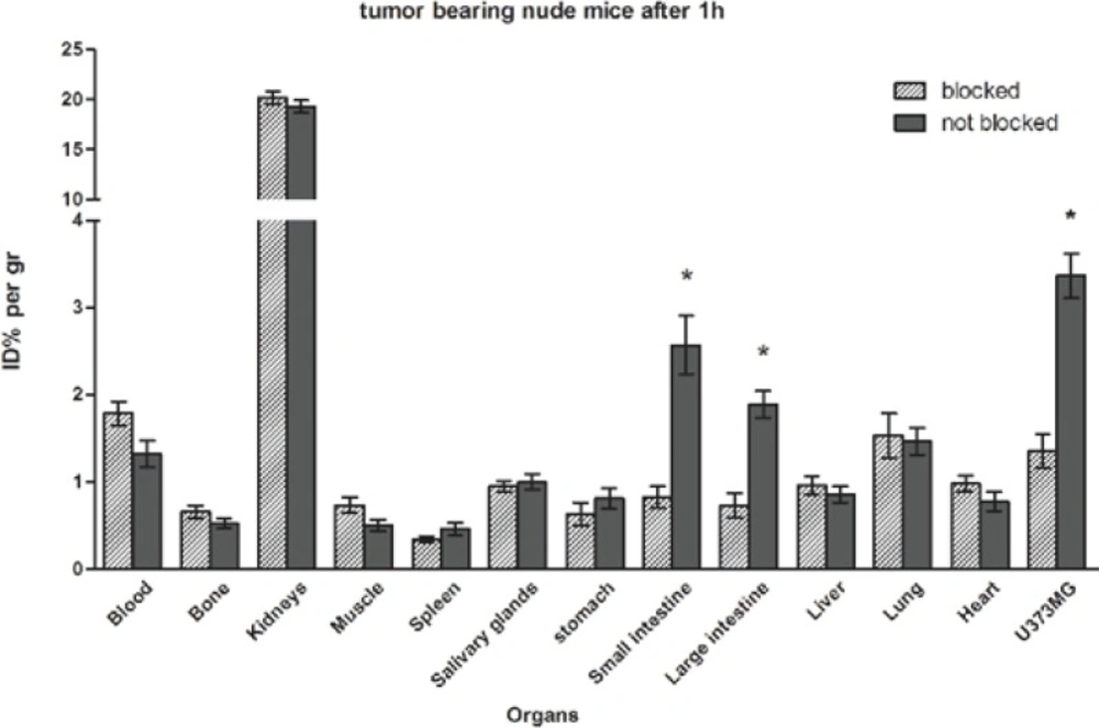
U373MG cells were grown and harvested by trypsinization. Cells were washed twice with fresh RPMI medium containing 20% FBS and centrifuged for 5 min at 200 g. Pellets were resuspended in RPMI medium containing 20% FBS. A 100 µL suspension of 4 × 10⁶ cells was subcutaneously injected on the right flank of each nude mouse. After four weeks, the size of inducted tumors was suitable for biodistribution study and mice were injected via the tail vein with radiolabeled peptide in two groups in the presence and absence of block. They sacrificed at 1 h and 4 h time points to assay the biodistribution. They were dissected and % ID/g of tissues was measured as described above. Finally, tumor-to-organ ratios were calculated.
Statistical analysis
Means and standard deviations for internalization study were computed on Microsoft Excel. Student’s t-test by GraphPad Prism was used to determine statistical significances for internalization and biodistribution study. Associated analyzing method by GraphPad Prism was accomplished to calculate the Kd and Bmax. The 95% level was considered for confidence intervals and significant differences as the default setting.


![ESI-Mass spectra of HYNIC-Tyr<sup>8</sup>-Met(O)<sup>11</sup>-SP obtained by LC-MS analysis. Isolated [M+2H]<sup>2+</sup> molecular ion at 758.1 m/z confirms the 1514.7 molecular mass of the HYNIC-Tyr<sup>8</sup>-Met(O)<sup>11</sup>-SP](https://brieflands.com/journals/ijpr/articles/125290/figures/ijpr-14-097-g001-preview.webp)