General
Pure compounds were isolated by column chromatography using silica gel 60 (63–200 µm, Merck, Germany), MN-polyamide SC-6 (Macherey-Nagel, Germany) and thin layer chromatography (Silica gel 60 F254, Merck, Germany). The structure elucidations were done using different spectral methods, including 1H-NMR, 13C-NMR (BB and DEPT), and EI-MS. The NMR spectra were obtained with Bruker AV-500. The EI-MS spectra were taken with Varian MAT 312 spectrometer.
Plant material
S. Mirzayanii was collected from Kerman, Iran. It was identified by plant taxonomist in the Kerman Faculty of Pharmacy, Kerman University of Medical, Sciences, Kerman, Iran and a herbarium voucher specimen was deposited there.
Extraction and isolation
The air dried plant material (2 Kg) was macerated in methanol (10 L) at room temperature for 3 days. The extraction was repeated three times. The extract was concentrated under reduced pressure to achieve a green gummy residue. Methanol extract was suspended in water and defatted with petroleum ether. The defatted part was then partitioned between ethyl acetate and water. The ethyl acetate fraction was chromatographed on normal phase silica gel column using gradient mixtures of dichloromethane: methanol (0 → 100%) to afford several fractions. Lymphocyte proliferation inhibitory assay of the resulted fractions was compared
in-vitro on peripheral blood lymphocytes (
12). The most active fraction with positive reaction with natural product reagent on its TLC profile was subjected separately to polyamide-SC6 column, using chloroform: methanol (0→20%) as the solvent system (
13). Obtained fractions, were purified more by recycling HPLC using C-18 YMC column (MeOH/H
2O, 7:3) to yield compounds 1-3 as the pure compounds.
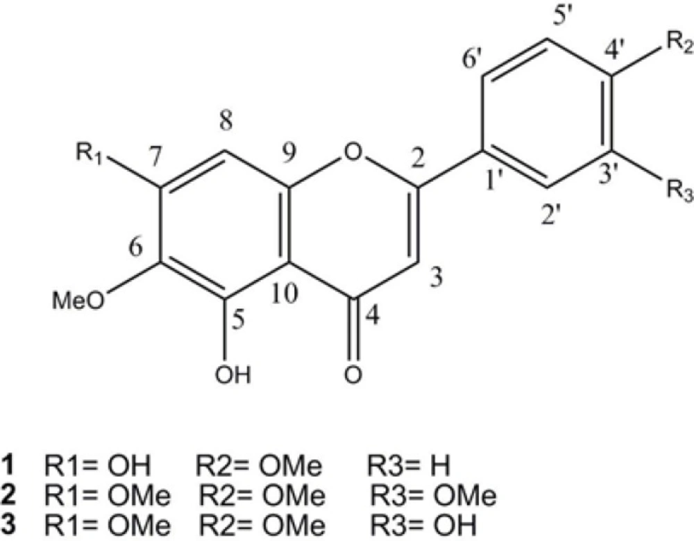
5,7-dihydroxy,6,4'–dimethoxyflavone (1). Light yellow powder. 1H-NMR (500 MHz, DMSO-d6), δH: 3.72, 3.91 (each 3H, s, OMe), 6.83 (1H, s, H-3) , 6.91 (1H, s, H-8), 6.93 (2H, d, J = 7.2 Hz, H-3', H-5'), 7.96 (2H, d, J = 7.2 HZ , H-2' ,6' ), 10.37 (7-OH), 12.91 (5-OH). 13C-NMR (125 MHz, DMSO-d6), δC: 56.4 (4'-OMe), 59.9 (6-OMe), 91.5 (C-8), 102.6 (C-3), 105.0 (C-10), 115.9 (C-3',5'), 121.1 (C-4'), 128.4 (C-2',6'), 131.9 (C-6), 152.0 (C-9), 152.5 (C-5), 158.5 (C-7), 161.2 (C-4'), 164.0 (C-2), 182.1 (C-4). EI-MS m/z (%): 314 (100), 299 (86), 285 (21), 271 (32), 268 (20), 254 (5), 181 (18), 153 (37), 135 (5), 119 (14), 69 (15).
5-hydroxy,6,7,3',4'–tetramethoxyflavone (2). Light yellow powder. 1H-NMR (500 MHz, DMSO-d6), δH:3.73, 3.85, 3.88, 3.93 (each 3H, s, OMe), 6.97 (1H, s, H-3) , 7.04 (1H, s, H-8), 7.13 (1H, s, J = 8.5 Hz, H-5'), 7.59 (1H, d, J = 2.0 HZ , H-2'), 7.72 (1H, dd, J = 8.5, 2.0 HZ , H-6'), 12.89 (5-OH). 13C-NMR (125 MHz, DMSO-d6), δC:55.7 (7-OMe), 55.9 (3'-OMe), 56.4 (4'-OMe), 59.9 (6-OMe), 91.6 (C-8), 103.6 (C-3), 105.1 (C-10), 109.6 (C-2'), 111.7 (C-5'), 120.1 (C-6'), 122.8 (C-1'), 131.9 (C-6), 149.0 (C-3'), 152.0 (C-4'), 152.3 (C-9), 152.6 (C-5), 158.6 (C-7), 163.6 (C-2), 182.2 (C-4). EI-MS m/z (%): 358 (100), 357 (21), 343 (92), 329 (21), 315 (25), 312 (24), 181 (14), 163 (13), 153 (25), 148 (6), 136 (6), 69 (10).
5,3'-dihydroxy,6,7,4'–trimethoxyflavone (3). Light yellow powder. 1H-NMR (500 MHz, DMSO-d6), δH:3.72, 3.86, 3.92 (each 3H, s, OMe), 6.81 (1H, s, H-3) , 6.90 (1H, s, H-8), 7.08 (1H, d, J = 8.5 Hz, H-5'), 7.47 (1H, d, J = 2.0 HZ , H-2'), 7.57 (1H, dd, J = 8.5, 2.0 HZ , H-6'), 9.42 (3'-OH), 12.89 (5-OH). 13C-NMR (125 MHz, DMSO-d6),δC:55.8 (7-OMe), 56.4 (4'-OMe), 59.9 (6-OMe), 91.5 (C-8), 103.3 (C-3), 105.1 (C-10), 112.1 (C-5'), 113.1 (C-2'), 118.7 (C-6'), 122.9 (C-1'), 131.9 (C-6), 146.8 (C-3'), 151.2 (C-4'), 151.9 (C-9), 152.6 (C-5), 158.6 (C-7), 163.8 (C-2), 182.1 (C-4). EI-MS m/z (%): 344 (100), 343 (36), 329 (100), 315 (32), 301 (34), 298 (30), 181 (17), 153 (29), 149 (14).
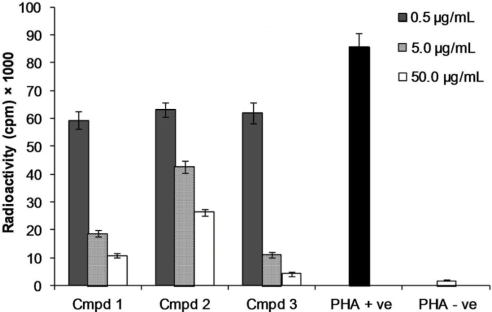
Lymphocyte proliferation assay
Human peripheral blood lymphocytes were incubated using three concentrations (0.5, 5, and 50 µg/mL) of the flavones 1-3 in triplicates in supplemented RPMI-1640 containing 5.0 µg/mL phytohemagglutinin (PHA) at 37 ºC in CO
2 environment for about 72 hours. Incubation for more eighteen hours after the addition of [
3H]-thymidine was done. Then cells were harvested with cell harvester and proliferation percent was determined by the radioactivity count as CPM reading by the Beta-scintillation counter (
14).
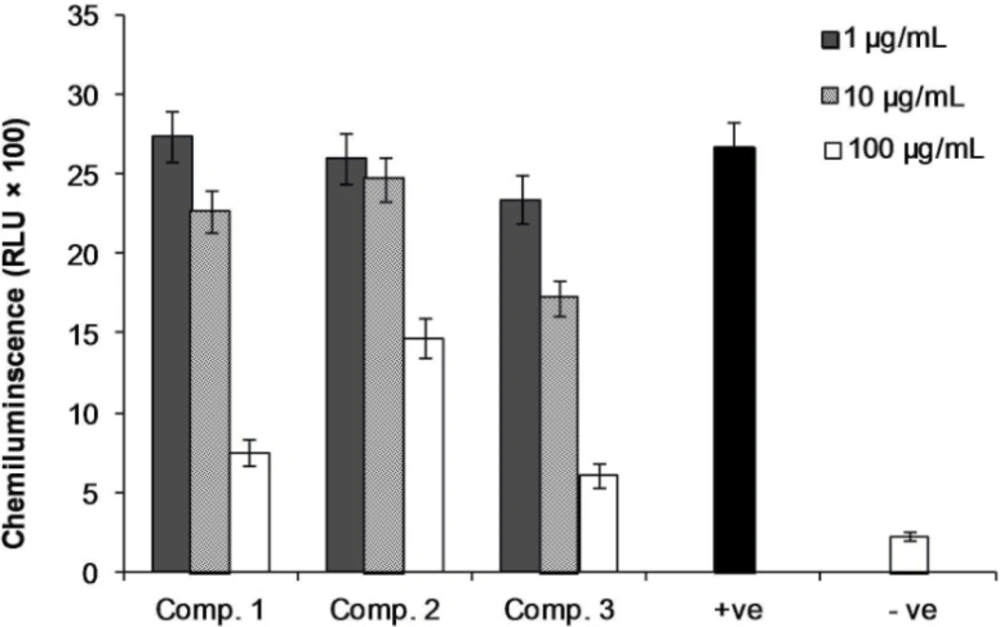
Phagocyte chemiluminscence assay
The reactive oxidants (ROS formation) in whole blood during the phagocytosis oxidative burst processes were measured by the luminol-enhanced chemiluminescence assay procedure (
14). Briefly, whole blood diluted in Hank's buffered salt solution was incubated with three concentrations (1, 10, and 100 μg/mL) of compounds 1-3 in triplicate for 30 minutes at 37 ºC. Then, 25 μL of 20 mg/mL Zymosan (Sigma Chemical Co, USA), followed by 25 μL of 0.07 mM luminol (Sigma Chemical Co., USA) were added to make a final volume of 100 µL. Positive and negative controls were included in the assay. The ROS chemiluminescence kinetic was monitored with a luminometer (Lab systems Luminoskan RS, Helsinki, Finland) for 50 minutes in the repeated scan mode. The peak and total integral chemiluminescence readings were recorded in the relative light unit (RLU).