Materials
Maize starch was procured from Meru Chem Pvt. Ltd., India. Hydroxypropyl Methylcellulose K100M (HPMC K100M) was procured from Colorcon Asia Pvt. Ltd. and solvents of analytical grade were supplied by Merck Ltd., Germany. Gift sample of salbutamol sulphate was received from FDC Ltd., Mumbai, India. 99mTc was provided as a gift sample from Spect Lab Nuclear Medicine Services, Pune, India.
Synthesis of acetylated starch and graft copolymers [Starch grafted poly (methyl methacrylate) (St-g-PMMA) & Acetylated starch grafted poly (methyl methacrylate) (Ast-g-PMMA)]
Acetylated starch and graft copolymers were synthesized and characterized by the methods previously described in our recent published paper (
13). Here on starch back bone, methyl methacrylate was grafted via redox reaction. Wherein, Ce(IV) ion was reduced to Ce(III) ion and made an active site on starch back bone for grafting of methyl methacrylate. Synthesized samples were used for further study.
Preparation of blends
HPMC K100M polymer was chosen for comparison with graft copolymers for its controlled release properties. Various combinations were selected and blend was prepared for salbutamol sulphate using starch/acetylated starch/St-g-PMMA/Ast-g-PMMA/HPMC K100M, spay dried lactose and magnesium stearate. All ingredients were blended in Conta blender until a homogenous mixture was obtained with batch formula shown in
Table 1. No more additives were included in order to get intrinsic information of the graft copolymeric material itself.
| S. No. | Ingredients | Formulation code
|
|---|
F1
| F2
| F3
| F4
| F5
|
|---|
| Qty (mg/tablets) |
|---|
| 1 | Salbutamol Sulphate | 8 | 8 | 8 | 8 | 8 |
| 2 | Maize Starch | 159 | --- | --- | --- | --- |
| 3 | Acetylated Starch (Ast) | --- | 159 | --- | --- | --- |
| 4 | St-g-MMA | --- | --- | 159 | --- | --- |
| 5 | Ast-g-MMA | --- | --- | --- | 159 | --- |
| 6 | Hypromellose (HPMC K 100M PREMIUM) | --- | --- | --- | --- | 159 |
| 7 | Spray dried lactose | 30 | 30 | 30 | 30 | 30 |
| 8 | Magnesium stearate | 3 | 3 | 3 | 3 | 3 |
| | Total Weight | 200 | 200 | 200 | 200 | 200 |
Preformulation study
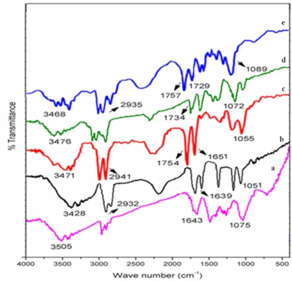
Drug–excipient compatibility study by FT-IR spectroscopy
FT-IR analysis was performed using physical mixture (1:1 w/w) of salbutamol sulphate with starch/acetylated starch/St-g-PMMA/Ast-g-PMMA.
Angle of repose
Angle of repose is an indicator of flowability of the material and it was determined by the fixed funnel and free standing cone method (
14). In this method, blends of drug and excipients were poured through a funnel which was fixed at certain height (h) above the graph paper that was placed on a flat horizontal surface. Different blends were poured until the apex of conical pile just touched the tip of the funnel. Radius of the conical pile (r) was measured and the angle of repose (θ) was calculated by using below mentioned formula:
θ= tan-1 (h/r)
Formulation of tablets
Tablets were prepared by direct compression method. The homogenous mixtures of different blends with suitable flow properties were shifted through #80 sieves, manually fed into the die and compressed in hydraulic press using a 8 mm flat faced beveled edge punch with the crushing force of 70-80 N. The average weight of tablets was 200 mg. Two hundred tablets of each batch were prepared and evaluated for various physicochemical parameters.
Evaluation of tablets
The physical testing on tablets of each batch was done after a relaxation period of at least 24 h. Weight variation test was performed on 20 individually weighed (Citizen CY204 Analytical Balance, Minnesota, USA) tablets according to the official method of United State Pharmacopoeia (USP/NF-32). The thickness and diameter of 10 tablets were measured individually using Vernier Caliper (Edutek instrumentation, Ambala, India) and an average value of thickness and diameter were calculated. The crushing strength (Kg/cm2) of prepared tablets was established using Monsanto hardness tester (MHT-20, Campbell Electronics, Mumbai, India). Tablets friability was determined by Roche friabilator (C-FT-20, Pharma Chem Machineries, Mumbai, India), and it was calculated as the percentage of weight loss in 20 tablets resulting from shock and attrition due to the revolution of plastic chamber operated for 4 minutes at 25 rpm.
Drug content uniformity
Ten tablets were weighed individually and crushed into a fine powder with a mortar and pestle. Crushed powder equivalent to 8 mg of the drug was weighed and transferred to a 50 mL volumetric flask. Distilled water was poured into a volumetric flask up to the mark of 50 mL and flask was further subjected to sonication. Drug was extracted from 50 mL solutions prepared previously using bath sonicator, with sonication time of 2 minutes. The actual drug content was determined using a UV–visible spectrophotometer (Shimadzu UV-1700 spectrophotometer, Japan) at 277 nm and the drug concentration was determined using constructed standard calibration curve, covering the drug solution concentration from 5.0-50.0 µg/mL.
Radiolabelling the tablets with Technetium-99 (99mTc)
Tablets were radiolabelled by drilling a small hole (1 mm) through the center of the tablet with an electric driller and introducing a radioactive solution (
15). Technetium (
99mTc) was chosen for radiolabelling of the tablets because of its short half-life of 6 h along with optimum electron emission. Diethylenetriamine-pentaacetic acid (DTPA) was used to provide the necessary
99mTc species for radiolabelling (
16). In a vial, 100 µL of
99mTc-labeled DTPA was taken and 1 mg of stannous chloride dihydrate (1 mg/mL in 10% acetic acid) was added. The pH was adjusted to 7.5 using 0.5 M sodium bicarbonate solution and mixture was stirred for 5 minutes. Afterwards 4–7 µL of aqueous solutions of
99mTc-DTPA of known radioactivity (100 MBq) was instilled in to the drilled holes, avoiding contact with the surface of the tablets. The radioactive solution was left to diffuse for 15 minutes and then dried to assure uniform dispersion of radioactive material within the matrix. Ethanolic solutions of graft copolymers and lactose (9:1) were used to fully seal the drilled holes. The strength of radioactive label (MBq) was determined by CAPINTEC CRC-15R detector (Pittsburgh, U.S.A).
Stability of radiolabelled tablets
Stability of 99mTc-labeled graft copolymer matrix tablets (F3 & F4) was tested in standard buffer solutions of pH 1.2, 6.8 and 7.4 in order to confirm that the activity would not leach out from the tablets during transit time of the formulation through GI tract. Dissolution tests for the release of the radioactive material was performed by placing radiolabelled tablets in USP type II paddle type dissolution apparatus containing different standard buffers solutions (900 mL), and it was operated at rotation speed of 50 rpm at 37 ± 0.5 °C for 6 h. At each 30 min interval, 5 mL of sample was withdrawn from dissolution vessel and withdrawn volume of dissolution media was replaced with fresh buffer solution. At the end of the dissolution testing, tablets were recovered from the dissolution medium and were further blotted using tissue paper. Activity of the test solutions was determined using a CRC-15R detector. The amount of radioactive strength remained in the tablets was estimated by dissolution testing.
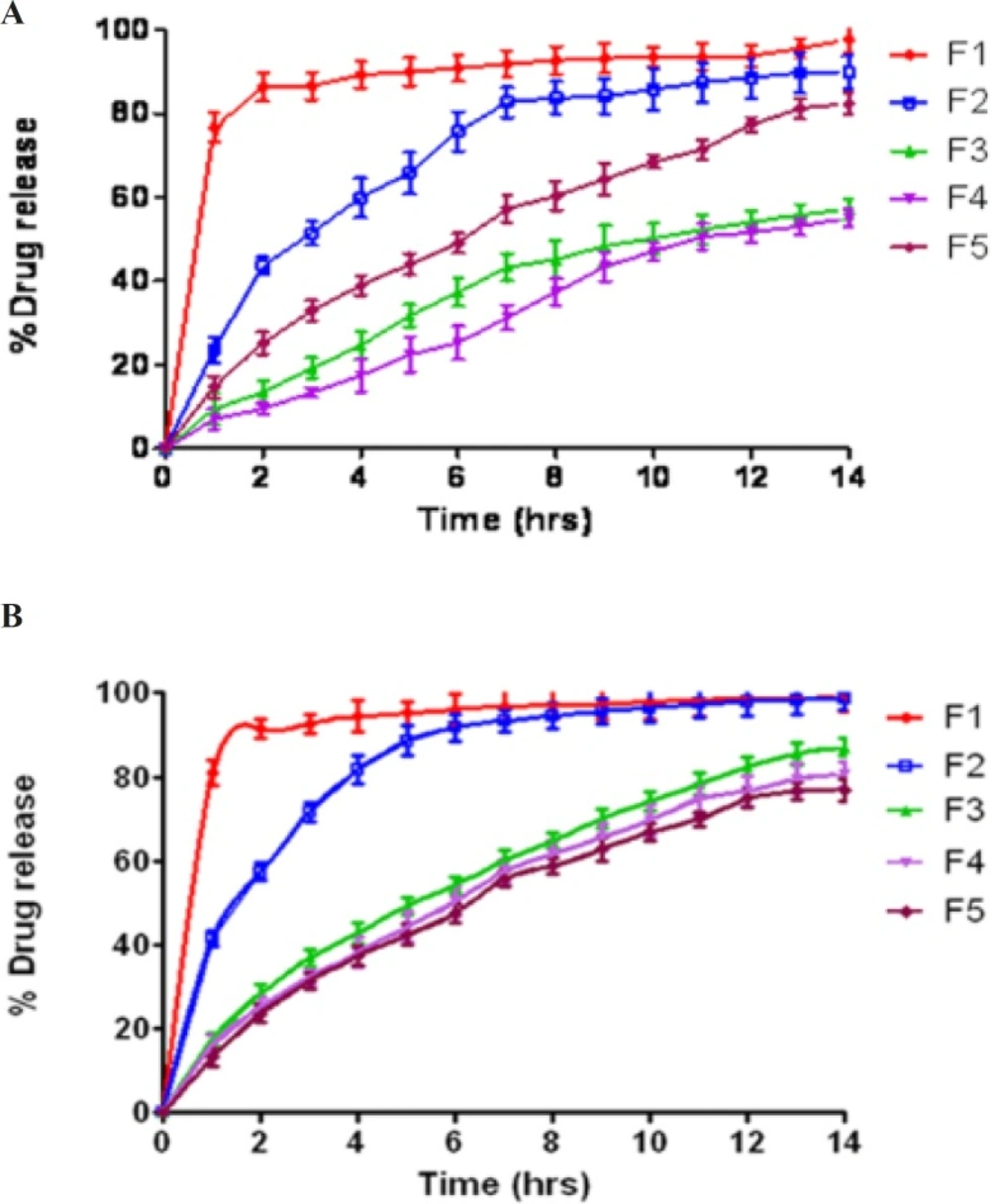
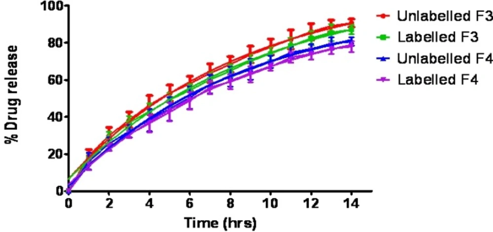
In-vitro drug release studies
Drug release studies were conducted on
99mTc-DTPA labeled tablets and compared with unlabelled tablets to determine whether the labeling process affects the kinetics of drug release from the tablets. Drug release study was carried out using USP dissolution type II apparatus with paddle rotating at 50 rpm
. Dissolution study was performed in simulated gastric fluid (0.1N HCl) and in simulated intestinal fluid (PBS pH 6.8) for 14 h
. The temperature of dissolution medium (900 mL) was maintained at 37 ± 0.5ºC throughout the study. Tablets were placed in different baskets. At a predetermined time intervals of 0, 1, 2, 3, 4, 5, 6, 8, 10, 12 and 14 h, 5 mL sample was withdrawn using a syringe fitted with 0.45 μm filter, and withdrawn volume was replaced with the fresh medium to maintain sink condition
. Samples were analyzed at 277 nm, using UV-visible spectrophotometer. The corresponding cumulative percent of drug released was determined using the constructed standard calibration curve at this wavelength covering the range for the assay. Mean release of three tablets was used to evaluate the drug release for each of the formulations (
17).
Kinetics of drug release
To study the mechanism of drug release from the optimized formulation of matrix tablets, in-vitro release profiles were correlated with various kinetic models like zero order (cumulative amount of drug released vs. time), first order (log cumulative percentage of drug remaining vs. time), Higuchi model (cumulative percentage of drug released vs. square root of time) and Korsmeyer-Peppas (log cumulative percentage of drug released vs. log time) release equations.
In-vivo Pharmacokinetic studies on rabbits
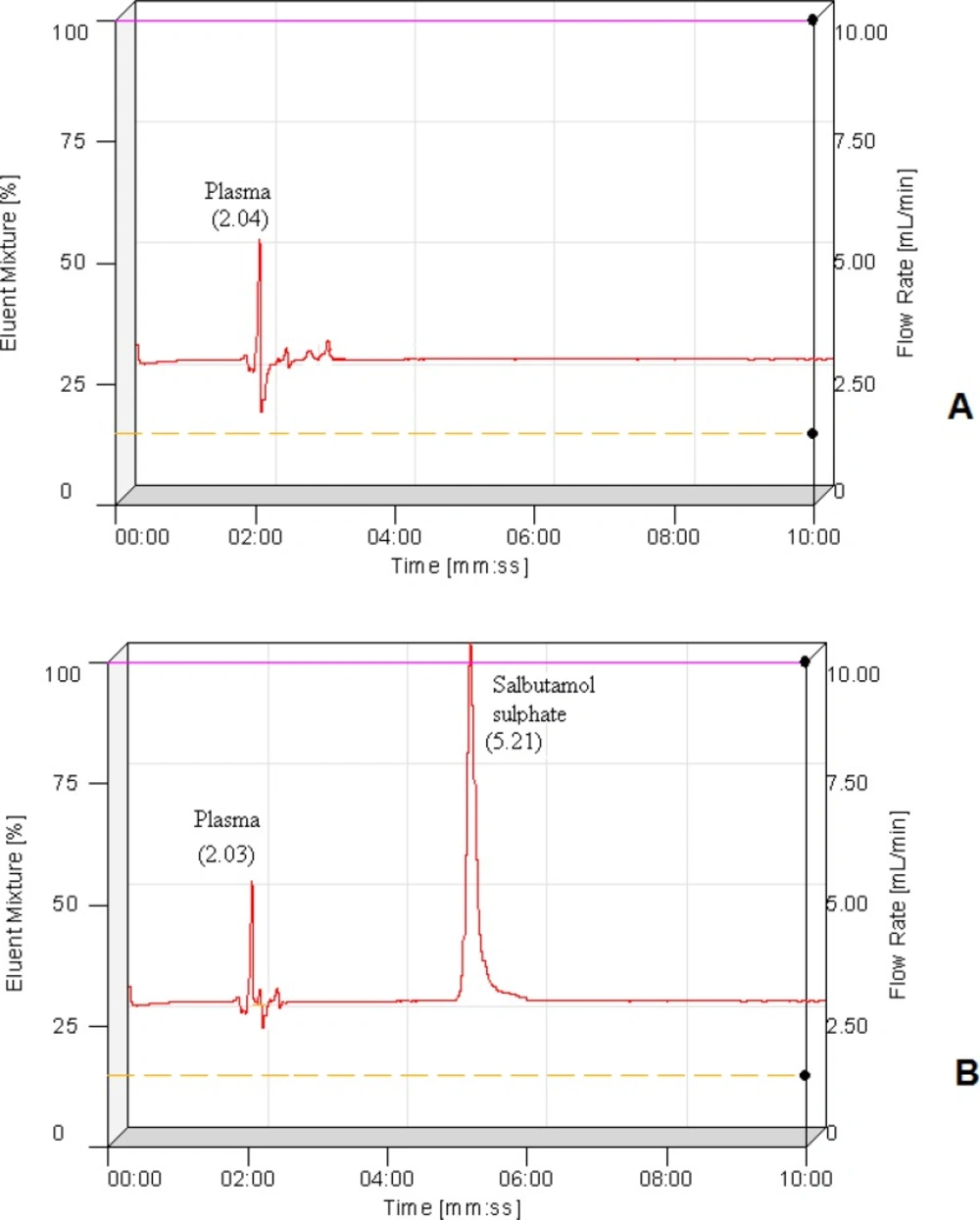
Bio-analytical method development
A high performance liquid chromatography system of Adept series CECIL CE 4201 with UV/Visible detector was used for analysis. The data were recorded by using the software “Power Stream”. The column used for separation was octadecylsilane (C18) with length 250 mm, internal diameter 4.6 mm (Phenomenex) and particle size 5 μm. Chloramphenicol was used as an internal standard. In 0.5 mL of plasma sample, 20 μL of internal standard (100 μg/mL) was added. Drug was extracted from plasma using 5 mL methanol. Mobile phase used for analysis contains acetonitrile, methanol and water in the ratio of 60:20:20 (v/v), and pH adjusted to 2.8 with orthophosphoric acid, with a flow rate of 0.5 mL/min. Sample of 20 μL was injected manually and chromatogram was recorded. Quantification of salbutamol sulphate was performed by plotting peak area ratio of SS to the internal standard as a function of its concentration.
Assay method validation
The developed method was validated following bioanalytical guidelines (
18). Bioanalytical method validation required the determination of selectivity, linearity, LOD, LOQ, accuracy, recovery and precision respectively.
Animal experimental protocol for pharmacokinetic study
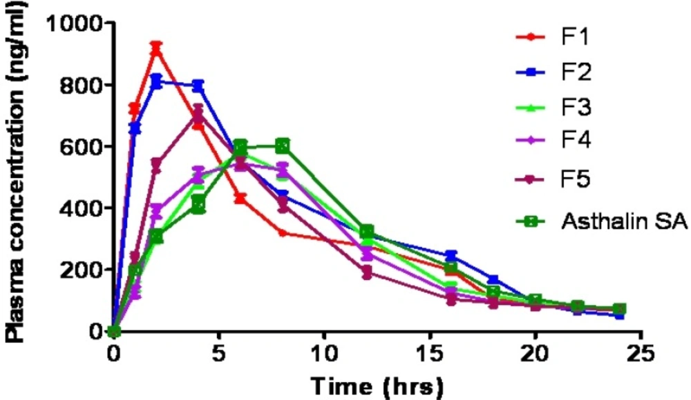
In-vivo studies were performed as per the guidelines of the Council for the Purpose of Control and Supervision of Experiments on Animals, Ministry of Social Justice and Empowerment, Government of India. Pharmacokinetic parameters of graft copolymer formulations (F3 and F4) were compared with tablets prepared by native starch (F1), acetylated starch (F2), HPMC K100M (F5) and commercial sustained release tablets (Asthalin SA-8 mg).
Male albino rabbits weighing 2.5-3.0 kg were randomly selected for the bioavailability study. The animals were subdivided into six groups and each group was comprised of three rabbits. Each group received of the tested formulas namely F1, F2, F3, F4, F5 and Asthalin SA-8 mg. The animals were fasted over night before tablet administration and during the experiment all rabbits had free access to water. Tablets were put behind the tongue to avoid their destruction due to biting. Blood samples (about 1 mL from each animal) were collected from the orbital sinus, before dosing (zero time) and at different intervals after dosing viz. 1, 2, 3, 4, 5, 6, 8, 10, 12, 14, 16, 18, 20, 22, and 24 h. Samples were collected in micro centrifuge tubes containing 50 µL of 10% w/w of disodium EDTA as anticoagulant. The collected samples were immediately centrifuged at 10000 rpm for 10 minutes and plasma were separated and stored at –20 °C.
Statistical analysis
Statistical significance was determined by one way ANOVA (Analysis of variance) (Graph Pad Instat software v 3.06, CA, USA). Significant differences between formulations were analyzed using student newmann-keuls multiple comparison test and p-values of <0.05 were considered to be statistically significant.
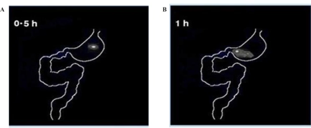
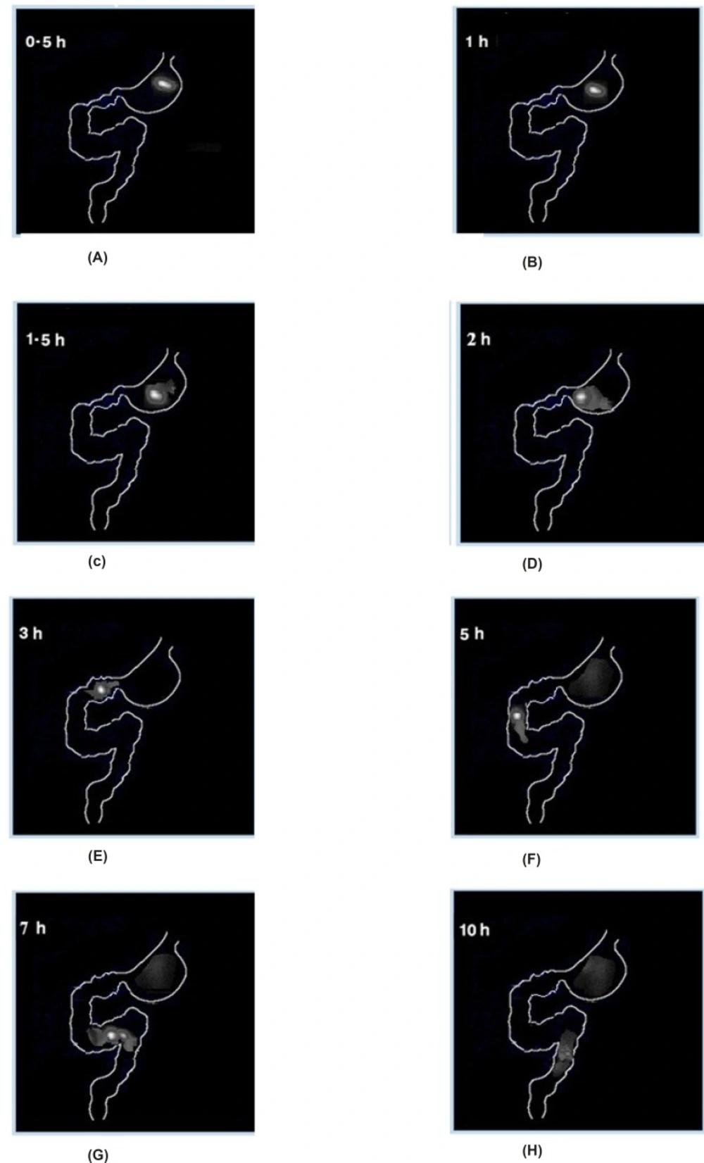
In-vivo imaging and animal study protocol for gamma scintigraphy
The whole experiment was approved by “Institutional Animal Ethical Committee” according to the rules of “Committee for the Purpose of Control and Supervision of Experiments on Animals” (CPCEA, Registration number Dean/10-11/57) for the care and use of laboratory animals were strictly followed throughout the experiment. Gastrointestinal transit of tablets was obtained by imaging studies using 99mTc as a radioactivity marker. Male albino rabbits weighing 2.5-3.0 Kg were randomly selected for gamma scintigraphic study. Rabbits were divided into 3 groups, having 3 animals in each and were fasted for 12 h prior to the gamma scintigraphic study. Group-I received radiolabelled starch matrix tablet (F1), group-II & III received graft copolymer containing matrix tablets F3 and F4 respectively; followed by sufficient amount of drinking water. Animals were anaesthetized with diethyl ether and serial scintigraphic examination was done at 0.5, 1, 1.5, 2, 3, 5, 7 and 10 h to visualize the transit of tablets in GIT, using a large field view gamma camera equipped with a high-resolution, parallel-hole collimator. The 140 keV gamma rays emitted by 99mTc were imaged. Gamma images were recorded using an online computer system and analyzed to determine the distribution of radioactivity in the stomach, intestine and colonic region.