Chemical and biological materials
Bovine serum albumin (BSA, fraction V, minimum 98%), glutaraldehyde 8% aqueous solution, N-succinimidyl 3-(2-Pyridyl Dithio) Propionate (SPDP), 5-fluorouracil, 3, 3’, 5, 5’ tetramethylbenzidine (TMB) substrate and Methylthiazolyldiphenyl-tetrazolium bromide (MTT) were obtained from Sigma (Steinheim, Germany). 2-iminothiolane (2-IT, Traut’s reagent) and 5,5’ -dithio-bis (2-nitro-benzoic acid) (Ellman’s reagent) were purchased from Pierce (IL, USA). Maleimide-poly (ethylene glycol)-Succinimidyl carbonate with the average molecular weight of 5000 Da (Mal-PEG5000-NHS) was obtained from Jen Kem Technology (Texas, USA). Recombinant extracellular part of HER2 was purchased from eBioscience (CA, USA). The production of 1F2 mAb was reported elsewhere (
16). Horse-radish peroxidase (HRP)-conjugated sheep anti-mouse Ig and fluorescein isothiocyanate (FITC)-conjugated sheep anti-mouse immunoglobulin were produced in our laboratory. All other reagents were of analytical grade and used as received.
The SKBR3 and MCF7 cell lines were obtained from National Cell Bank of Iran (NCBI, Pasteur Institute of Iran, Tehran, Iran). The SKBR3 cells were grown in DMEM culture media containing 10% fetal bovine serum (FBS) (Gibco Invitrogen, Carlsbad, CA), 100 U/mL penicillin (Gibco Invitrogen), 1000 U/mL leukemia inhibitory factor (LIF) (Millipore, USA), and 1% glutamine (Gibco Invitrogen). The MCF7 cells were cultured in RPMI 1640 medium (Gibco Invitrogen) containing 15% FBS, 10 µg/mL insulin (Exir Co., Boroojerd, Iran), 100 µg/mL streptomycin, and 100 U/mL penicillin. Both cell lines were incubated at 37 C in a humidified atmosphere containing 5% CO2.
Preparation and separation of 5-FU-loaded BSA nanoparticles
5-FU-loaded BSA nanoparticles were prepared by a well-known desolvation technique as previously described (
18,
20). Briefly, 0.2 g BSA in 2.0 mL of 2 mg/mL 5-FU aqueous solution titrated to pH 8.2 and converted to nanoparticles by continuous addition of ethanol by a syringe pump at the rate of 1.0 mL/min under constant stirring (550 rpm). Subsequently, 120 µL of 8% glutaraldehyde aqueous solution was added drop-wise to induce particles cross-linking. The cross-linking process continued overnight keeping the suspension under stirring. The produced nanoparticles were separated from the solution by three times of centrifugation (25,000 g, 20 min), followed by washing and redispersion in 10 mM NaCl pH 8 to the original volume.
Thiolation of 1F2 mAb
A crucial step in the attachment process of mAb to nanoparticles is the introduction of free sulfhydryl groups (thiolation) on mAb structure. Primary amino groups of the mAb can react with 2-IT, leading to creation of active groups suitable for conjugation to nanoparticles. Thiolation of 1F2 mAb was performed under an optimized condition (
17). In principle, 1F2 solution (500 µL, 1.2 mg/mL) in phosphate buffer saline (PBS, pH 8.0) and containing 5 mM EDTA was incubated with 100-fold molar excess of 2-IT solution for 1 h at 20
°C. Afterward, the thiolated 1F2 was purified by dialysis against 10.0 mL PBS pH 8.0 for 4 hours with 10 times buffer replacement. The amount of thiol groups produced was quantified with Ellman's reagent based on molar absorptivity method as previously described (
17).
Conjugation of 1F2 mAb to BSA nanoparticles using Mal-PEG5000-NHS
Thiolated 1F2 mAb was conjugated to 5-FU-loaded BSA nanoparticles through Mal-PEG5000-NHS. PEGylation of BSA nanoparticles was performed under optimum conditions (
18). Briefly, 5 mg/mL of 5-FU-loaded BSA nanoparticles in phosphate buffer (pH 7.0) was incubated with a concentration of Mal-PEG5000-NHS equal to 10-fold molar excess of superficial free amino groups for 30 min at 27 °C under constant shaking. Then, PEGylated nanoparticles were separated by centrifugation and redispersed as mentioned before. For 1F2 coupling, 1 mL of the sulfhydryl-reactive 5-FU-loaded nanoparticle suspension was incubated with 50 μL of the thiolated 1F2 for 12 hours at 20 °C under constant stirring (600 rpm). Then, the 1F2-coupled nanoparticles were separated from unreacted 1F2 mAb by centrifugation and their 1F2 content was measured by indirect ELISA to determine the amount of the conjugated 1F2 mAb to drug-loaded nanoparticles.
Conjugation of 1F2 mAb to BSA nanoparticles using SPDP
SPDP is a short-chain crosslinker for amine-to-sulfhydryl conjugation. The amine-reactive NHS ester will react with lysine residues to form a stable amide bond. The other end of the spacer arm, pyridyldithiol reactive groups will react with sulfhydryls to form a disulfide bond. In order to investigate the effect of chain length of the crosslinker on conjugation efficiency, thiolated 1F2 mAb was also coupled to 5-FU-loaded BSA nanoparticles by SPDP under optimum conditions obtained after some pre-experiments. 120 μL of SPDP solution (20 mM) was added to 1 mL of the 5-FU-loaded BSA nanoparticle suspension (5 mg/mL). The resulting solution was kept at room temperature under constant stirring for 1 hour and then, SPDP modified nanoparticles were separated by centrifugation followed by redispersion in phosphate buffer pH 7. SPDP modified nanoparticles were incubated with 50 µL of thiolated 1F2 for 12 h at room temperature. The 1F2-coupled 5-FU-loaded BSA nanoparticles were separated from unreacted 1F2 molecules by centrifugation and their 1F2 content was assessed by indirect ELISA.
Measurement of the level of monoclonal antibody-coupled to drug-loaded nanoparticles by indirect ELISA
All reactions were carried out in sealed microtiter polystyrene plates (Maxisorp, Nunc, Roskilde, Denmark) in a volume of 50 µL. Plates were washed three times after each incubation with PBS (0.15 M, pH 7.2) containing 0.05% Tween-20 (Sigma, Germany) (PBS-T). Initially the plate was coated with 0.5 µg/mL recombinant extracellular part of HER2 in PBS and incubated at 4 °C overnight. After washing, the plate was blocked using a blocking buffer (PBS containing 3% non-fat skim milk) at 37 °C for 1.5 h. Then, nanoparticles modified with 1F2 mAb through Mal-PEG5000-NHS and SPDP were added to the plate at different dilutions and kept for 1.5 h at 37 °C. Different concentrations of 1F2 mAb (1-100 ng/mL) were also added to obtain a standard curve. The washing step was repeated and HRP-conjugated sheep anti-mouse Ig was added and the plate was incubated for another 1.5 h at 37 °C. Following the final washing, the reaction was revealed with the TMB substrate. Finally, the reaction was terminated by 20% H2SO4 and the optical density (OD) measured by a multiscan ELISA reader (Organon Teknika, Turnhout, Belgium) at 450 nm. The amount of conjugated 1F2 to BSA nanoparticles with different crosslinkers was determined by the use of the standard curve. The study was carried out in triplicate and the results are reported as mean ± standard deviation (SD).
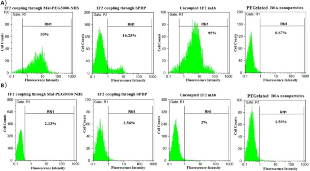
Cellular binding assessment of nanoparticles by flow cytometry
MCF7 and SKBR3 cells were stained indirectly at surface level. After harvesting the cells by trypsinization and washing with a washing buffer for two times (PBS, 0.1% BSA, 0.1% NaN3, pH 7.4), 106 cells were incubated with 100 µL of 1F2-conjugated to 5-FU-loaded BSA nanoparticles (1 mg/mL) through Mal-PEG5000-NHS and SPDP at 4 °C for 1 hour. 1F2 mAb (2.5 µg/mL) and BSA nanoparticles were also involved as positive and negative controls, respectively. After incubation, the cells were washed twice with the washing buffer and then incubated with FITC conjugated sheep anti-mouse immunoglobulin at 4 °C for 1 hour. The cells were scanned with a flow cytometer (Partec, Nuremberg, Germany) after washing. The Flomax software (Partec) was used for data analyses.
Physicochemical characterization of nanoparticle formulations.
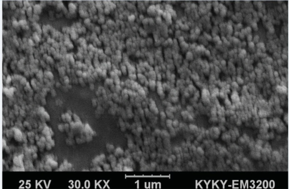
Particle size, polydispersity index (PDI) and zeta potential of BSA nanoparticles 5-FU-loaded BSA nanoparticles, 5-FU-loaded PEGylated BSA nanoparticles and 1F2-coupled 5-FU-loaded BSA nanoparticles were evaluated using a dynamic light scattering (DLS) apparatus (Malvern instrument Ltd., UK). The samples were diluted with distilled water before the analyses. In addition, the morphology of 1F2-coupled 5-FU-loaded BSA nanoparticles was analyzed using a scanning electron microscope (SEM) (KYKY-EM3200 model, China).
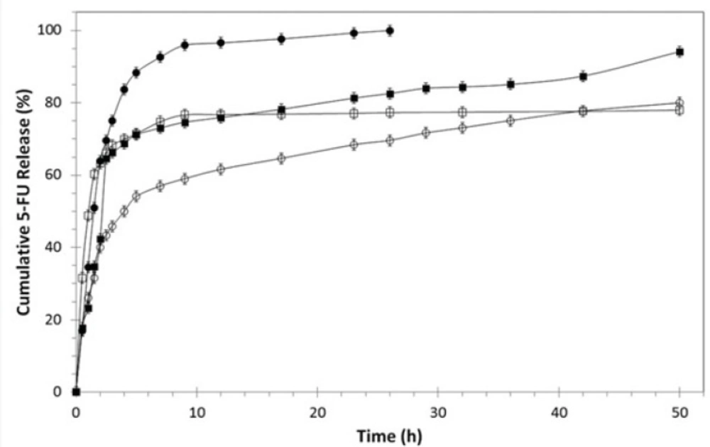
In-vitro drug release study
In-vitro cumulative release behavior of 5-FU from BSA nanoparticles, PEGylated BSA nanoparticles and 1F2-coupled BSA nanoparticles was evaluated during a period of 50 hours using dialysis method. The freeze-dried drug-loaded nanoparticle formulations with equal amount of 5-FU (1 mg) were suspended in separate dialysis tube bags and kept in 10 mL of PBS pH 7.4 at 37 °C in shaking water bath at 100 rpm. At predefined time intervals, PBS samples containing the released drug were taken and analyzed spectrophotometerically at 266 nm and then poured back into the release medium.
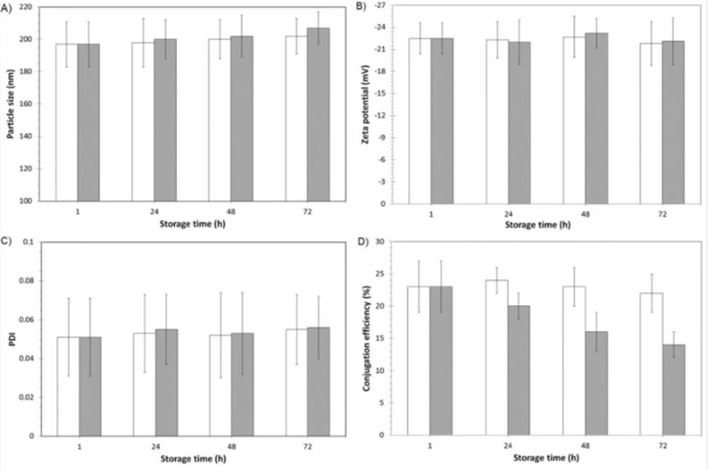
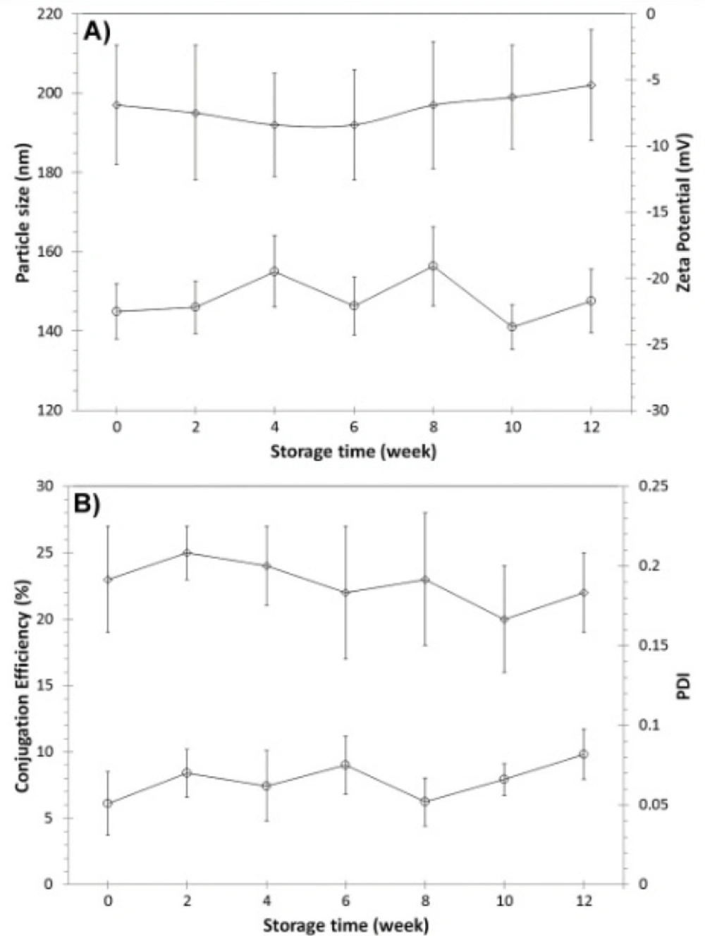
Short and long-term storage stability investigation of mAb-modified drug-loaded nanoparticles
The short time stability of the physicochemical properties and biological reactivity of 1F2-coupled 5-FU-loaded BSA nanoparticles were evaluated by DLS and ELISA during 72 hours of storage (time intervals of 1, 24, 48 and 72 hour after production) at 4 and 37 °C. In addition, the physicochemical and biological stability of 1F2-modified drug-loaded nanoparticles were assessed every two weeks during three months of storage at room temperature. In this regard, samples of nanoparticles containing 5% trehalose as preservative (
17,
21) were freeze-dried, stored at room temperature and resuspended in distilled water before the analyses.
In-vitro cytotoxicity evaluation
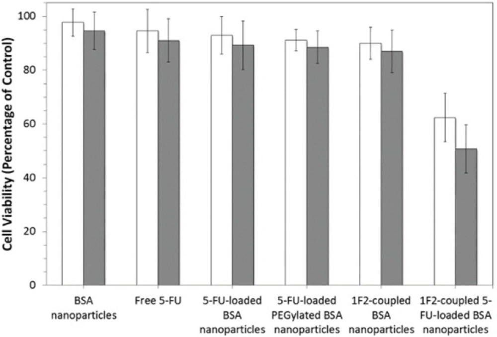
In-vitro specificity and cytotoxicity effect of 1F2-coupled 5-FU-loaded BSA nanoparticles was evaluated on HER2-positive SKBR3 and compared with five other systems consisting of BSA nanoparticles, free 5-FU, 5-FU-loaded BSA nanoparticles, 5-FU-loaded PEGylated BSA nanoparticles and 1F2-coupled BSA nanoparticles. Briefly, cells (1 10
4) were transferred into 96-well plates and incubated at 37 °C for 48 hours. After complete attachment of the cells, the supernatant was substituted with 100 µL of fresh media containing the mentioned systems with equal IC
30 concentration of 5-FU (2 mM) (
22) and nanoparticles (20 mg/mL). In addition, wells with no treatment were considered as control. In order to investigate the effect of contact time on cell specific attachment and cytotoxicity of the systems, cells were incubated with the nanoparticle formulations for 1 and 5 hours at 37
°C. Our some pretests revealed that incubation time more than 5 hours did not increase the cytotoxicity of the systems and therefore, we considered 5 hours as the higher contact time. Then, the supernatant media were removed, fresh media was added to all wells and the cells were further incubated for 72 hours at 37
°C. After the end of the incubation time, the cell viability was assessed by MTT assay. The medium was replaced by a mixture of fresh DMEM medium and MTT solution (5 mg/mL in PBS), followed by 2 hours incubation at 37
°C. After dissolution of MTT with dimethylsulfoxide (DMSO, Sigma), the absorbance of the resulting solution was measured using a Microplate reader (Awareness Technology, USA) at a wavelength of 540 nm. The cell viability ratio was evaluated through comparing absorbance of treated cells against the untreated controls.
For control experiment, HER2 weakly expressing MCF7 cells were used. MCF7 cells were incubated with the systems at the same concentration of 5-FU and nanoparticles for 1 and 5 hours at 37 °C. After washing, the cells were further incubated for 72 hours at 37 °C. The cell viability assay was performed as described before.