Size, zeta potential and lipid recovery
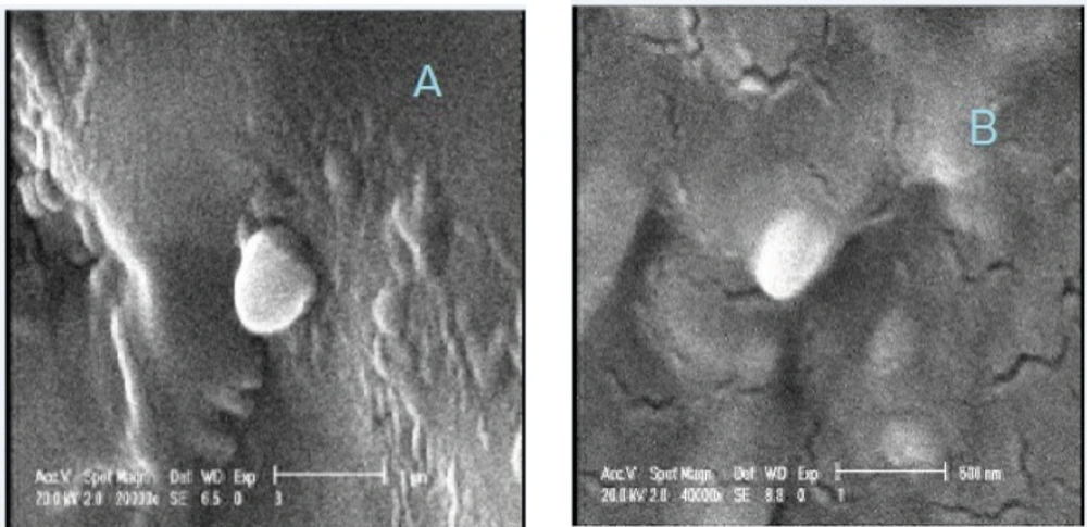
SLN particles showed a size of 130 ± 1.39 nm (mean ± SD, n=3) and their surfaces carried a negative charges with a zeta potential of – 44 ± 2.1 mV (mean ± SD, n=3). Under SEM, a dense solid sphere was observed for freshly prepared SLNs (
Figure 1). Size increase in SLNs was observed in SEM investigation due to partial melting of some nanoparticles and aggregation of them.
Scanning electron micrograph of freshly prepared plain solid lipid nanoparticle (A) and nanoparticles incorporated into hydrogel (B).
Phospholipid content of SLN was calculated based on calibration curve to be 0.045 ± 0.6 mg per one mL of SLN dispersion, indicating a recovery around 91%. Our results also showed that phospholipid content does not change (P ˃ 0.05) after purification by centrifugal filtration by Vivaspin 2, MWCO: 5000.
Gel capacity and SLN integrity in Poloxamer gel
One of the most important factors in ability of Poloxamer 407 to form a gel at physiological temperature is its concentration. Based on previous studies (-), it is essential to choose a minimum concentration of polymer at which it forms a gel (at body temperature) to avoid side effects. Poloxamer concentration of 20 % (w/v) was selected for plain (without SLN) system here, based on the manufacturer suggestion (
33). Maximum capacity of gel for incorporation of SLNs was evaluated through gel consistency and gel clarity studies on SLN-containing systems at 37 °C, as described above. Results (
Table 1 ) showed that up to 50% of water can be replaced with SLN dispersion with minimum changes in clarity or consistency of the obtained gel at 20 % Poloxamer.
| SLN dispersion (mL) | Cold water (mL) | Gel clarity* | Gel consistency* |
| 0 | 80 | + | + |
| 20 | 60 | + | + |
| 30 | 50 | + | + |
| 40 | 40 | + | + |
| 50 | 30 | + | +/- |
| 60 | 20 | _ | _ |
| 80 | 0 | _ | _ |
: +: Acceptable; +/-: Moderate;-: Not acceptable
The integrity of SLNs incorporated into sol-gel system was evaluated by scanning electron microscopy. Results (
Figure 1) showed that particles keep their spherical morphology in hydrogel system. Moreover, particle size of SLN-containing gels after gel erosion at different time intervals (
Table 2) increased slightly from 130 ± 1.39 in freshly prepared free SLN dispersion to 138 ± 1.82 (after 4 h) and 142 ± 2.1 (after 8 h) (P˂0.05). There were no differences between sizes of SLNs at 4 h and 8 h after gel erosion. Changes in particle size might be related to changes in hydration shell due to polymer accumulation on the surface of particles. Plus, obtained results indicated that SLN particles stay intact in the gel and are released from the Poloxamer hydrogel as particles.
| Plain SLN | Erosion time (h)
|
| 4 h | 8 h |
| 130 ± 1.39 | 138±1.82 | 142 ± 2.1 |
Different mechanisms have been suggested for gelation of Poloxamer 407 such as ordering of micelles to a cubic structure, dehydration of poly (propylene oxide) (PO) core, and micelles entanglements (
37,
38). It is generally accepted that spherical micelles in Poloxamer 407 solution consist of a PO core with an poly (ethylene oxide) (EO) water swollen shell based on their lipophilicity (
35) and proportion of EO which is approximately 70 % by weight (
39). As a result, SLN would be loaded in PO core to thermodynamically stabilize the combined system.
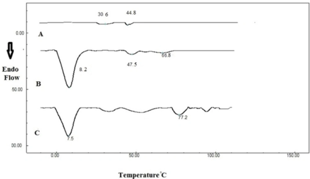
Differential scanning calorimetry (DSC)
Figure 2 shows DSC thermograms of dried 20 % Poloxamer 407 solution, indicating a transition at 30.6 °C. Addition of SLNs changed this transition temperature from 30.6 to 28.5 °C. This peak has a low enthalpy and can be detected at low scanning rates such as 0.5 °C/min (
40). Enthalpy of thermogelation is reported to be very low (
41). Therefore the sol-gel transition of Poloxamer 407 was determined here by the stirring method and rheological study, as described later.
Second transition temperature in Poloxamer 407 thermogram was measured to be 44.8 and 47.5 °C before and after SLN incorporation respectively. These transitions might be related to Poloxamer melting point, which is stated to be around 50 °C (
39). Another transition temperature (66.8 °C) was also observed in SLN-containing system which was attributed to stearic acid melting which is in the range of 65 to 70 °C (
42).
Differential scanning calorimetry thermograms of A: Poloxamer 407 (20% w/v), B: Poloxamer 407 (20% w/v) containing 20 mL SLN dispersion and C: SLN dispersion
A high-enthalpy transition was also observed in SLN system around 8 °C that was not affected by Poloxamer 407.
Sol-gel transition temperature
Stirring magnet bar studies showed that Poloxamer 20% solution (sol) has a sol-gel transition temperature of 24 ± 1.2 °C. After SLN incorporation, gelling temperature increased to 27 ± 0.9 °C, which is closer to body temperature. Higher gelling temperature results in lower amount of polymer required for gel formation
in-vivo (
43).
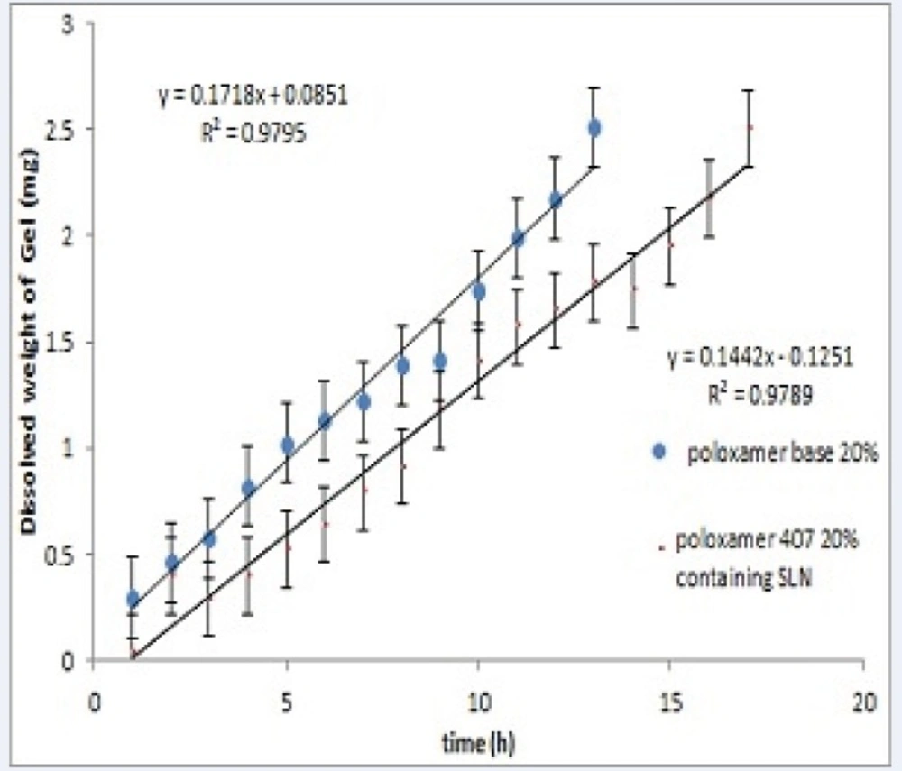
Erosion test
Figure 3 shows the erosion (dissolution) of Poloxamer gel for plain and SLN-containing systems over 20 hours. Both systems showed a linear profile of amount dissolved versus time (R
2> 0.98). Results also show that incorporation of SLNs into Poloxamer solution decreases polymer erosion rate from 0.17 to 0.14 mg/h (P<0.05).
In-vitro erosion profile of in-situ forming systems
Rheological properties of sol-gel systems
Evaluation of rheological properties is very useful in physicochemical characterization of gel formulations and help understanding their process of thermal gelling in-situ.
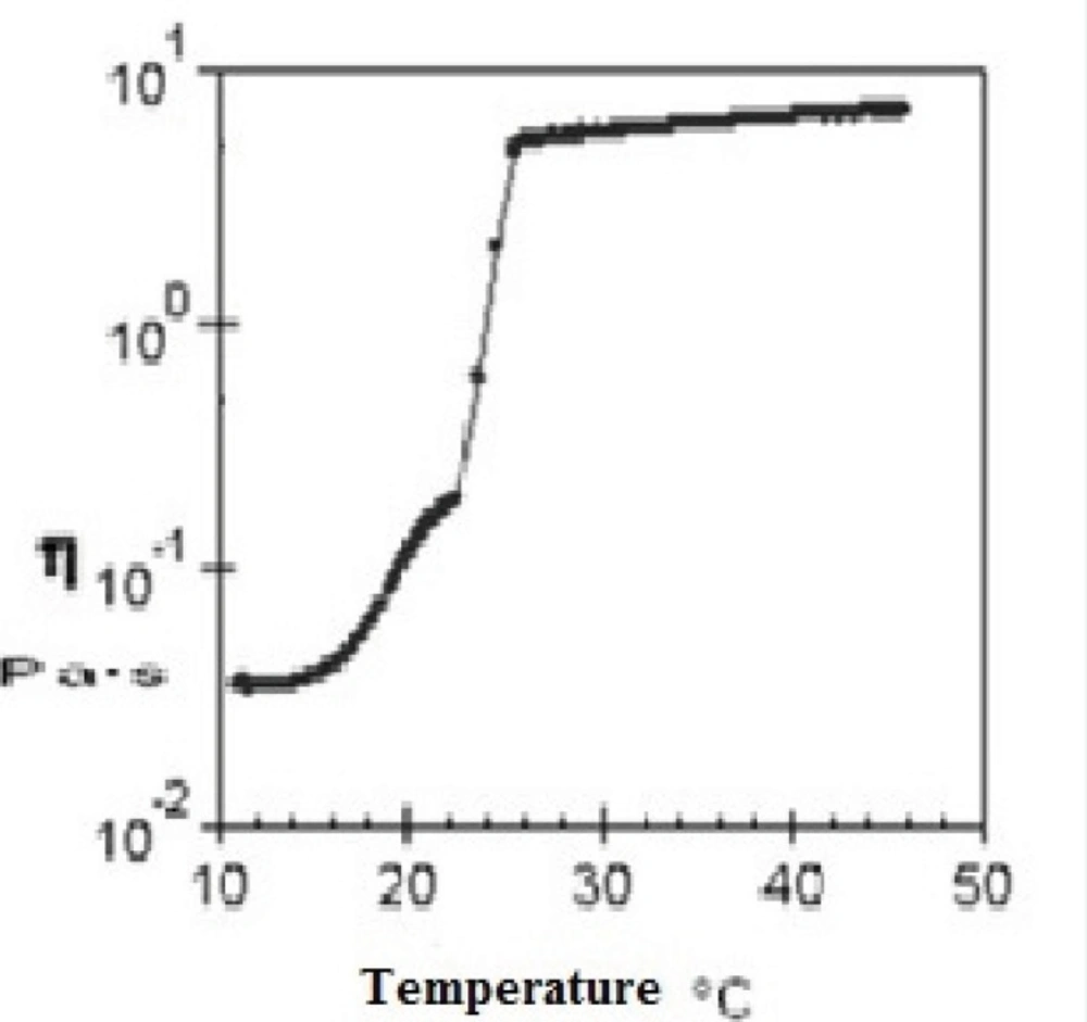
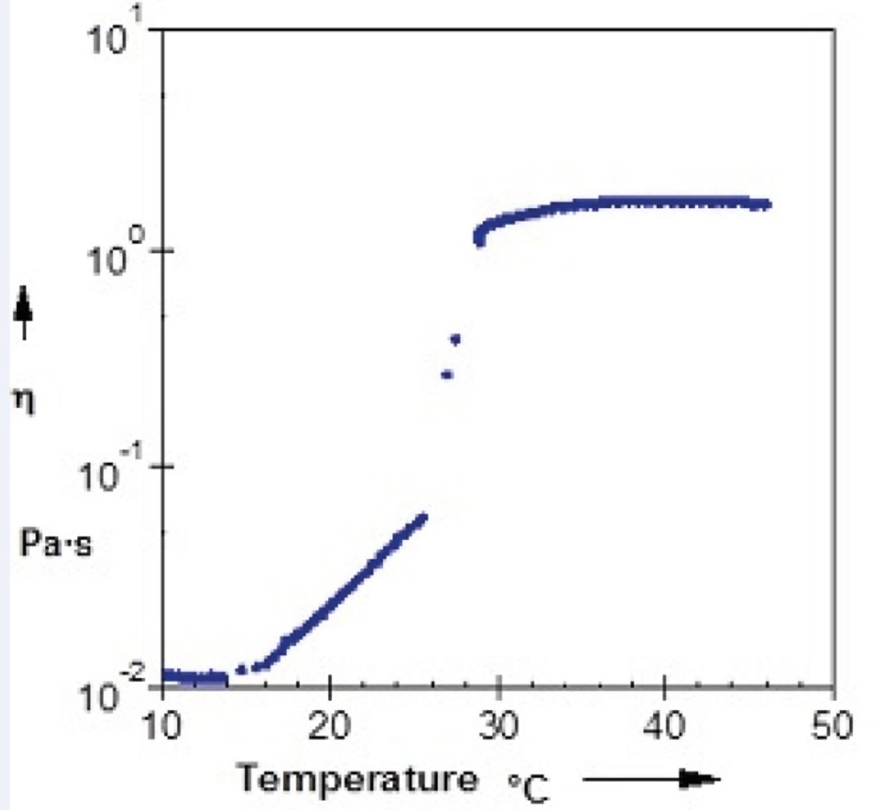
Figure 4 and
Figure 5 show viscosity of Poloxamer 407 solutions as a function of temperatures for plain and SLN-containing systems respectively. Both systems showed a constant viscosity upto about 15 °C, but their viscosities increased as the temperature elevated. After gel formation (sol-gel transition) viscosity became constant again. Based on these results, the sol-gel transition temperature of plain and SLN-containing system, which is equal to maximum viscosity of the system, was measured to be 26 °C and 29 °C respectively which shows good agreement with stirring test results.
Relationship between the viscosity of a 20% (w/v) solution of Poloxamer 407 and temperature
Relationship between the viscosity and temperature for 20% (w/v) solution of Poloxamer 407 containing 20 mL SLN dispersion
The viscosity of Poloxamer 407 aqueous solutions (15–30%) against temperatures (15-35 °C) has been investigated and it has been found that an exponential relationship exists between viscosity and temperature, with the slopes depending on Poloxamer concentration (
44). It has also been shown that while a sol solution of Poloxamer 407 shows a Newtonian behavior (linear relationship between shear rate and shear stress), the system changes to a gel with non-Newtonian behavior when concentration or temperature is increased (
45).
Poloxamer 407 gels can show viscoelastic properties. They have an elastic or storage modulus, G', which represents the amount of energy stored and recovered per cycle of deformation of solid-like component of material. The viscous or loss modulus, G", is characteristic of liquid part (
46). The most common way to determine viscoelastic properties of dispersion is oscillatory rheological study as a function of temperature within the linear viscoelastic region under shear stresses which are tolerable for gel structure.
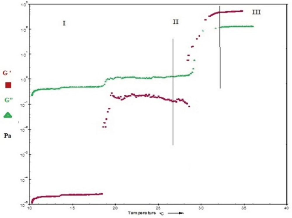
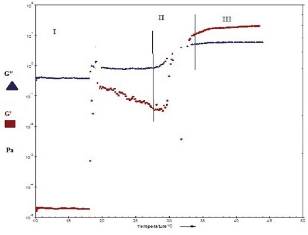
Figure 6 and
Figure 7show the profile of oscillatory parameters versus temperature for plain Poloxamer and SLN-included Poloxamer systems respectively. This profile shows three distinct phases of sol, gel, gel stabilization; designated as phases I, II and III here. In phase I, that is prior to the gelling point, the elastic modulus, G', shows low values and the samples are characterized as a viscous sol system with higher G". In the second phase and at gelling temperature (phase II), drastic increase in elastic modulus, G', was observed indicating formation of gels with elastic behavior.
Figures 6 and
7 show that while G' is lower than G" in phases I and II, it becomes higher than G" in later stages of phase II and phase III. G'>G" is said to be indicator of a well-built structure of a soft gel, possibly due to physical entanglements of polymeric chains (
47). Final phase (phase 3) states the stabilization of the elastic modulus, G', above the transition temperature.
Temperature dependency of the dynamic moduli, G" and G' of a freshly prepared 20% (w/v) Poloxamer 407 solution.
Temperature dependency of the dynamic moduli, G" and G' of 20% (w/v) Poloxamer 407 containing 20 ml SLN dispersion.
Our results also reveal that both plain and SLN-containing systems show similar behavior in terms of changes in G' and G" against temperature, indicating that SLN incorporation at low concentrations does not change the gel structure.
The gelation temperature (sol-gel transition) can be calculated from G′ and G" changes versus temperature and it is considered to be the temperature at which storage (G′) and loss modulus
(G″) become equal,
i.e. the crossover point of G′ and G″, that is usually halfway in phase II (
21,
48,
49). By evaluating crossing point of G′ and G″ in
Figure 6 and
Figure 7, the gelation temperatures were calculated to be approximately 26 ± 0.8 °C for plain Poloxamer and 29 ± 0.7 °C for SLN- containing system. These results are in agreement with stirring magnetic bar results. In this direction, it has been shown that inclusion of cyclodextrin in Poloxamer 407 gel increases sol gel transition temperature, possibly by disturbing the micellar packing and entanglements of Poloxamer 407 (
30,
43).
The gelation temperature has been considered to be suitable in the range of 25-37 °C for in-situ forming gels. Lower gelling point causes gelling at normal room or laboratory conditions and causes difficulties in manufacturing, handling and administrating (
50) especially in injectable formulations (
51).
Another considerable parameter in injectable in-situ forming systems is gelation time or kinetic of gelation. This is the time required for an injectable in-situ forming system to turn from liquid state to gel (
52) and it is equal to the time that take for the storage modulus (G′) to became higher than the loss modulus (G") in oscillatory parameters versus temperature diagrams (
21,
53). In this study, as is seen in
Figure 6 and
Figure 7, gelling time is short (approximately one minute) in Poloxamer 407 solution and does not change after incorporation of SLN into this system. Such a fast gelation time reduces the risk of burst release due to lower possibility of dilution and drainage at the site of application (
53,
54).
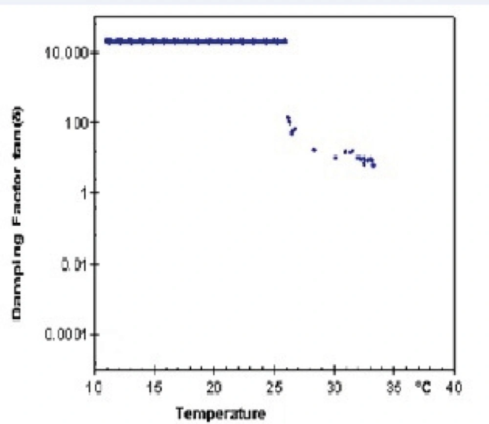
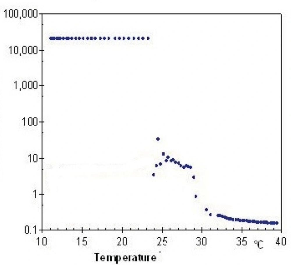
Lag phase or damping factor is obtained by dividing G" to G' and is an indicator of stability of polymeric structure based on their interactions.
Figure 8 and
Figure 9 show damping factor changes as a function of temperature in Poloxamer and combined system with nanoparticles. At low temperatures, the measured damping factor of all samples was higher than one which represents smaller interaction of internal structure in sol state. By elevation of temperature, the damping factors decreased to values less than one which shows stronger chain interaction in gel and confirm elastic behavior of gel formation. SLN incorporation decreased the damping factor (
Figures 8 and
9), indicating a more stable gel.
Temperature dependency of damping factor (G"/G') of 20% (w/v) freshly prepared Poloxamer 407 solution.
Temperature dependency of damping factor (G"/G') of 20% (w/v) Poloxamer 407 system congaing 20 mL SLN dispersion.