Materials
Peptide P5 (ELAAWCRWGFLLALLPPGIAG, Purity > 95%) was synthesized by Peptron Co. (Daejeon, South Korea). Dimiristoylphosphatidylcholine (DMPC) and Dimiristoylphosphoglycerol (DMPG) were purchased from Avanti Polar Lipid (Alabaster, USA). Cholesterol, L- ascorbic acid, ammonium molybdate, Tris and Tricine were from Sigma-Aldrich (Steinheim, Germany). Methanol and acetonitrile (LiChrosolv®, gradient grade), ethanol and isopropanol (Emsure®), dimethyl sulfoxide (DMSO), Trifluoroacetic acid (TFA) and acrylamide were purchased from Merck KGaA (Darmstadt, Germany). HEPES buffer was from Invitrogen Co. (Scotland, UK).
Encapsulation of P5 into liposomes
P5 was encapsulated in liposomes following three different methods based on film-hydration procedure. For all formulations, lipids were used at a molar ratio of 15:2:3 (DMPC, DMPG, Chol) respectively and liposomes were prepared to contain 200 μg/mL P5 peptide and 40 mM of total lipid.
Method A
Lipids were dissolved in chloroform while P5 was dissolved in DMSO (10 μg/μL) and then they were combined in glass tubes and dried to a thin film by rotary evaporation (Heidolph, Germany) under reduced pressure. The lipid film was freeze-dried (VD-800F; Taitech, Japan) overnight to remove the solvents completely. The lipid film was then hydrated in HEPES buffer (10 mM, pH 7.2) containing 5% dextrose by intermittent vortexing and bath sonication under argon for a short time (approximately 30 sec at 25 ºC ) to disperse completely the lipids into the buffer. The resulting multilamellar vesicles (MLVs) were extruded 5 times through 400 nm and 11 times through 100 nm polycarbonate membranes at 25 ºC using a mini extruder (Avestin, Canada) to form 100 nm small unilamellar vesicles (SUVs) with a uniform size. To remove free peptide, 1 mL of liposomal dispersion was filled into the dialysis membrane (Cut off = 12-14 kDa) and dialyzed for 24 h at 4 ºC against 3 L of HEPES-dextrose buffer to allow the free peptide diffuse out. Vesicle size, polydispersity index and zeta potential of liposomes were determined by dynamic light scattering (Malvern Instruments, Malvern, UK). Liposomes were stored at 4 ºC under argon.
Method B
All the steps were the same as method A except that the lipid film was hydrated in HEPES-dextrose buffer containing 10% (v/v) of DMSO and peptide P5.
Method C
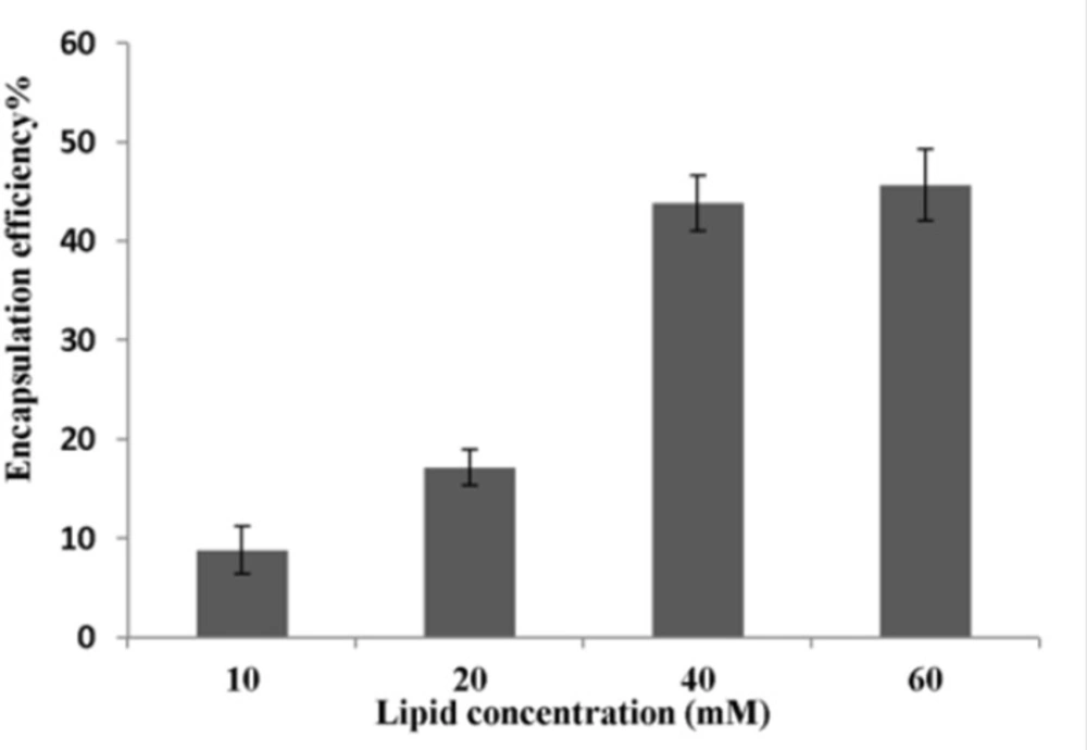
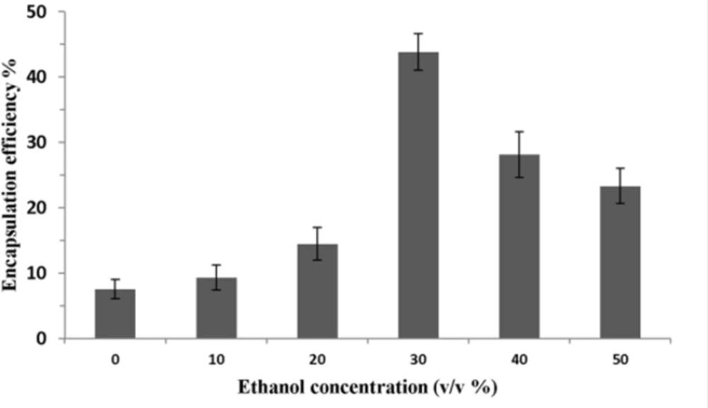
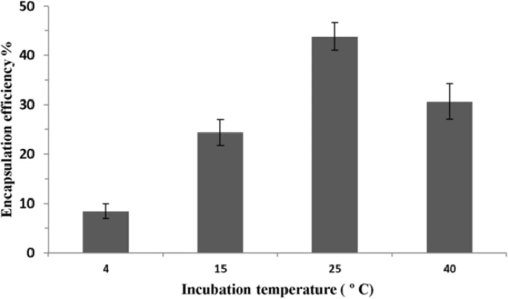
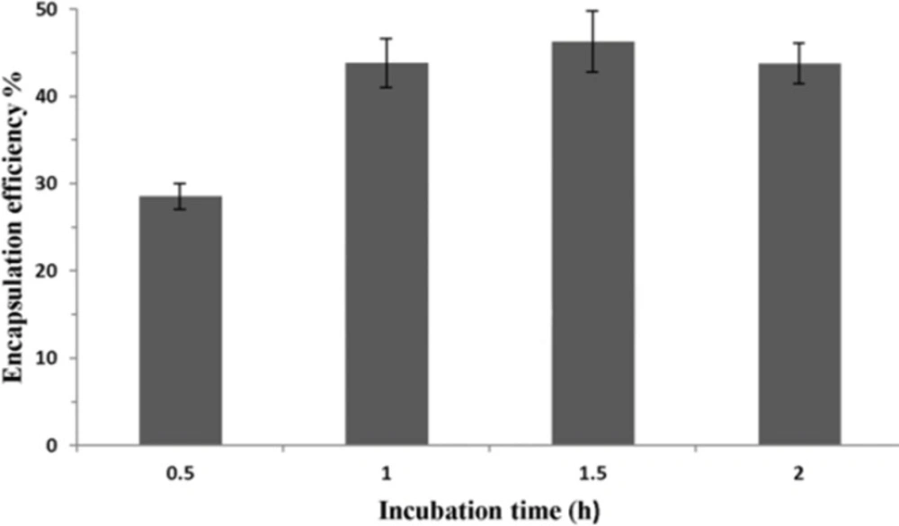
Lipid film was prepared as described in method A. The lipid film was dissolved in 300 μL ethanol and 700 μL HEPES-dextrose buffer containing 10% (v/v) of DMSO. The resulting dispersion was sonicated for about 15 s and extruded as explained in method A. 20 μL of P5 solution in DMSO was slowly added to preformed liposomes while vortexing. Subsequently, the ethanolic mixture of liposome and P5 was incubated at 25 ºC for 1 h and then dialyzed as described above to remove unencapsulated peptides, ethanol and DMSO. The effects of operating parameters of this method such as lipid concentration (10, 20, 40 and 60 mM), ethanol concentration (0, 10, 20, 30, 40 and 50% v/v), incubation temperature (4, 15, 25 and 40 ºC) and incubation time (0.5, 1, 1.5 and 2 hrs) on encapsulation efficiency were investigated.
RP-HPLC analysis
The analysis of peptide content of liposomes and their encapsulation efficiency were conducted by injection of different samples into a KNAUER smart line HPLC (Berlin, Germany). The RP-HPLC was equipped with a Nucleosil C18, 5 μm, 150 × 4.6 mm, 100 Aº column (KENAUER) and an UV detector (KENAUER S2600) set at 220 nm. The mobile phases employed were A (water + 0.1% TFA) and B (acetonitrile + 0.1% TFA). Elution program was a gradient starting with 100% A and increasing to 30% B in 2 min, 60%B in 12 min and 90% B in 2 min. The flow rate was set to 1 mL/min. The peptide concentration in all samples was determined using a standard curve generated by known concentrations of peptide dissolved in isopropanol containing 0.1% TFA.
Determination of peptide recovery from liposomes using different solvents
All experiments were set up in glass tubes in triplicate. Each tube was contained 10 μL of peptide solution and 500 μL of empty liposome containing 30% (v/v) ethanol. The mixture was incubated for 1 h at 25 ºC and dialyzed (Cut off =1000 Da) for 24 h at 4 ºC against 1.5 L of HEPES-dextrose buffer to remove ethanol and DMSO. Five hundred μL of methanol, ethanol, or isopropanol or 500 and 1000 μL isopropanol containing 0.1% TFA was added to 500 μL liposomes mixed with peptide in each tube. The tubes were vortexed and incubated at 40 ºC for 10 min. Three injections of 20 μL from each tube were made to the RP-HPLC. The amount of peptide in each injection was determined using a standard curve. For each extraction media, the average of peptide amount in 9 injections was put in equation 1 to calculate peptide recovery.
REC% = (P Recovered / P Added) × 100 equation (1)
Where REC% is the percent recovery, P Recovered is the amount of peptide recovered from liposomes and P Added is the amount of peptide added to empty liposomes.
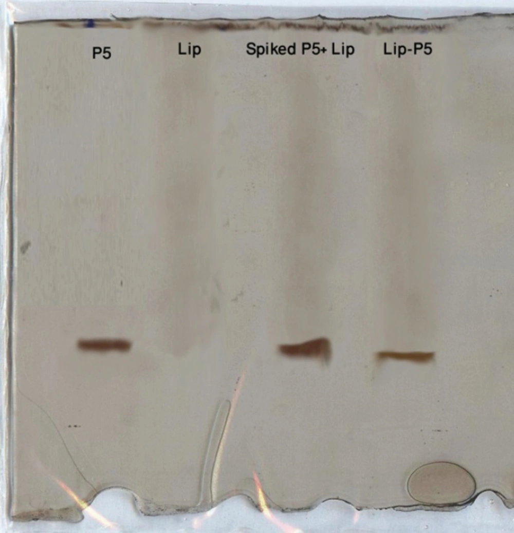
SDS-PAGE analysis of Lip-P5
The analytical SDS-PAGE was carried out to determine qualitatively whether Lip-P5 contains P5 after purification. The gel consisted of running gel (16% (w/v) acrylamide / 6M urea), stacking gel (4% (w/v) acrylamide) and spacer gel (10% (w/v) acrylamide). The gel thickness was 0.7 mm. The anode buffer was 0.1 M Tris, pH 8.9 and cathode buffer was 0.1 M Tris, 0.1 M Tricine, 0.1% SDS, pH 8.25. Electrophoresis was carried out with an initial voltage of 30 V, which was gradually increased to 300 V at the end of the run. After electrophoresis the gels were stained for peptide with silver (
23).
Determination of encapsulation efficiency
All the measurements were performed in triplicate in microcentrifuge tubes. Liposome suspensions (50 μL) were added to the tubes. Based on the P5 peptide recovery experiments, the optimized volume of the best extraction medium was added to the liposomes. The tubes were then vortexed and incubated at 40 ºC in a water bath for 10 min. Samples (20 μL; n=3) were injected to RP-HPLC. The peptide content in the injected volumes was determined by comparison with a standard curve for the peptide. Encapsulation efficiency (EE) was calculated using equation 2.
EE% = (PLLiposome / PL Total) × (1/REC%) × 100 equation (2)
Where EE% is the encapsulation efficiency of the peptide in liposomes, PL liposome is the peptide to phospholipids ratio (w/w) in liposomes and PL Total is the peptide to phospholipids ratio (w/w) used for formulation.
The amount of phospholipids was determined by using a phosphorus analysis spectrophotometric method (
24,
25). Briefly, liposome samples (μL) containing theoretically 80 ± 50 nmol of phosphorus were placed into glass tubes. To each tube, 400 μL of a 10 N sulfuric acid solution was added and heated in an aluminum block at 200 ºC for 1 h. After cooling, 100 μL of a 0.1 mg/mL hydrogen peroxide solution was added and heated again for 10 min. The tubes were cooled and 500 μL of a 0.1 mg/mL ascorbic acid solution and 470 μL of a 2.2 mg/mL ammonium molibdate solution added to them. After vortexing, the solution was heated at 100 ºC in bath water for 10-20 min. The analyses were performed by reading the absorbance at 800 nm (Spekol 1500, analytikjena, Germany).