Animals
Adult male C57BL/6 mice weighing 20-25 g (8–10 weeks) were purchased from Pasteur Institute (Karaj, Iran). The animals were maintained three per cage under 12 h light/dark cycles under controlled temperature (23 ± 2 °C). Food and water were freely available. All experiment procedures were designed based on international guidelines for animal studies and approved by the ethical committee for animal research, Tarbiat Modares University, Tehran. Efforts were made to minimize both the number of animals used and their suffering.
Interventions
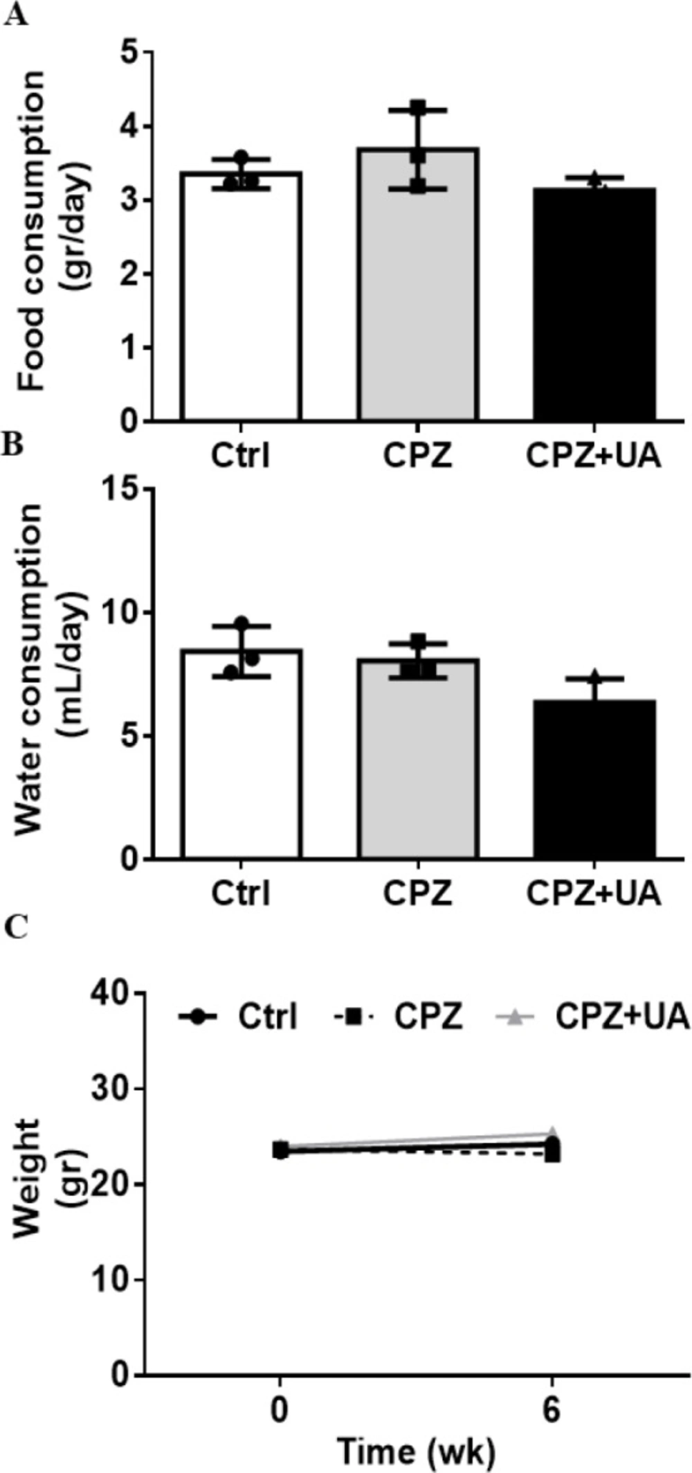
For induction of demyelination, the male C57BL/6 mice were fed with a diet containing 0.2% cuprizone (oxalic acid bis (cyclohexylidene hydrazide); Sigma-Aldrich Inc., St. Louis, MO, USA) mixed into their normal chow for 6 weeks. The mice were divided into three groups including control with normal water, cuprizone diet with normal water, and cuprizone diet with water containing UA (1 mg/mL) (Enzo Life Sciences). The animals’ brains were collected at 6 weeks post cuprizone. The animal’s weight and food intake were recorded every other day.
Y-maze behavioral test
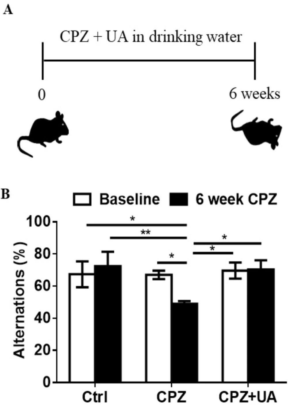
Y-maze test was performed to confirm the presence of behavioral impairment due to demyelination in their brains. Spontaneous alternations within the Y-maze were calculated prior to induction of demyelination and 6 weeks after feeding with cuprizone diet. The Y-maze arms were labeled as A, B, and C. The number of overlapping entrance sequences (e.g., ABC, BCA) was defined as the number of spontaneous alternations. The percentage of alternations was calculated as follows.
Processing and sectioning of brain tissues
The animals were perfused with phosphate buffer saline. The brains were removed and the hemispheres were dissected. The right hemispheres were post-fixed in 4% paraformaldehyde 24 h, then in sucrose 30% (2-3 days), and embedded in optimal cutting temperature (OCT) for long term preservation in -80 °C. Ten micrometer thick sections were cut using a cryostat (Histoline, Italy) and mounted on superfrost plus slides for later evaluations. The sections were stored at -20 °C until final staining.
Luxol Fast Blue and Cresyl Fast Violet staining
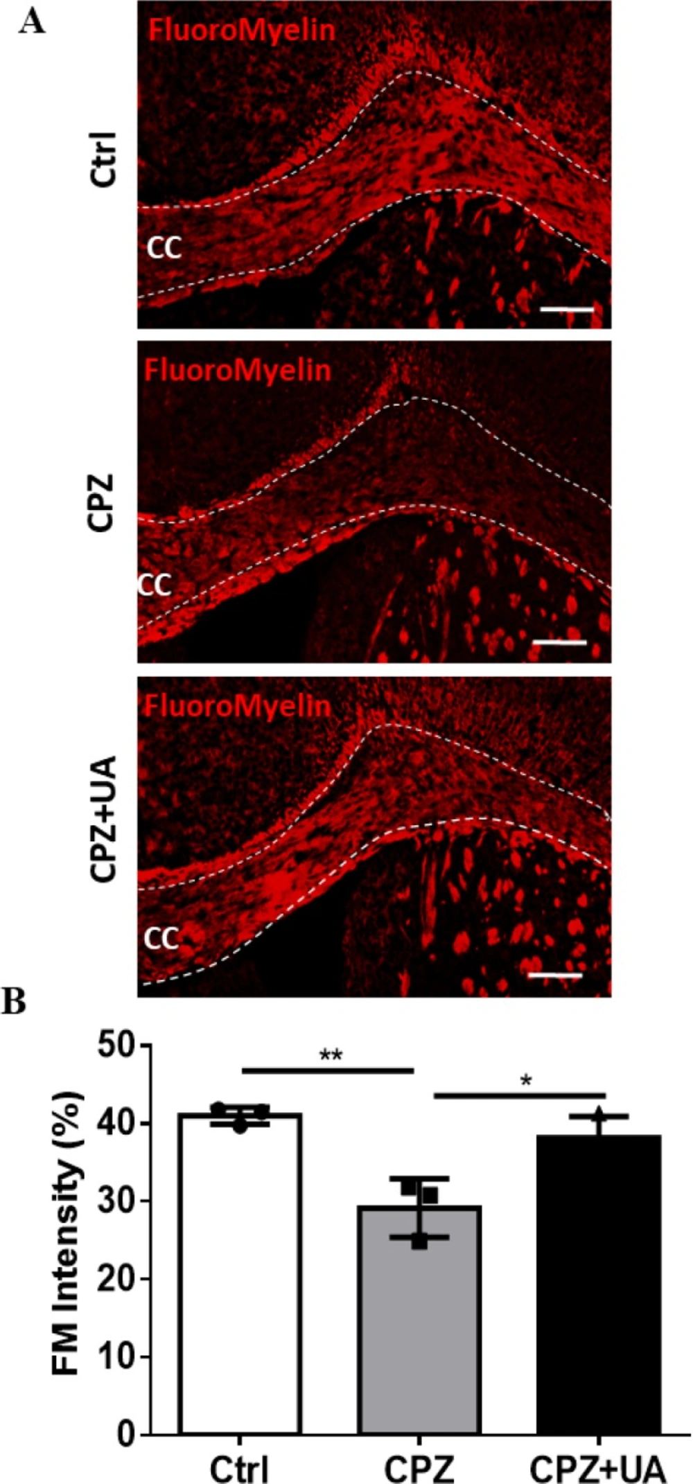
To evaluate the myelination intensity and the extent of demyelination in corpus callosum, the sections with 10-μm thick were selected for myelin staining. For FluoroMyelin (FM) staining, the sections were incubated with red FM (Invitrogen F34652) for 20 min at room temperature. FM solution were prepared by diluting FM 1:300 in PBS.
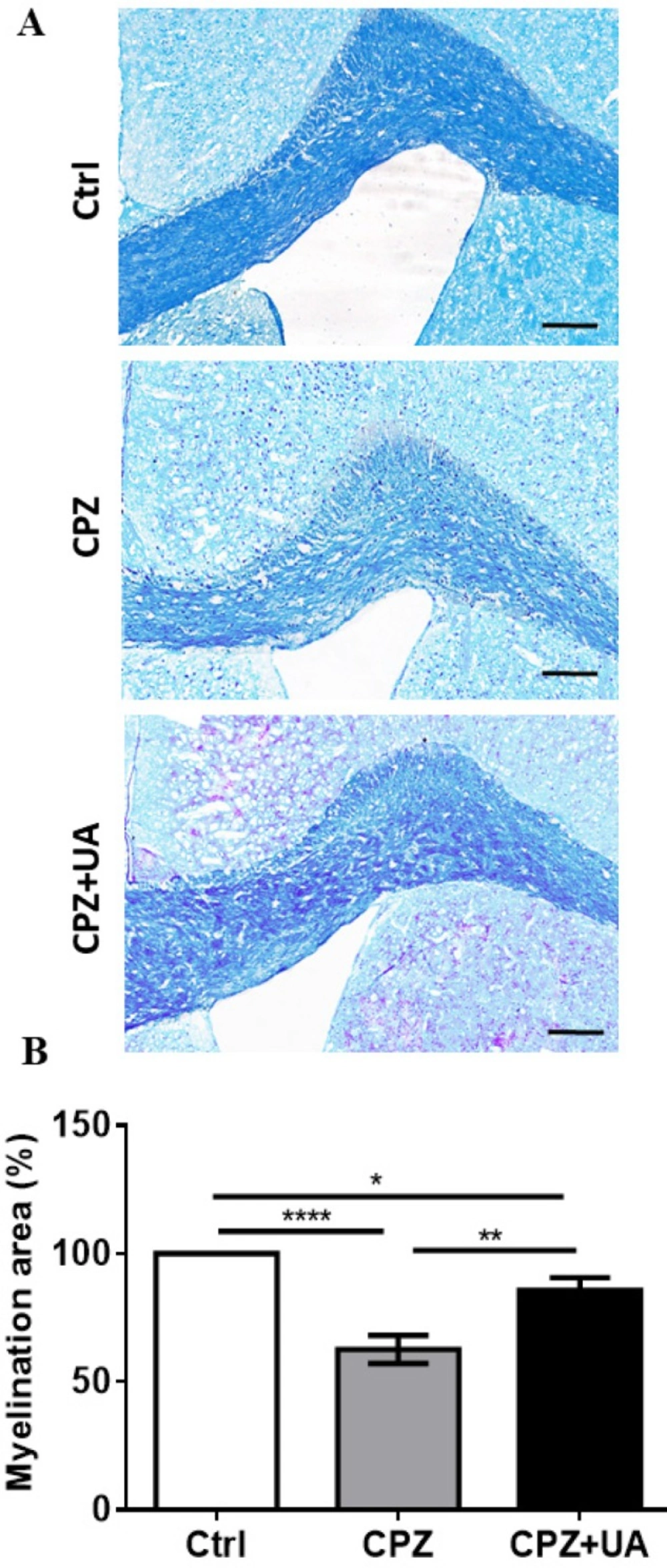
For Luxol fast blue (LFB) staining the sections were stained with 0.1% LFB solution (British Drug House, London, UK) at 60 °C for 1 h. Adequate contrast was made by rapid immersion of preparations in 0.05% lithium carbonate followed by several changes of 70% alcohol. The sections were washed with distilled water, and then counterstained with 0.1% Cresyl Fast Violet (Merck, Germany) for 4 min. After washing with distilled water, the sections were dehydrated in a graded series of alcohols, cleared in xylene, coverslipped, and screened for demyelination and subsequent quantitative analysis. For each section, the extent of demyelination was assessed by using Image J software as the percentage of whole corpus callosum. The extent of demyelination was averaged for 8 sections obtained from each animal and then averaged for each animal group (n = 3 for each data point).
Immunostaining
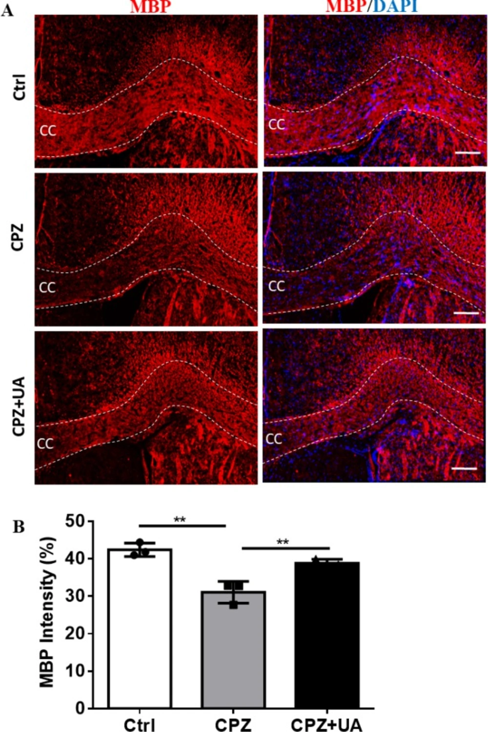
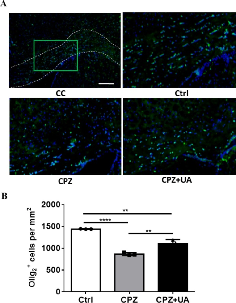
To perform the immunofluorescence staining against myelin antigens, the sections were washed three times with PBS containing 0.05% Tween 20, then incubated in blocking solution for 1 h at room temperature. Blocking solution consisted of 0.1% Triton X-100 (Sigma-Aldrich, T8532), 1% bovine serum albumin, and 5% normal serum in PBS. Then, the samples were incubated at 4 °C for overnight with primary antibodies against Olig
2 and MBP. Afterward, the sections were washed three times with PBS and incubated with corresponding secondary antibody for 1 h at room temperature. The list of antibodies used in this experiment is provided in
Table 1. To counterstain and visualize cell nuclei, the samples were treated with 4´,6-diamidino-2-phenylindole (DAPI; Sigma-Aldrich; D-8417), and again washed with PBS for three times. The sections were coverslipped and then the images were obtained using an Olympus BX 51 Microscope and DP-72 camera for consequent analysis. The Analysis was performed on 8 to 12 sections, which were randomly chosen per animal. Areas corresponding to corpus callosum in one-half of the brain were analyzed for myelination intensity.
Data Analysis
Each animal group included 3-6 mice. Statistical analyses were conducted using GraphPad Prism (GraphPad Software, La Jolla, CA, USA). One-way analysis of variance ANOVA, followed by Tukey post-test was used for comparing the experimental groups. The results were expressed as mean ± SEM. The differences were considered significant when p-value was less than 0.05.