Materials
APAP, nicotinamide adenine dinucleotide (NADH), antimycin A, ubiquinone 1, sodium succinate, rotenone, 2,6-dichloroindophenol sodium, phospho(enol)pyruvic acid monopotassium salt, lactate dehydrogenase (LDH), ATP and oligomycin A were obtained from Sigma-Aldrich (St. Louis, MO, USA). All other chemicals with analytical grade were purchased from Sinopharm Chemical Reagent Co., Ltd (Shanghai, China). EGCG (>98% pure) was provided by Shanghai Ronghe Pharmacy Co., Ltd. (China).
Animals
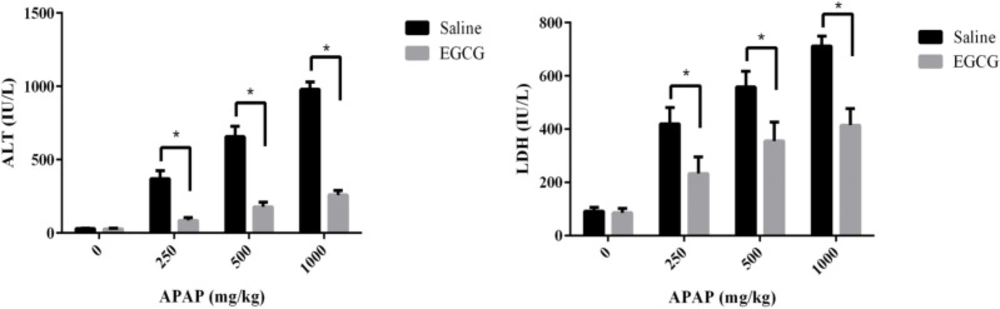
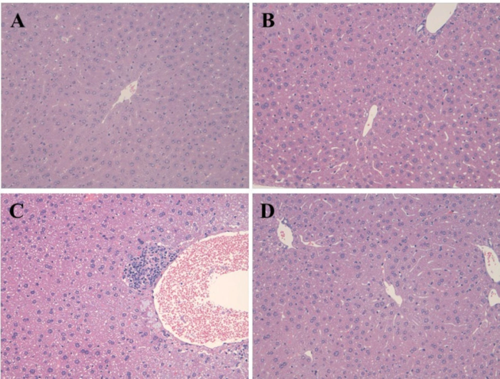
Male Sprague-Dawley (SD) rats (230-280 g, Shanghai Lingchang Biotechnology Co., Ltd. China) with one group of six were used for this study, and the experimental design consisted of eight groups. In four groups of them, the 100 mg/kg EGCG as daily dose was administered to rats by gavage for five consecutive days, and the control group was fed with normal saline. Subsequently, three groups with the treatment of EGCG and another three groups without the treatment of EGCG were administered with 250, 500 and 1000 mg/kg APAP, respectively, by intraperitoneal injection. After 8 h, all rats were killed by CO2. Blood samples from the postcaval vein were collected into coded tubes containing anticoagulant EDTA and then were used for the extraction of plasma by centrifugation. The liver tissues were isolated and stored at -80 C. Partly liver samples were fixed in 10% neutral buffered formalin and then processed histological assessment with H-E staining. All experimental protocols employed herein were approved by the Committee on the Care of Laboratory Animal Resources, College of Biological Science and Engineering, Fuzhou University.
Methods
Measurement of alanine aminotransferase (ALT) and LDH activity
ALT and LDH in plasma were measured by ALT assay kit (Beckman Co., Ltd, German) and LDH assay kit (Shanghai Shenneng Biotechnology Co., Ltd, China), respectively.
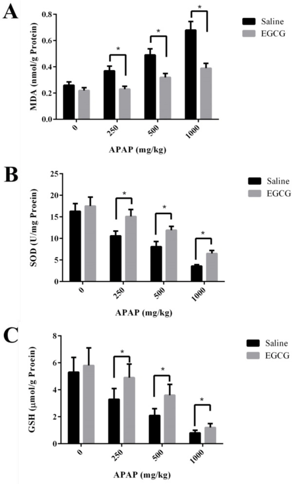
Measurement of MDA, SOD and GSH activity
The levels of oxidative stress -malondialdehyde (MDA) and biomarkers for antioxidant- superoxide dismutase (SOD) and GSH were measured using a commercially available kit (Nanjing Jiancheng Bioengineering Institute, Nanjing, China). In brief, partly liver of rats was homogenized in ice-PBS, then the samples were centrifuged at 3000 g for 10 min at 4 C. The supernatants were used for the measurement of MDA, SOD, and GSH activities according to the manufacturers’ instructions.
Isolation of rat hepatic mitochondria
Rat liver mitochondria were prepared as the description of the previous report with minor modification (
30). After being washed by isolation buffer (70 mM sucrose, 190 mM mannitol, 20 mM HEPES, 0.2 mM EDTA, pH 7.5), the liver was homogenized using a 100 ml Dounce type homogenizer. Then the concentrations of mitochondrial proteins were determined by the Bradford kit of Bio-Rad (Hercules, CA, USA).
Treatment of isolated liver mitochondria from untreated rats
After liver mitochondria from untreated rats were isolated according to the methods mentioned above, mitochondrial preparations (1 mg/mL) were incubated with 0, 3.75, 7.5, 15, 30, 45, 60, and 100 µM EGCG at room temperature for 30 min. On the other hand, APAP was dissolved in DMSO and added to final concentrations of 0, 20, 40, 60, 80, 100 mM in mitochondria preparations. The final DMSO concentration was 5‰. Before it was treated with 60 mM APAP for 5min, the mitochondria were incubated with 30, 60 and 100 μM EGCG for 30 min prior to downstream experiments.
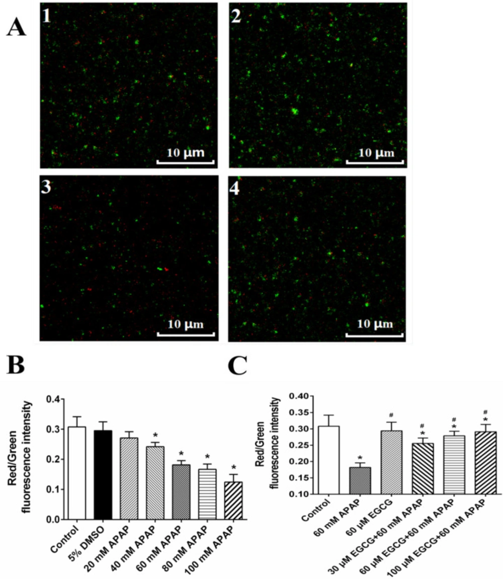
Detection of mitochondrial membrane potential (MMP)
The analysis of MMP in liver tissues was determined by the Sigma-Aldrich (St. Louis, MO, USA) JC-10 Assay Kit according to the kit’s instructional manual, and the excitation wavelength was 488 nm, and the emission wavelengths were 530 nm and 590 nm. The fluorescence intensity ratio between 590 nm and 530 nm was calculated to reflect changes in MMP. At the same time, the changes of the membrane potential of mitochondria dyed by JC-10 were observed by a confocal fluorescence microscope (CTR6500, Leica) as described previously (
31).
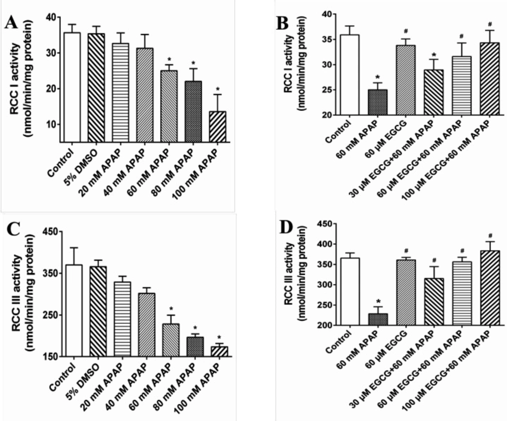
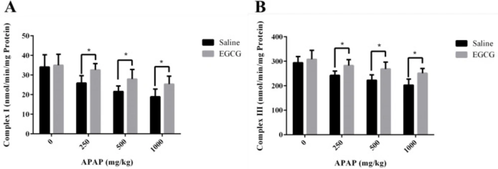
Measurement of respiratory chain complexes (RCCs) activities
Mitochondria treated with EGCG and/or APAP were centrifuged and re-suspended in 25 mM potassium phosphate (pH 7.2) and subjected to three cycles of freezing-and-thawing. The liver mitochondria treated with EGCG and/or APAP
in-vitro and administered with EGCG and APAP
in-vivo were used for the measurement of RCC I–V activities. The method is carried out according to a reported protocol (
32), except that a smaller reaction volume of 0.2 mL was used.
Statistical analysis
SPSS statistical package (SPSS, Chicago, USA) was used to perform statistical analysis. Effect of APAP exposure on LDH, ALT, MDA, SOD, GSH, complex I and complex III in rats was performed using one-way analysis of variance (ANOVA). Student’s t-test was applied for comparisons between averages of two samples. For the histopathological data, the significance of a decreased incidence of centrilobular hypertrophy and granuloma of livers in the APAP-treated groups pre-incubated with EGCG, when compared to the corresponding APAP-treated groups without incubation of EGCG, was evaluated by Fisher’s exact test. The minimal of level of significance chosen was p < 0.05.