Cytotoxic Activity and Phytochemical Analysis of Artemisia haussknechtii Boiss

authors:

avatar Sajjad Nasseri ORCID 1 , avatar Mohammad-Reza Delnavazi 2 , avatar Farshad H. Shirazi 3 , avatar Faraz Mojab ORCID 1 , *

Department of Pharmacognosy, School of Pharmacy, Shahid Beheshti University of Medical Sciences, Tehran, Iran
Department of Pharmacognosy, Faculty of Pharmacy, Tehran University of Medical Sciences, Tehran, Iran
Department of Toxicology, School of Pharmacy, Shahid Beheshti University of Medical Sciences, Tehran, Iran

how to cite: Nasseri S, Delnavazi M, Shirazi F H, Mojab F. Cytotoxic Activity and Phytochemical Analysis of Artemisia haussknechtii Boiss. Iran J Pharm Res. 2022;21(1):e126917. https://doi.org/10.5812/ijpr-126917.

Abstract

Cytotoxic activity of crude extract and fractions (petroleum ether, dichloromethane, and n-butanol) of Artemisia haussknechtii aerial parts was investigated by MTT assay. Dichloromethane fraction showed the highest cytotoxic effect on MCF-7 cell line (IC50 = 297.17 ± 7.99 µg/mL). Phytochemical analysis of the most effective fraction was carried out using normal phase column chromatography (CC) to get eight sub-fractions (A-H). Thin-layer chromatography (TLC) and high-performance liquid chromatography (HPLC) were used for further purification. Four known compounds with cytotoxic effects on cancer cell lines were isolated from the most active fraction, including 5-Hydroxy-3',4',6,7-tetramethoxyflavone (eupatilin 7-methyl ether), 5-hydroxy 3,3',4',6,7-pentamethoxy-flavone (artemetin), 6-methoxy-7-hydroxycoumarin (scopoletin), and methyl caffeate. Structure elucidation of isolated compounds was done using spectroscopic techniques, including ESIMS and 1D-NMR (1H and 13C). Cytotoxic activity of A. haussknechtii is probably due to coumarin and flavonoid compounds.

1. Background

The genus Artemisia, with more than 500 species, is the largest member of the Compositae (Asteraceae) family, distributed in different areas of Asia, Europe, and North America (1). The inhibitory effects of essential oils, crude extracts, and different fractions of Artemisia against the growth of cancer cell lines have been reported (2). The studies have demonstrated that secondary metabolites, mainly flavonoids, terpenoids, and coumarins, are responsible for the cytotoxic effects in the genus Artemisia (3-6). Essential oil of A. persica and A. turcomanica inhibited the MCF-7 cell line time and dose-dependently (7). Essential oil of A. annua showed cytotoxic activity on SMMC-7721 cells via inducing apoptosis (8). Essential oil of A. lavandulaefolia exhibited antiproliferative activity against HeLa cells by activating caspase 3 (9). Essential oil of A. vulgaris induced apoptosis in HL-60 leukemic cells and showed anticancer activity with cytochrome C releasing and activating caspases-3, 8, and 9 (10). Dichloromethane extract of A. biennis showed the most effective cytotoxic activity on K562 and HL-60 cancer cell lines by apoptosis induction (11). Similar results have been obtained for A. ciniformis, A. turanica, and A. armeniaca on human cancer cell lines (12-14). Select fractions obtained from A. ciniformis and A. biennis were assessed for their cytotoxic activity on B16/F10, PC3, and MCF-7 cells and revealed promising results as potential sources of cytotoxic phytochemicals (15). A study on extracts of three samples of A. khorassanica for their cytotoxic activity against AGS, HELA, HT-29, and MCF-7 exhibited that dichloromethane extract was most effective on cancer cell lines. Furthermore, MCF-7 was the most sensitive among other cell lines (16).

Artemisia haussknechtii Boiss., with Persian names Dermaneh Zagrosi and Dermaneh Sakhreie, is one of the 34 species of Artemisia that grows in the west of Iran (17). A study on its crude extracts and essential oils has demonstrated in vitro antifungal (18), antibacterial, antioxidant (19), and insecticidal activities (20). Also, camphor and 1,8-cineole have been reported as significant constituents in the volatile oil. Other components included cis-davanone, 4-terpineol, linalool, beta-fenchyl alcohol, and borneol (21, 22).

2. Objectives

Due to the importance of the Artemisia genus for inhibiting the growth of different cancer cell lines, this study investigated the cytotoxicity effect of their crude extract and fractions. In addition, the purification and structure elucidation of compounds responsible for cytotoxic effects of the most effective fraction were done.

3. Methods

3.1. Plant Material

The aerial parts of A. haussknechtii Boiss. were collected from Kurdistan province (Salavat Abad mountain pass), Iran, in November 2018. The sample was identified by Alireza Dolatyari. The voucher specimen (No. 320) was deposited in the Herbarium of the Iranian Biological Research Center, Tehran, Iran.

3.2. Extraction and Fractionation

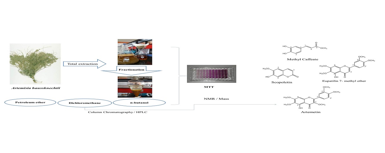
Shade-dried and grained aerial parts of A. haussknechtii (800 g) were macerated by 70% MeOH/H2O (4 × 8 L × 3 days). The crude extract (100 g) was fractionated via liquid-liquid extraction. The crude extract was dispersed in H2O, and three solvents (petroleum ether (40:60), dichloromethane, and n-butanol) were used to yield 3, 11, and 65 g of each fraction, respectively. All samples were concentrated using a rotary evaporator under reduced pressure and temperature below 45°C.

3.3. General Experimental Procedures

The chromatographic process was performed using an analytical HPLC system (Shimadzu Lc 10A, Tokyo, Japan) equipped with a photodiode array detector (PDA) and an Ascentis® column (250 × 10 mm i. d., 5 μm). Pre-coated TLC aluminum sheets (silica gel 60 F254, Merck, Germany) were applied for monitoring all subfractions under a UV cabinet (254 and 365 nm) using an anisaldehyde sulfuric acid reagent to combine similar subfractions. The NMR spectra were recorded on a Varian INOVA (500 MHz) spectrometer using CDCl3 and DMSO-d6 as the solvent. The ESIMS data were obtained from Triple Quad LC/Mass (Agilent technologies 6410, USA). All solvents used in the extraction and purification process were purchased from Merck (German) and Chem lab (Belgium) with laboratory and HPLC grades.

3.4. Cell Culture Conditions

Human breast adenocarcinoma MCF-7 cell line (ATCC No. HTB-22) was purchased from the Pasteur Institute (Tehran, Iran) and maintained at 37ºC in a humidified atmosphere (90%) containing 5% CO2. The cells were seeded in the RPMI-1640 medium with 10% (v/v) heat-inactivated fetal bovine serum, 100 U/mL penicillin, and 100 mg/mL streptomycin (Gibco, USA).

3.5. Cytotoxicity Assay

The cytotoxic effects of crude extract and fractions on the MCF-7 cell line were evaluated by MTT assay. First, 180 µL of medium containing MCF-7 cells was seeded on 96-well plates (approximately 5 × 10³ cells/well). Each well was treated with a specific concentration of samples (20 µL) after 24 h incubation (37°C and 5% CO2). All samples and media were removed 48 h later. Then, 20 µL MTT (5 mg/mL) was added to each well, and the plates were further incubated for 3 h at 37°C. Finally, 100 µL of dimethyl sulfoxide (DMSO) was added to each well to dissolve formazan crystals. All experiments were repeated three times for each concentration. The optical density (OD) was measured on a microplate reader (BioTek Instruments, USA) at 570 nm. The inhibitory rate of cell proliferation was calculated according to the following formula: Growth inhibition (%) = (Acontrol – Atreated) / Acontrol × 100

Where Acontrol is the absorbance of the control group (containing no samples) and Atreated indicates absorbance of the sample at 570 nm. The results were reported for each sample as IC50 (µg/mL).

3.6. Purification and Structure Elucidation

The chromatographic process of the most effective fraction based on cytotoxic results was performed with column chromatography (CC) (L = 100 cm and D = 5.5 cm). A portion of dichloromethane fraction (10 g) moved on silica gel normal phase (230 - 400 mesh ASTM, Merck, Germany) column and eluted with the increasing amount of ethyl acetate in chloroform (1:9 - 10:0) to get eight subfractions (A-H) (Figure 1).

Isolation scheme of compounds

Further purification of C was performed on TLC sheets and mobile phase chloroform: ethyl acetate (9:1) to get compounds 1 and 2 (Rf = 0.65, 5 mg, and Rf = 0.72, 2 mg, respectively). Purification of D by analytical HPLC (mobile phase: 0 - 40, MeOH from 20% to 80% in H2O; 40 - 45 min, 80% MeOH in H2O; 45 - 46 min, 80% MeOH in H2O to 100% MeOH; 46 - 50 min, MeOH 100%; 50 - 52 min, MeOH 100% back to 20% MeOH in H2O) yielded compound 3 (7 mg, Rt = 13.2 min). Also, purification of F performed by the same gradient solvent system yielded compound 4 (4 mg, Rt = 25 min) (Figure 1). Structure elucidation of the isolated compounds was carried out by spectrum data obtained from 1D-NMR (13C and 1H-NMR) and mass (ESIMS). All data were in good agreement with those reported in the literature. The chemical structures of isolated compounds are shown in Figure 2.

4. Results and Discussion

This study evaluated the cytotoxic effects of crude extract and three fractions on MCF-7 cells by MTT assay. The results showed that the dichloromethane fraction had the most effective cytotoxic effect (Table 1).

Table 1.

IC50 Values (µg/mL) for Cytotoxic Effects of Crude Extract and Fractions on MCF-7 Cell Line (Mean ± SD, n = 3)

Total ExtractPetroleum EtherDichloromethanen-Butanol
987.98 ± 4.23377.18 ± 3.36297.17 ± 7.991094.85 ± 9.24

Nonpolar and semipolar extracts and fractions can be good candidates for cytotoxicity studies of the Artemisia genus. The polarity of the solvent plays a vital role in the extraction process. Dichloromethane is an organic solvent with a low polarity that can extract secondary metabolites with similar polarities, like terpenoids, coumarins, alkaloids, and polymethoxylated flavonoids. These types of phytochemical compounds can be responsible for cytotoxic effects on cancer cell lines (11, 12, 23, 24). Also, some mechanisms have been proposed for exerting cytotoxic effects of dichloromethane fractions and their isolated compounds, such as increasing the level of Bax protein, cleavage of PARP protein, and formation of DNA fragments (11, 14). Phytochemical analysis of the fraction with the most cytotoxic effects led to the isolation of eupatilin 7-methyl ether (1), artemetin (2), scopoletin (3), and methyl caffeate (4).

4.1. Spectroscopic Data of Isolated Compounds

Compound (1) 5-Hydroxy-3',4',6,7-tetramethoxyflavone (eupatilin 7-methyl ether): ESI-MS (m/z): 359.3 [M+H]+, yellow needle crystals, molecular formula C19H18O7; 1H NMR (500 MHz, CDCl3) δ 12.75 (1H, s, 5-OH), 7.52 (1H, dd, J = 8.5, 2.2 Hz, H-6'), 7.34 (1H, d, J = 2.1 Hz, H-2'), 6.98 (1H, d, J = 8.6 Hz, H-5'), 6.59 (1H, brs, H-8), 6.55 (1H, brs, H-3), 3.99 (3H, s, 7-OMe), 3.98 (3H, s, 3'-OMe), 3.96 (3H, s, 4'-OMe), 3.93 (3H, s, 6-OMe). 13C NMR (75 MHz, CDCl3) δ 182.78 (C-4), 164.14 (C-2), 158.91 (C-7), 153.40 (C-9), 153.24 (C-5), 152.47 (C-4'), 149.52 (C-3'), 132.84 (C-6), 123.97 (C-1'), 120.26 (C-6'), 111.32 (C-5'), 108.97 (C-2'), 106.33 (C-10), 104.78 (C-3), 90.77 (C-8), 61.05 (6-OMe), 56.50 (3'-OMe), 56.28 (4'-OMe), 56.04 (7-OMe), (25-29).

Compound (2) 5-hydroxy 3,3',4',6,7- pentamethoxy-flavone (artemetin): ESI-MS (m/z): 389.3 [M+H]+, yellow crystals, molecular formula C20H20O8; 1H NMR (500 MHz, CDCl3) δ 12.61 (1H, brs, 5-OH), 7.73 (1H, dd, J = 8.5, 2.1, H-6'), 7.69 (1H, d, J = 2.1, H-2'), 6.99 (1H, d, J = 8.6, H-5'), 6.50 (1H, s, H-8), 3.97 (3H, s, 4'-OMe), 3.97 (3H, s, 3'-OMe), 3.96 (3H, s, 7-OMe), 3.92 (3H, s, 6-OMe), 3.87 (3H, s, 3-OMe). 13C NMR (75 MHz, CDCl3) δ 178.89 (C-4), 158.78 (C-7), 155.94 (C-2), 152.8 (C-5), 152.33 (C-9), 151.46 (C-4'), 148.85 (C-3'), 138.81 (C-3), 132.35 (C-6), 123.09 (C-1'), 122.32 (C-6'), 111.36 (C-2'), 110.85 (C-5'), 106.63 (C-10), 90.32 (C-8), 60.9 (6-OMe), 60.22 (3-OMe), 56.33 (7-OMe), 56.30 (3'-OMe),56.01 (4'-OMe), (30-32).

Compound (3) 6-methoxy-7-hydroxycoumarin (scopoletin): ESI-MS (m/z): 193.1 [M+H]+, beige needle crystal, molecular formula C10H8O4; 1H NMR (500 MHz, CDCL3) δ 7.59 (1H, d, J = 9.5 Hz, H-4), 6.92 (1H, s, H-5), 6.85 (1H, s, H-8), 6.27 (1H, d, J = 9.5 Hz, H-3), 6.11 (1H, brs, 7-OH), 3.96 (3H, s, 6-OMe). 13C NMR (75 MHz, CDCl3) δ 161.55 (C-2), 150.44 (C-7), 149.82 (C-9), 144.14 (C-6), 143.38 (C-4), 113.63 (C-5), 111.64 (C-10), 107.62 (C-3), 103.18 (C-8), 56.58 (6-OCH3), (33, 34).

Compound (4) methyl caffeate: ESI-MS (m/z): 193.1 [M-H]-, yellow needle crystals, molecular formula C10H10O4; 1H NMR (500 MHz, DMSO-d6) δ 7.48 (1H, d, J = 16 Hz, H-7), 7.05 (1H, J = 2.2 Hz, H-2), 7.00 (1H, dd, J = 8.1, 2.0 Hz, H-6), 6.76 (1H, d, J = 8.1 Hz, H-5), 6.27 (1H, d, J = 15.9 Hz, H-8), 3.68 (3H, s, OCH3). 13C NMR (75 MHz, DMSO-d6) δ 167.08 (C-9), 148.48 (C-4), 145.62 (C-3), 145.19 (C-7), 125.50 (C-1), 121.4 (C-6), 115.73 (C-5), 114.84 (C-2), 113.71(C-8), 51.24 (OCH3) (35, 36).

Chemical structures of isolated compounds

Polymethoxyflavones (PMFs) belong to flavonoid compounds with two or more methoxy groups in the structure, imposing anti-inflammatory and anticancer pharmacological effects. Acetylation and hydroxylation of PMFs have been shown to reduce lipophilicity and improve their pharmacological effects. The in vitro and in vivo studies have revealed potent cytotoxic activity of these compounds in all three stages of cancer cells formation (initiation, promotion, and progression) (37). Eupatilin 7-methyl ether and artemetin are polymethoxyflavones that have been found in various Artemisia species (31, 38-43). Eupatilin 7-methyl ether has shown moderate cytotoxic effects against Hep-G2, MCF-7, and HeLa cell lines with IC50 values of 28.3, 9.5, and 10.1 µg/mL, respectively (29). While up to 100 µM of this compound had no cytotoxic effect, 200 and 300 µM have exhibited significant cytotoxic effects against human normal fibroblast cells (BJ-1) (44). Eupatilin 7-methyl ether isolated from A. argyi has shown a significant protective effect on contrast-induced nephrotoxicity by iodixanol in LLC-PK1 cells (38). Scopoletin belongs to coumarin compounds and has been found in A. annua and A. incisa (45, 46). Many pharmacological effects have been reported for scopoletin, such as antibacterial, antifungal, antioxidant, and antiproliferative (47). Scopoletin, isolated from the ethyl acetate extract of A. argyi, showed outstanding antiproliferative activity against CCRF-CEM leukemia cells with an IC50 of 2.6 μM (48). Methyl caffeate has been reported from A. integrifolia, A. argyi, and A. annua (49-51). In vitro studies have shown significant cytotoxic activity of cinnamic acid derivatives, like methyl caffeate, on different cancer cell lines (IC50 ≤ 5 µg/mL) (52).

4.2. Conclusions

To the best of our knowledge, this is the first report on cytotoxic activity and phytochemical analysis of total extract and fractions of A. haussknechtii, which resulted in the isolation of compounds with cytotoxic potential on cancer cell lines. Various compounds isolated from this plant, especially polymethoxyflavones, can be considered for further studies.

Acknowledgements

References

  • 1.

    Watson LE, Bates PL, Evans TM, Unwin MM, Estes JR. Molecular phylogeny of Subtribe Artemisiinae (Asteraceae), including Artemisia and its allied and segregate genera. BMC Evol Biol. 2002;2:17. [PubMed ID: 12350234]. [PubMed Central ID: PMC130036]. https://doi.org/10.1186/1471-2148-2-17.

  • 2.

    Taleghani A, Emami SA, Tayarani-Najaran Z. Artemisia: a promising plant for the treatment of cancer. Bioorg Med Chem. 2020;28(1):115180. [PubMed ID: 31784199]. https://doi.org/10.1016/j.bmc.2019.115180.

  • 3.

    Hosseinzadeh L, Shokoohinia Y, Arab M, Allahyari E, Mojarrab M. Cytotoxic and Apoptogenic Sesquiterpenoids from the Petroleum Ether Extract of Artemisia aucheri Aerial Parts. Iran J Pharm Res. 2019;18(1):391-9. [PubMed ID: 31089373]. [PubMed Central ID: PMC6487405].

  • 4.

    Hajdu Z, Hohmann J, Forgo P, Mathe I, Molnar J, Zupko I. Antiproliferative activity of Artemisia asiatica extract and its constituents on human tumor cell lines. Planta Med. 2014;80(18):1692-7. [PubMed ID: 25295671]. https://doi.org/10.1055/s-0034-1383146.

  • 5.

    Hong L, Ying SH. Ethanol extract and isolated constituents from artemisia dracunculus inhibit esophageal squamous cell carcinoma and induce apoptotic cell death. Drug Res (Stuttg). 2015;65(2):101-6. [PubMed ID: 25076224]. https://doi.org/10.1055/s-0034-1372647.

  • 6.

    Yuan H, Lu X, Ma Q, Li D, Xu G, Piao G. Flavonoids from Artemisia sacrorum Ledeb. and their cytotoxic activities against human cancer cell lines. Exp Ther Med. 2016;12(3):1873-8. [PubMed ID: 27602097]. [PubMed Central ID: PMC4998194]. https://doi.org/10.3892/etm.2016.3556.

  • 7.

    Nikbakht MR, Sharifi S, Emami SA, Khodaie L. Chemical composition and antiprolifrative activity of Artemisia persica Boiss. and Artemisia turcomanica Gand. essential oils. Res Pharm Sci. 2014;9(2):155-63. [PubMed ID: 25657784]. [PubMed Central ID: PMC4311293].

  • 8.

    Li Y, Li MY, Wang L, Jiang ZH, Li WY, Li H. [Induction of apoptosis of cultured hepatocarcinoma cell by essential oil of Artemisia Annul L]. Sichuan Da Xue Xue Bao Yi Xue Ban. 2004;35(3):337-9. Chinese. [PubMed ID: 15181829].

  • 9.

    Zhang LM, Lv XW, Shao LX, Ma YF, Cheng WZ, Gao HT. [Essential oil from Artemisia lavandulaefolia induces apoptosis and necrosis of HeLa cells]. Zhong Yao Cai. 2013;36(12):1988-92. Chinese. [PubMed ID: 25090687].

  • 10.

    Saleh AM, Aljada A, Rizvi SA, Nasr A, Alaskar AS, Williams JD. In vitro cytotoxicity of Artemisia vulgaris L. essential oil is mediated by a mitochondria-dependent apoptosis in HL-60 leukemic cell line. BMC Complement Altern Med. 2014;14:226. [PubMed ID: 25002129]. [PubMed Central ID: PMC4227289]. https://doi.org/10.1186/1472-6882-14-226.

  • 11.

    Tayarani-Najaran Z, Makki FS, Alamolhodaei NS, Mojarrab M, Emami SA. Cytotoxic and apoptotic effects of different extracts of Artemisia biennis Willd. on K562 and HL-60 cell lines. Iran J Basic Med Sci. 2017;20(2):166-71. [PubMed ID: 28293393]. [PubMed Central ID: PMC5339657]. https://doi.org/10.22038/ijbms.2017.8242.

  • 12.

    Tayarani-Najaran Z, Hajian Z, Mojarrab M, Emami SA. Cytotoxic and apoptotic effects of extracts of Artemisia ciniformis Krasch. and Popov ex Poljakov on K562 and HL-60 cell lines. Asian Pac J Cancer Prev. 2014;15(17):7055-9. [PubMed ID: 25227790]. https://doi.org/10.7314/apjcp.2014.15.17.7055.

  • 13.

    Tayarani-Najaran Z, Sareban M, Gholami A, Emami SA, Mojarrab M. Cytotoxic and apoptotic effects of different extracts of Artemisia turanica Krasch. on K562 and HL-60 cell lines. Sci World J. 2013;2013:628073. [PubMed ID: 24288497]. [PubMed Central ID: PMC3830890]. https://doi.org/10.1155/2013/628073.

  • 14.

    Mojarrab M, Lagzian M, Emami SA, Asili J, Tayarani-Najaran Z. In vitro anti-proliferative and apoptotic activity of different fractions of Artemisia armeniaca. Rev Bras Farmacogn. 2013;23(5):783-8. https://doi.org/10.1590/s0102-695x2013000500010.

  • 15.

    Ramazani E, Tayarani-Najaran Z, Shokoohinia Y, Mojarrab M. Comparison of the cytotoxic effects of different fractions of Artemisia ciniformis and Artemisia biennis on B16/F10, PC3 and MCF7 Cells. Res Pharm Sci. 2020;15(3):273-80. [PubMed ID: 33088327]. [PubMed Central ID: PMC7540818]. https://doi.org/10.4103/1735-5362.288434.

  • 16.

    Mahmoudi M, Zamani S, Rabe T, Ahi A, Emami SA. Evaluation of the Cytotoxic Activity of Different Artemisia Khorasanica Samples on Cancer Cell Lines. Pharmacologyonline. 2009;2:778-86.

  • 17.

    Mozaffarian V. Assadi M, Masoumi AA, editors. Flora of Iran. Compositae: Anthemideae and Echinopeae. 59. Tehran: Research Institude of Forests and Rangelands; 2008. 448 p.

  • 18.

    Khanahmadi M, Ghasemi S, Bahraminejad S, Sheikholeslami M, Abdolmaleki M. Antifungal activity of Artemisia haussknechtii against Fusarium oxysporum and Bipolaris sorokiniana. Indian J Agric Sci. 2012;82(8):727-30.

  • 19.

    Khanahmadi M, Rezazadeh S, Shahrezaei F, Taran M. Study on Chemical Composition of Essential oil and Anti-oxidant and Anti Microbial Properties of Artemisia haussknechtii. J Med Plant. 2009;8(31):132-41.

  • 20.

    Hashemi SM, Safavi SA. Toxicity of essential oil from Artemisia haussknechtii (Boiss), to larvae and adults of Tribolium confusum (Jacquelin du Val). Biharean Biol. 2013;7(2):57-60.

  • 21.

    Rustaiyan A, Tabatabaei-Anaraki M, Kazemi M, Masoudi S, Makipour P. Chemical Composition of Essential Oil of ThreeArtemisiaSpecies Growing Wild in Iran:Artemisia kermanensisPodl.,A. kopetdaghensisKrasch., M.Pop et Lincz. ex Poljak., andA. haussknechtiiBoiss. J Essent Oil Res. 2009;21(5):410-3. https://doi.org/10.1080/10412905.2009.9700205.

  • 22.

    Jalali Heravi M, Sereshti H. Determination of essential oil components of Artemisia haussknechtii Boiss. using simultaneous hydrodistillation-static headspace liquid phase microextraction-gas chromatography mass spectrometry. J Chromatogr A. 2007;1160(1-2):81-9. [PubMed ID: 17612552]. https://doi.org/10.1016/j.chroma.2007.05.096.

  • 23.

    Sahu N, Meena S, Shukla V, Chaturvedi P, Kumar B, Datta D, et al. Extraction, fractionation and re-fractionation of Artemisia nilagirica for anticancer activity and HPLC-ESI-QTOF-MS/MS determination. J Ethnopharmacol. 2018;213:72-80. [PubMed ID: 29109061]. https://doi.org/10.1016/j.jep.2017.10.029.

  • 24.

    Gul MZ, Chandrasekaran S, K M, Bhat MY, Maurya R, Qureshi IA, et al. Bioassay-Guided Fractionation and In Vitro Antiproliferative Effects of Fractions of Artemisia nilagirica on THP-1 cell line. Nutr Cancer. 2016;68(7):1210-24. [PubMed ID: 27618154]. https://doi.org/10.1080/01635581.2016.1205900.

  • 25.

    Erenler R, Telcİ İ, ElmastaŞ M, AksİT H, GÜL F, TÜFekÇİ AR, et al. Quantification of flavonoids isolated from Mentha spicata in selected clones of Turkish mint landraces. Turk J Chem. 2018;42(6):1695-705. https://doi.org/10.3906/kim-1712-3.

  • 26.

    Li S, Pan MH, Lai CS, Lo CY, Dushenkov S, Ho CT. Isolation and syntheses of polymethoxyflavones and hydroxylated polymethoxyflavones as inhibitors of HL-60 cell lines. Bioorg Med Chem. 2007;15(10):3381-9. [PubMed ID: 17391969]. https://doi.org/10.1016/j.bmc.2007.03.021.

  • 27.

    El-Bassossy T, Ahmed F, El-Mesallamy A. Phytochemical Analysis and Biological Evaluation of Forsskaolea Viridis Aerial Parts. Acta Pol Pharm. 2019;76(5):815-23. https://doi.org/10.32383/appdr/108519.

  • 28.

    Zhao HY, Yang L, Wei J, Huang M, Jiang JG. Bioactivity evaluations of ingredients extracted from the flowers of Citrus aurantium L. var. amara Engl. Food Chem. 2012;135(4):2175-81. [PubMed ID: 22980787]. https://doi.org/10.1016/j.foodchem.2012.07.018.

  • 29.

    Awad BM, Habib ES, Ibrahim AK, Wanas AS, Radwan MM, Helal MA, et al. Cytotoxic activity evaluation and molecular docking study of phenolic derivatives from Achillea fragrantissima (Forssk.) growing in Egypt. Med Chem Res. 2017;26(9):2065-73. https://doi.org/10.1007/s00044-017-1918-6.

  • 30.

    Huo C, Li Y, Zhang M, Wang Y, Zhang Q, Qin F, et al. Cytotoxic flavonoids from the flowers of Achillea millefolium. Chem Nat Compd. 2013;48(6):958-62. https://doi.org/10.1007/s10600-013-0438-y.

  • 31.

    Lan JE, Li XJ, Zhu XF, Sun ZL, He JM, Zloh M, et al. Flavonoids from Artemisia rupestris and their synergistic antibacterial effects on drug-resistant Staphylococcus aureus. Nat Prod Res. 2021;35(11):1881-6. [PubMed ID: 31303068]. https://doi.org/10.1080/14786419.2019.1639182.

  • 32.

    Qadir M, Dangroo NA, Agnihotri VK, Shah WA. Isolation, characterisation, antifungal activity and validated UPLC/MS/MS method for quantification of novel compound from Artemisia tournefortiana Reichb. Nat Prod Res. 2021:1-11. https://doi.org/10.1080/14786419.2021.1915310.

  • 33.

    Kamau RW, Juma BF, Baraza LD. Antimicrobial compounds from root, stem bark and seeds of Melia volkensii. Nat Prod Res. 2016;30(17):1984-7. [PubMed ID: 26517430]. https://doi.org/10.1080/14786419.2015.1101104.

  • 34.

    Gu X, Bai B, Chen Y, Wang M, Dong Y, Yuan C, et al. Chemical Constituents from the Tubers of Kosteletzkya virginica. Chem Nat Compd. 2016;52(2):356-8. https://doi.org/10.1007/s10600-016-1644-1.

  • 35.

    Lima TC, Ferreira AR, Silva DF, Lima EO, de Sousa DP. Antifungal activity of cinnamic acid and benzoic acid esters against Candida albicans strains. Nat Prod Res. 2018;32(5):572-5. [PubMed ID: 28423912]. https://doi.org/10.1080/14786419.2017.1317776.

  • 36.

    Phan Duc T, Nguyen Thien TV, Jossang A, Nguyen Kim PP, Grellier P, Jaureguiberry G, et al. New wedelolides, (9 R ) - eudesman-9,12-olide δ-lactones, from Wedelia trilobata. Phytochem Lett. 2016;17:304-9. https://doi.org/10.1016/j.phytol.2016.06.001.

  • 37.

    Tung Y, Chou Y, Hung W, Cheng A, Yu R, Ho C, et al. Polymethoxyflavones: Chemistry and Molecular Mechanisms for Cancer Prevention and Treatment. Curr Pharmacol Rep. 2019;5(2):98-113. https://doi.org/10.1007/s40495-019-00170-z.

  • 38.

    Lee D, Kim CE, Park SY, Kim KO, Hiep NT, Lee D, et al. Protective Effect of Artemisia argyi and Its Flavonoid Constituents against Contrast-Induced Cytotoxicity by Iodixanol in LLC-PK1 Cells. Int J Mol Sci. 2018;19(5). [PubMed ID: 29735908]. [PubMed Central ID: PMC5983776]. https://doi.org/10.3390/ijms19051387.

  • 39.

    Kikhanova ZS, Iskakova ZB, Dzhalmakhanbetova RI, Seilkhanov TM, Ross SA, Suleimen EM. Constituents of Artemisia austriaca and their Biological Activity. Chem Nat Compd. 2013;49(5):967-8. https://doi.org/10.1007/s10600-013-0796-5.

  • 40.

    Seo JM, Kang HM, Son KH, Kim JH, Lee CW, Kim HM, et al. Antitumor activity of flavones isolated from Artemisia argyi. Planta Med. 2003;69(3):218-22. [PubMed ID: 12677524]. https://doi.org/10.1055/s-2003-38486.

  • 41.

    Tang HQ, Hu J, Yang L, Tan RX. Terpenoids and flavonoids from Artemisia species. Planta Med. 2000;66(4):391-3. [PubMed ID: 10865468]. https://doi.org/10.1055/s-2000-8538.

  • 42.

    Xiao J, Jiao F, Zhao X, Zhong W, Duan D, Wang L, et al. Rupestonic acids H and I, two new sesquiterpenes from the flowers of Artemisia rupestris L. Phytochem Lett. 2018;27:78-81. https://doi.org/10.1016/j.phytol.2018.06.008.

  • 43.

    Ortet R, Prado S, Regalado EL, Valeriote FA, Media J, Mendiola J, et al. Furfuran lignans and a flavone from Artemisia gorgonum Webb and their in vitro activity against Plasmodium falciparum. J Ethnopharmacol. 2011;138(2):637-40. [PubMed ID: 21982788]. https://doi.org/10.1016/j.jep.2011.09.039.

  • 44.

    Awad HM, Abd-Alla HI, Mahmoud KH, El-Toumy SA. In vitro anti-nitrosative, antioxidant, and cytotoxicity activities of plant flavonoids: a comparative study. Med Chem Res. 2014;23(7):3298-307. https://doi.org/10.1007/s00044-014-0915-2.

  • 45.

    Li KM, Dong X, Ma YN, Wu ZH, Yan YM, Cheng YX. Antifungal coumarins and lignans from Artemisia annua. Fitoterapia. 2019;134:323-8. [PubMed ID: 30822508]. https://doi.org/10.1016/j.fitote.2019.02.022.

  • 46.

    Rashid MU, Alamzeb M, Ali S, Shah ZA, Naz I, Khan AA, et al. A new irregular monoterpene acetate along with eight known compounds with antifungal potential from the aerial parts of Artemisia incisa Pamp (Asteraceae). Nat Prod Res. 2017;31(4):428-35. [PubMed ID: 27187805]. https://doi.org/10.1080/14786419.2016.1185718.

  • 47.

    Firmansyah A, Winingsih W, Manobi JDY. Review of Scopoletin: Isolation, Analysis Process, and Pharmacological Activity. Biointerface Res Appl Chem. 2021;11(4):12006-19. https://doi.org/10.33263/briac114.1200612019.

  • 48.

    Adams M, Efferth T, Bauer R. Activity-guided isolation of scopoletin and isoscopoletin, the inhibitory active principles towards CCRF-CEM leukaemia cells and multi-drug resistant CEM/ADR5000 cells, from Artemisia argyi. Planta Med. 2006;72(9):862-4. [PubMed ID: 16881019]. https://doi.org/10.1055/s-2006-947165.

  • 49.

    Wang MJ, Wang JL, Wang D, Shi ZC, Li J, Zhao M, et al. Study on chemical constituents of Artemisia integrifolia (II). Zhong Cao Yao. 2019;50:5411-8.

  • 50.

    Kim KO, Lee D, Hiep NT, Song JH, Lee HJ, Lee D, et al. Protective Effect of Phenolic Compounds Isolated from Mugwort (Artemisia argyi) against Contrast-Induced Apoptosis in Kidney Epithelium Cell Line LLC-PK1. Molecules. 2019;24(1). [PubMed ID: 30621054]. [PubMed Central ID: PMC6337708]. https://doi.org/10.3390/molecules24010195.

  • 51.

    Wang Q, Hou GM, Li DY, Li ZL. A new coumarin glycoside isolated from Artemisia annua. Zhong Cao Yao. 2018;49:2953-8.

  • 52.

    Nam NH, You YJ, Kim Y, Hong DH, Kim HM, Ahn BZ. Syntheses of certain 3-aryl-2-propenoates and evaluation of their cytotoxicity. Bioorg Med Chem Lett. 2001;11(9):1173-6. [PubMed ID: 11354370]. https://doi.org/10.1016/s0960-894x(01)00165-2.