


1. Background
2. Objectives
3. Patients and Methods
3.1. Patient Population
3.2. Doppler Ultrasound
3.3. MRI Protocol
3.4. MRI Analysis
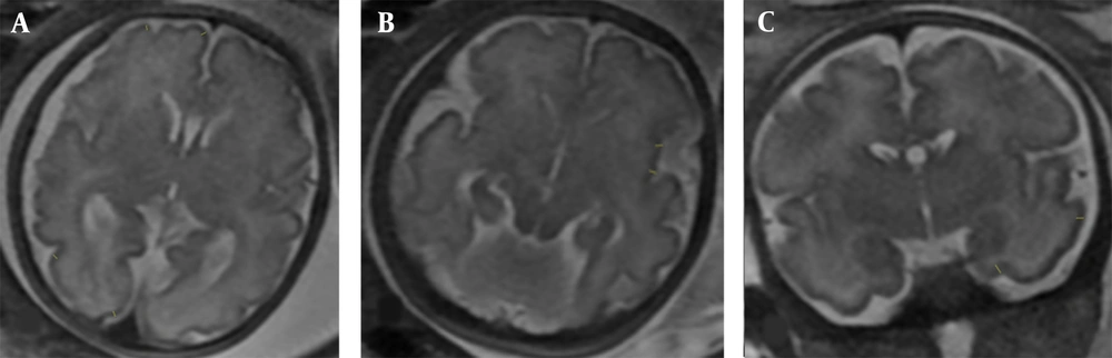
T2-weighted magnetic resonance images (MRI) show the brain cortical thickness assessment. A, Frontal and occipital lobe cortical thickness measurements (in the axial plane, at the level of the cavum septum pellucidum); B, Insular cortical thickness measurements (in the axial plane, right below the plane of the cavum septum pellucidum); C, Temporal lobe cortical thickness measurement (in the coronal plane, just anterior cut to the pons level). The two parts were measured from the inner side to the outer side.
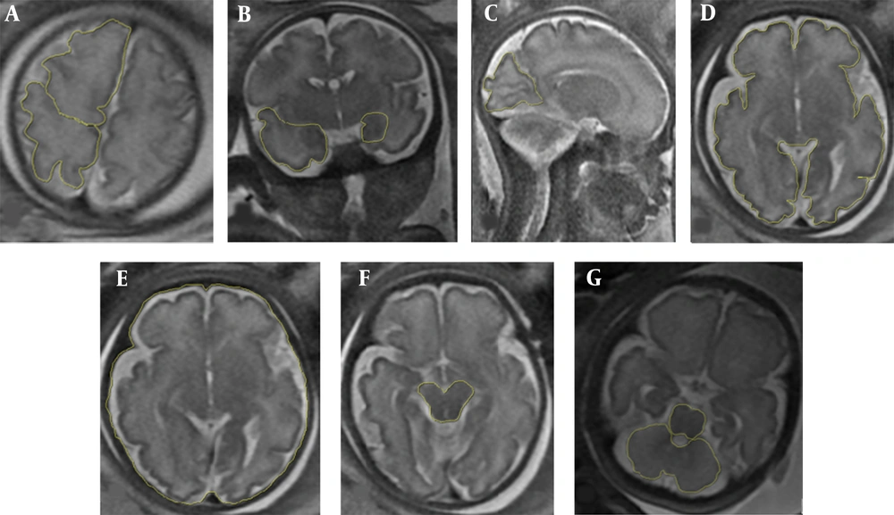
T2-weighted magnetic resonance images (MRI) demonstrate different brain lobe areas. A, Frontal and parietal lobe areas (in the axial plane, right above the cavum septum pellucidum and lateral ventricles); B, Temporal lobe and hippocampus areas (in the coronal plane, right anterior to the pons level); C, Occipital lobe area (in the first para-sagittal plane, near the falx, posterior to the parieto-occipital sulcus); D, Whole brain area (WBA) (in the axial plane, at the level of the cavum septum pellucidum); E, Whole intracranial area (at the same level of WBA by tracing the cursor inside the bony calvarium); F, Midbrain area; G, Cerebellum and pons areas (in the axial plane, at the level of the middle cerebellar peduncle).
3.5. Data Analysis
4. Results
4.1. Demographics and Clinical Indicators
| Variables | IUGR subtype A (n = 24) | IUGR subtype B (n = 18) | P-value | IUGR (n = 42) | Control group (n = 28) | P-value |
|---|---|---|---|---|---|---|
| Maternal characteristics | ||||||
| Age (y) | 29.33 ± 5.53 | 30.39 ± 4.94 | 0.593 | 29.79 ± 5.25 | 29.81 ± 2.67 | 0.792 |
| Height (cm) | 162.04 ± 5.57 | 161.28 ± 4.35 | 0.768 | 161.71 ± 5.04 | 157.64 ± 14.63 | 0.470 |
| Weight (kg) | 77.37 ± 15.09 | 77 ± 12.20 | 0.932 | 77.21 ± 13.77 | 73.43 ± 12.52 | 0.247 |
| BMI (kg/m2) | 29.44 ± 5.47 | 29.63 ± 4.75 | 0.903 | 29.52 ± 5.11 | 28.15 ± 4.41 | 0.250 |
| Preeclampsia | 4 (16.7) | 1 (5.6) | 0.02 | 5 (11.9) | 0 (0) | 0.001 |
| Fetal characteristics | ||||||
| Age (weeks) | 33 + 6 | 34 + 1 | 0.690 | 34 + 1 | 34 | 0.98 |
| Weight (g) | 1704.83 ± 487.7 | 1829.1 ± 562.8 | 0.448 | 1758 ± 517.8 | 1954 ± 662 | 0.157 |
| Sex (female) | 12 (50) | 10 (55.6) | 0.721 | 22 (52.4) | 6 (21.4) | 0.010 |
| HC < 5% | 13 (54.2) | 4 (22.2) | 0.0002 | 17 (40) | 0 (0) | < 0.0001 |
| Perinatal outcomes | ||||||
| Birth weight (g) | 2194.76 ± 544.98 | 2616.3 ± 363.4 | 0.011 | 2377. ± 514.6 | 2965.7 ± 733.13 | < 0.0001 |
| GA at birth (weeks) | 36 + 6 | 37 + 2 | 0.330 | 37 + 1 | 37 + 2 | 0.978 |
| NICU admission | 8 (38.1) | 3 (18.8) | 0.012 | 11 (29.7) | 8 (28.6) | 0.89 |
| Intrauterine stillbirth | 1 (5) | 0 (0) | 1 | 1 (2.9) | 0 (0) | 1 |
Abbreviations: BMI, body mass index; HC, head circumference; GA, gestational age; IUGR, intrauterine growth restriction; NICU, neonatal intensive care unit.
a Values are expressed as mean ± SD or No. (%) unless otherwise indicated.
4.2. MRI Examination
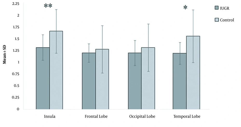
The polynomial contrast analysis demonstrates a difference in the corrected cortical thickness of different brain regions between the Intrauterine growth restriction (IUGR) and control groups. The bars show mean ± SD for each group, and asterisks indicate P < 0.05.
| Variables | IUGR group (n = 42) | Control group (n = 28) | P-value |
|---|---|---|---|
| Cortical thickness (mm) | |||
| Insula | 0.034 ± 0.007 | 0.043 ± 0.012 | 0.001 |
| Frontal lobe | 0.031 ± 0.005 | 0.033 ± 0.013 | 1 |
| Occipital lobe | 0.031 ± 0.007 | 0.034 ± 0.013 | 0.250 |
| Temporal lobe | 0.036 ± 0.007 | 0.047 ± 0.027 | 0.010 |
| Area (cm2) | |||
| Frontal lobe | 0.214 ± 0.028 | 0.209 ± 0.029 | 0.374 |
| Temporal lobe | 0.086 ± 0.024 | 0.086 ± 0.022 | 0.611 |
| Occipital lobe | 0.105 ± 0.024 | 0.100 ± 0.016 | 0.274 |
| Hippocampus | 0.017 ± 0.008 | 0.0125 ± 0.003 | < 0.001 |
| Cerebellum | 0.147 ± 0.021 | 0.130 ± 0.019 | 0.001 |
| Midbrain | 0.043 ± 0.015 | 0.043 ± 0.007 | 0.983 |
| Pons | 0.031 ± 0.014 | 0.028 ± 0.003 | 1 |
| WBA | 0.806 ± 0.170 | 0.812 ± 0.027 | 0.028 |
Abbreviations: IUGR, intrauterine growth restriction; WBA, whole brain area.
| Variables | IUGR subtype A (n = 24) | IUGR subtype B (n = 18) | P-value |
|---|---|---|---|
| Cortical thickness (mm) | |||
| Insula | 0.032 ± 0.005 | 0.036 ± 0.009 | 0.104 |
| Frontal lobe | 0.029 ± 0.003 | 0.032 ± 0.007 | 0.104 |
| Occipital lobe | 0.030 ± 0.006 | 0.032 ± 0.007 | 0.562 |
| Temporal lobe | 0.037 ± 0.006 | 0.036 ± 0.007 | 0.415 |
| Area (cm2) | |||
| Frontal lobe | 0.208 ± 0.029 | 0.222 ± 0.024 | 0.169 |
| Temporal lobe | 0.085 ± 0.015 | 0.088 ± 0.034 | 0.668 |
| Occipital lobe | 0.105 ± 0.021 | 0.105 ± 0.028 | 0.627 |
| Hippocampus | 0.015 ± 0.003 | 0.019 ± 0.012 | 0.334 |
| Cerebellum | 0.146 ± 0.022 | 0.148 ± 0.019 | 0.791 |
| Midbrain | 0.045 ± 0.019 | 0.041 ± 0.007 | 0.368 |
| Pons | 0.028 ± 0.011 | 0.034 ± 0.017 | 0.44 |
| WBA | 0.785 ± 0.220 | 0.831 ± 0.049 | 0.566 |
Abbreviations: IUGR, intrauterine growth restriction; WBA, whole brain area.
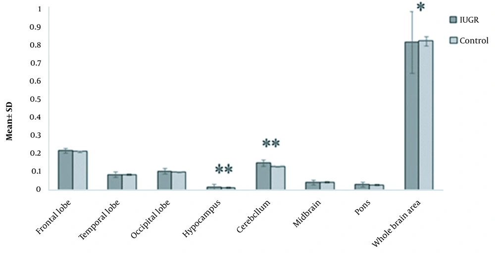
The polynomial contrast analysis demonstrates a difference in the corrected brain areas in different lobes between the Intrauterine growth restriction (IUGR) and control groups. The bars show mean ± SD for each group, and asterisks represent P < 0.05.


