


1. Background
The prevalence of unruptured intracranial aneurysm is 3.2% among adults (1). Generally, these aneurysms remain asymptomatic until the point of rupture, however many might present with non-specific symptoms such as severe headache or cranial nerve palsy. According to a long-term cohort study, the annual risk of rupture reaches 1.3% (2). The decision on proper management and treatment indications of unruptured aneurysms remains challenging and not clearly defined (3). The rationale for treatment of unruptured aneurysms is to prevent rupture, subarachnoid hemorrhage (SAH) and subsequent neurologic complications. Since the introduction of intravascular coils, treatment of unruptured intracranial aneurysms (UIAs) has been evolving. There are a variety of intravascular treatment options; however, the treatment choice for many variations of UIAs remains the subject of debate. Flow-diverters (FDs) devices have emerged as a new tool in treating UIAs, especially those who are difficult to manage with older intravascular techniques including giant, wide-necked, fusiform variants of UIAs and those with disadvantageous relation of the aneurysm neck and parent artery.FDs are tubular stent-like implants that immediately decrease inflow and outflow of the aneurysm and promote neo-endothelization of the parent vessel (4). Various FDs have been employed with pipeline embolization device (PED) showing high durability and safety for selected UIAs. A number of studies on intravascular treatment of unruptured intracranial aneurysms have reported more than 90% mid-term success rate for pipeline embolization devices with acceptable safety, morbidity and mortality (5-8).
2. Objectives
This study was conducted to present the mid-term outcome following endovascular treatment of unruptured intracranial aneurysms in 20 patients using PED. We aimed to measure the midterm success rate and assess for possible complications.
3. Patients and Methods
3.1. Patient Population
This prospective study was conducted on a total of 20 patients harboring a single intracranial wide neck aneurysm who were referred to advanced diagnostic and interventional imaging center of Imam Khomeini Hospital between July and October 2010
The diagnosis was confirmed on the base of four-vessel cerebral digital subtraction angiography (DSA). Patients with single wide-neck (> 4 mm or neck-to-sac ratio < 1:2) aneurysms were included in this study. The exclusion criteria were as follows: Patients who were at high risk for general anesthesia or cardiovascular interventional procedures, patients with multiple coronary lesions, and patients who had a history of contrast agent hypersensitivity and individuals with multiple intracranial aneurysms.
3.2. Endovascular Procedure
The purpose of this procedure was to cover the maximum length of the aneurysm through insertion of the pipeline stent. The procedure of stent insertion was carried out on a flat unit of DSA.
All the procedures were performed under general anesthesia. All patients received a loading dose of 500mg of aspirin and 600 mg of clopidogrel a day before the procedure. Platelet function was assessed by means of bleeding time (BT). Before stent insertion, patients were administered 5000 units of bolus intravenous heparin as well as 500 mg of aspirin. All procedures were closely monitored by the project supervisor.
The main artery was catheterized with a 6 F guide catheter. Marksman microcatheter was then entered and advanced into the artery distal to the aneurysm using a micro-guidewire (0.016 inch). To prevent any vascular damage, the tip of the distal end of the microcatheter was placed in a straight segment in the main artery. PED was then directed by the microcatheter to the location of the aneurysm. Whilst progressing forward with the insertion wire, the microcatheter was withdrawn and the PED was expanded at the site of the aneurysm.
DSA was carried out following stent deployment to assess the position of the stent immediately after stent insertion. If the stent was not placed appropriately within the aneurysm, then the outcome was recorded as failed. Successful stent insertion was further categorized into three groups based on the amount of contrast passing between the stent and the wall of the aneurysm. These categories included excellent, good, and poor stagnation. A higher amount of contrast stasis (higher stagnation) within the aneurysm is associated with facilitated intra-aneurysmal thrombus formation. After the procedure, daily dual therapy of 100mg of aspirin and 75mg of clopidogrel was initiated.
3.3. Follow-Up
Patients were followed 4 to 8 months after the procedure. Clinical follow-up was performed at months 1, 2, 4, and 6 post-procedure. DSA was carried out to assess stent integrity, displacement from its original position, stent occlusion and the presence of residual aneurysm. Procedure success was defined as complete occlusion of the aneurysm.
3.4. Data Analysis
Statistical analysis was carried out using SPSS software (version 16.0, SPSS Inc. Chicago, IL, U.S.A.). Non-parametric tests (Mann-Whitney and Kruskal Wallis) were used for analysis of variables. All statistical tests were 2-tailed and P value < 0.05 was considered statistically significant. All means are expressed as mean ± SD.
4. Results
Fifteen females and five males with a mean age of 51 (range, 20 - 78) years were enrolled. The frequency of each morphological type of aneurysm is shown in Table 1. In 18 patients (90%), the aneurysm was in the anterior circulation; whereas, in two individuals (10%) it involved the posterior circulation.
| Morphology | No. (%) |
|---|---|
| Saccular | 15 (75) |
| Dissecting | 3 (15) |
| Bilobal | 1 (5) |
| Fusiform | 1 (5) |
| Total | 20 (100) |
Frequency of Various Morphologic Types of Aneurysm
Out of 20 participants, only one patient (5%) required two stents and the rest (95%) were treated with a single stent. Due to technical errors in appropriate stent placement, the intervention attempt failed in one patient. Immediate DSA after intervention showed excellent result in 15, good stagnation in three and poor stagnation in one patient.
Two patients experienced transient acute symptoms following the procedure, which included transient hemi-paresthesis and transient altered level of consciousness. No acute hemorrhagic or thromboembolic event was observed.
Aneurysm morphology, location of the aneurysm, size of aneurysms and acute transient complication did not seem to affect the outcome of the procedure. However, the number of cases was too small to draw statistical analysis.
4.1. Mid-term Follow-Up
One patient died after three months due to subarachnoid hemorrhage (SAH). The mean duration of follow up by angiography was 6 months and 6 days in the remaining 18 patients with successful attempt. Follow-up DSA showed excellent outcome with complete occlusion in 14 patients (77.8%) and residual aneurysm in four patients (22.2%). Table 2 demonstrates the demographics of all 20 patients enrolled in this study.
| Age | Gender | Number of Stent | Morphology | Aneurysm Number | Acute Complication | Final Result | Aneurysm Location | Aneurysm Size | Anti-Coagulants | Stent Displacement | Neurological Dysfunction After Insertion | In-Stent Stenosis | Time to Angiography | Death | Angiography Outcome | |
|---|---|---|---|---|---|---|---|---|---|---|---|---|---|---|---|---|
| 1 | 71 | F | 1 | S | 1 | No | Exc. | Ant. | Small | Yes | No | No | No | 7 | Complete Occlusion | |
| 2 | 59 | M | 1 | S | 1 | No | Exc. | Ant. | Small | Yes | No | No | No | 7 | Complete Occlusion | |
| 3 | 56 | F | 1 | S | 1 | No | Exc. | Ant. | Giant | Yes | No | No | No | 6 | Residual Aneurysm | |
| 4 | 78 | M | 1 | D | 1 | No | Poor | Ant. | Small | Yes | No | Yes | Yes | 7 | Complete Occlusion | |
| 5 | 52 | F | 1 | S | 1 | No | Exc. | Ant. | Small | Yes | No | No | No | Yes | ||
| 6 | 20 | M | 1 | B | 1 | No | Exc. | Ant. | Small | Yes | No | No | No | 7 | Complete Occlusion | |
| 7 | 25 | F | 1 | S | 1 | No | Exc. | Ant. | Small | Yes | No | No | No | 7 | Complete Occlusion | |
| 8 | 51 | M | 1 | S | 1 | No | Exc. | Ant. | Small | Yes | No | No | No | 6 | Residual Aneurysm | |
| 9 | 39 | F | 1 | S | 1 | No | Good | Post. | Small | Yes | No | No | No | 7 | Complete Occlusion | |
| 10 | 52 | F | 1 | S | 1 | No | Exc. | Ant. | Small | Yes | No | No | No | 6 | Residual Aneurysm | |
| 11 | 70 | F | 1 | D | 1 | Yes | Good | Ant. | Giant | Yes | No | No | No | 7 | Complete Occlusion | |
| 12 | 25 | F | 1 | S | 1 | No | Good | Ant. | Giant | Yes | No | No | No | 8 | Complete Occlusion | |
| 13 | 66 | M | 2 | F | 1 | No | Exc. | Ant. | Giant | Yes | No | No | No | 6 | Complete Occlusion | |
| 14 | 55 | F | 1 | S | 1 | No | Exc. | Ant. | Small | Yes | No | No | No | 4 | Complete Occlusion | |
| 15 | 37 | F | 1 | S | 1 | No | Exc. | Ant. | Small | Yes | No | No | No | 7 | Complete Occlusion | |
| 16 | 57 | F | 1 | S | 1 | No | Exc. | Ant. | Small | Yes | No | No | No | 7 | Complete Occlusion | |
| 17 | 58 | F | 1 | S | 1 | No | Exc. | Ant. | Small | Yes | No | No | No | 7 | Complete Occlusion | |
| 18 | 33 | F | 1 | S | 1 | No | Exc. | Ant. | Small | Yes | No | No | No | 6 | Residual Aneurysm | |
| 19 | 68 | F | 1 | D | 1 | Yes | Exc. | Post. | Small | Yes | No | No | No | 6 | Complete Occlusion | |
| 20 | 45 | F | 1 | S | 1 | - | Failed | Ant. | Small | - | - | - | - |
In Detail Demographic, Clinical, Radiologic and Intervention Related Data of All Participants of the Study
Stent migration was not observed in any patients. 25% - 50% intra-stent stenosis (in-stent stenosis) was depicted in only one patient (5.5%). No chronic neurological symptoms were reported following stent insertion in any patients.
5. Discussion
PED has emerged as one of the most advanced choices for the treatment of intracranial aneurysms. This study showed that the use of PED brings favourable outcome and minimal mid-term complications when used for treating single unruptured intra-cerebral aneurysms.
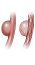
Flow diverter devices redirect blood flow from the aneurysm sac into the distal artery without occluding side branches. This leads to flow stasis within the aneurysm, which commences progressive intra-aneurysmal thrombosis. Furthermore, this type of stent promotes endoluminal reconstruction of the parent vessel, which finally leads to aneurysm occlusion (3). This event takes place over several months. Therefore, residual aneurysmal filling immediately after PED insertion is normally expected. Szikora et al. reported immediate occlusion of small (<5mm) aneurysms in 21% of patients treated with PED (9). The immediate result of PED insertion could be assessed by DSA as the amount of contrast stagnation inside aneurysm sac. The more contrast stagnation, the faster aneurysm occlusion is expected. The present study showed good or excellent results in all patients except one with poor stagnation of the contrast in whom follow-up DSA showed complete occlusion of the aneurysm.
PED is a self-expanding stent-like flow diverter with high metal-to-surface area. Nominal diameter varies from 2.5 to 5 mm with 0.25 mm increments and the nominal length range is between 10 and 35 mm with 2 mm increments. PED is delivered over a 0.027-inch microcatheter and carries a radiopaque platinum tip beyond the distal end of the stent (10). PED diameter should be roughly close to the target vessel diameter, while PED length must be at least 6 mm longer than the aneurysm neck size.
A number of studies have reported superior results when using multiple PEDs for a single aneurysm specially when favourable flow decline is not achieved by means of a single PED (11). According to Chalouhi et al. (12), there is no significant superiority for multiple over single PED. In one patient, due to highly persistent blood flow circulating within the aneurysm, we decided to use two stents. The midterm outcome of the procedure in this patient was excellent.
Chalouhi et al. compared the use of coiling vs. PED in a series of patients with large saccular aneurysms and found significantly higher aneurysmal occlusion and significantly less necessity for retreatment, while the morbidity rate did not differ between the two groups (13).
According to a meta-analysis of 29 studies, morbidity and mortality of flow diverter devices is 4% and 5%, respectively (14). The rate of post-procedural complications of PED seems to be less likely. A study on 251 aneurysms treated with PED demonstrated permanent morbidity and mortality of only 1% and 0.5%, respectively (15). Yu et al. reported a complete aneurysm occlusion rate of 84% in a multicenter study with peri-procedural death or stroke seen in 3.5% (16). They suggested PED to be the first line choice for treating un-ruptured intracranial aneurysms.According to a review of 210 patients treated with PED, mortality occurred in 1.9%, which is comparable to the mortality risk of coiling (10).
In the present study, mild in-stent stenosis was depicted in only one patient. There is no clear protocol to delineate the duration and dosage of antiplatelet therapy to decrease the risk of in-stent thrombosis and stenosis. Very few cases of in-stent thrombosis are reported after PED insertion with the majority being less than 50% (5, 8, 17). One major concern about PED is the risk of aneurysm rupture in the latency period before total aneurysm occlusion (18). Intracranial hemorrhage is reported in 3.8% of individuals after PED employment (10). We observed SAH in only one patient of this study leading to the patient’s death.
The present study showed a favourable midterm result in 77.8% of the participants. Lylyk et al. used PED for treatment of 63 intracranial aneurysms and achieved complete occlusion in 95% of the lesions in a 12-month follow-up (5). Meanwhile, according to the experience of Chan et al. with PED on 13 wide neck aneurysms of the internal carotid artery, a success rate of 69% was achieved after a mean follow-up of 14 months (19). Fischer et al. achieved 74% complete aneurysm exclusion in a 10-month follow-up of 49 aneurysms (17).
We used DSA for follow-up of treated UIAs. DSA is the gold standard for follow-up of UIAs after endovascular treatment (20). CT angiography and MR angiography can be utilized, however both are inferior to DSA for detection of residue or recurrence of the aneurysm (21).
This study was subject to several limitations, first the sample size was small and statistical analysis on the role of various demographic factors on the final outcome could not be measured. Second, we did not gather data about required coverage of the stent. Overall, further studies with a larger number of participants are necessary for these purposes.
In conclusion, the use of pipeline embolization device for the treatment of unruptured intracranial aneurysms appears encouraging with favorable mid-term clinical outcome and minimal complication. However, larger studies with longer follow-up durations are warranted.
