


1. Background
In recent years, due to the increase in the number of computed tomography (CT) examinations, concerns about radiation exposure have increased (1). Many studies showed that CT scans increase the incidence of cancer risk (2, 3). The dose of the superficial organs in CT scan examination is high and the breast is one of the organs that is directly irradiated through thoracic CT scans and it is also a sensitive organ to radiation (4). Breast cancer is one of the most important reasons causing death in women; therefore, the strategy of radiation protection must be used in order to reduce radiation dose to these organs (5). Normally, lead protective is used to protect the organs, but bismuth shields are used for protection against CT scan tests. Bismuth shields protect superficial radiosensitive organs such as breast by attenuating primary X-ray beams (6, 7).
Currently, many studies have focused on composite shields rather than lead shields (8). For example, Tappouni and Mathers showed that bismuth shield could reduce breast dose about 38% (9). Nonetheless, the use of bismuth shields will increase the noise of the images and influences the quality of the images (10). Einstein et al. showed that using bismuth shield decreased the contrast to noise ratio by 20.9% and it has caused increase in the image noise significantly (11).
There are many ways to improve shield efficiency in terms of dose reduction. One of them is to use nanoparticles against micro-particle in the construction of bismuth shields (12, 13). By using nanoparticles, the shielding efficiency for dose reduction could be increased (14, 15). Mehnati et al. used bismuth oxide (Bi2O3) nanoparticles to make a shield in order to be used for conventional radiography. They showed that using the nano-Bi2O3 shield, the dose reduction was equal to 42% and 31%, respectively related to 60 and 100 kVp (16). By increasing the thickness of the shield, dose reduction is improving. A study showed that bismuth shield with 0.4 mm thickness could reduce the dose about 36% in conventional radiography. Although when four layers of the shield were used, dose reduction was increased by 73% (17).
2. Objectives
Because the breast protection is considered important in chest CT scan tests, the main goal of this study was to synthesize Bi2O3 nanoparticles to prepare composite shields in different ratios as well as different thicknesses for reducing the received dose by the breast in chest CT examinations. Also, image quality was evaluated with and without the use of the shield in different conditions.
3. Materials and Methods
3.1. Preparation of Bi2O3 Nanoparticles
In order to synthesize the nanoparticles, 5 g of bismuth nitrate was mixed with nitric acid (67%) and stirred for two hours to make a homogeneous solution. Then, 25% of citric acid solution was added slowly to the first solution for 2 hours. A clear solution was obtained at high speed for 2 hours at 25ºC. At this stage, the pH of the solution was about 2 - 3. Then 60% of the polyethylene glycol solution was added slowly to the solution for 2 hours until the solution was obtained. The solution was heated at 90ºC for 3 hours until it became yellow and the color was yellow over time, which was washed 3 times with distilled water and ethanol through centrifugation. The sediment was dried at 350ºC for 3 hours and at 500ºC for 2 hours to obtain a Bi2O3 nanoparticle powder.
For analysis of the nanoparticles, the transmission electron microscope (TEM), X-ray diffraction (XRD), dynamic light scattering (DLS) methods were used to study the properties and size of nanoparticles.
3.2. Construction of Nano-Composite Shields
After synthesis, Bi2O3 nanoparticles and silicone rubber were used to prepare nano-bismuth shield. The shields were made relative to the proportion of Bi2O3 weight in 10% and 15%. The thicknesses of shields were equal to 0.5 and 1 mm, respectively and the size of the shields were equal to 20 × 20 cm2.
After adding bismuth nanoparticles to silicone, the mixture was blended by a mechanical stirrer for 1 hour until achieving the homogeneous distribution of Bi2O3 nanoparticles within the silicone matrix. The composite of Bi2O3 nanoparticle was placed in the mold and was dried in room temperature for one week. Scanning Electron Microscope (SEM)-MAP imaging was also used to evaluate the distribution of nanoparticles within the matrix in the nano-bismuth shield.
3.3. Dosimetry Condition
The female chest phantom (18) and the 16 multi-slice CT scan (TOSHIBA) were used. Dose measurements were done using 100 thermoluminescent dosimeters (TLDs: GR-200) (Hangzhou Freq control Electronic Technology Ltd., China). Scan conditions for routine adult chest CT protocols included 110 kVp, 105 mAs, pitch = 1 and 5-mm slice thickness. Every experiment was repeated three times. In the first time, with the presence of CT, the skin and glandular dose of normal and large breasts with and without the use of composite shields 10% and 15% 0.5 mm were evaluated. The second experiment, the skin and glandular dose of normal and large breasts in the state with and without the use of composite shields 10% and 15% 1 mm were assessed. In every session, to measure entrance skin dose at the level of the breast, four TLD chips were placed on each breast skin around the nipple. Also, to measure breast glandular dose, four TLDs were placed in the fourth layer of large and normal breast phantoms. Therefore, in addition, for each scan, sixteen TLDs were used. After reading each TLD, the element correction coefficient (ECC) was considered and the received dose of each TLD chip was obtained using the calibration curve. The average of the TLD doses was calculated and recorded.
In order to reduce artifact, 1 cm of foam was used below the shields. To calculate the image quality and image noise after using shields, the CT number and the standard deviation of region of interests (ROI) (with diameter 2 cm2) in the anterior, central and posterior region of the slice were used. Statistical analysis of the results was calculated using SPSS version 16.0 (IBM Co, Armonk, NY, USA) software and Wilcoxon test. All graphs were done using GraphPad Prism 6 (GraphPad Co, CA, USA).
4. Results
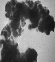
Figure 1 illustrates the TEM image of Bi2O3 nano-particle. The obtained Bi2O3 sample is the approximate spherical polycrystalline particles (Figure 1). The powder XRD analysis of the Bi2O3 nanoparticles has been shown in Figure 2A. The test was done in the diffraction angle (2θ) ranging from 4º to 70º. The place of the peaks revealed that the Bi2O3 nanoparticles were synthesized successfully and the card of joint committee on powder diffraction standards (JCPDS) by the number of 41-1449 is a standard card and it shows that the sample is exactly Bi2O3.
 images of nano-bismuth oxide nanoparticles. A, With less magnification B, With more magnification")
Transmission electron microscope (TEM) images of nano-bismuth oxide nanoparticles. A, With less magnification B, With more magnification
 analysis of the bismuth oxide (Bi<sub>2</sub>O<sub>3</sub>) nanoparticles. B, Dynamic light scattering (DLS) analysis of the Bi<sub>2</sub>O<sub>3</sub> nanoparticles")
A, X-ray diffraction (XRD) analysis of the bismuth oxide (Bi2O3) nanoparticles. B, Dynamic light scattering (DLS) analysis of the Bi2O3 nanoparticles
Also the DLS was used for testing the size of nanoparticles. Based on the results, the nanoparticle size ranged from 20 to 60 nanometers and most of the particles had the size of 24 nanometers (Figure 2B).
The distribution of nanoparticles in the matrix was calculated through SEM-MAP analysis as shown in Figure 3. The SEM-MAP analysis of nano-Bi2O3 shields at 10% and 15% of ratios showed that the nanoparticles have a good distribution in the base silicone matrix.
-MAP of bismuth oxide (Bi<sub>2</sub>O<sub>3</sub>) nanoparticles in silicone matrix. A, 10% nano-Bi<sub>2</sub>O<sub>3</sub> shield. B, 15% nano-Bi<sub>2</sub>O<sub>3</sub> shield")
Scanning electron microscope (SEM)-MAP of bismuth oxide (Bi2O3) nanoparticles in silicone matrix. A, 10% nano-Bi2O3 shield. B, 15% nano-Bi2O3 shield
The amount of the received dose in the skin and glandular layers of the breast was equal to 4.84 and 5.26 mSv, respectively. When nano-Bi2O3 shield (1 mm) was used at 10% and 15%, the dose was reduced significantly in both skin and glandular layers, but dose reduction for the skin layer was higher (Figure 4).
 shields with 0.5 thickness. B, Evaluation of the dose of the skin layer and the fourth layer of a large breast with and without using nano-Bi<sub>2</sub>O<sub>3</sub> shields with 1 mm thickness.")
A, Evaluation of the dose of the skin layer and the fourth layer of normal breast with and without using nano-bismuth oxide (Bi2O3) shields with 0.5 thickness. B, Evaluation of the dose of the skin layer and the fourth layer of a large breast with and without using nano-Bi2O3 shields with 1 mm thickness.
The statistical analysis of data in Figure 4A. showed that the reduction of the dose level by the use of 10% and 15% nano-Bi2O3 shields with 0.5 mm thickness was statistically significant compared to non-shield test mode (shield 10% P = 0.0460) and (shield 15% P = 0.0251). Comparison of dose reduction revealed that dose reduction in 10% and 15% shields was statistically significant (P = 0.0277) at the same 0.5 mm thickness.
In addition, statistical analysis of Figure 4B showed that the reduction of the dose level by the use of 10% and 15% nano-Bi2O3 shields with 1 mm thickness was statistically significant compared to non-shield test mode (shield 10% P = 0.0305) and (shield 15% P = 0.0083). Dose reduction comparisons were also statistically significant in the cases of 10% and 15% (P = 0.0227) at the same 1 mm thickness. Moreover, statistical results showed that in the same percentage of fillers, with increasing thickness, dose reduction was not significant (shield 10%, P = 0.1038) and (shield 15%, P = 0.1112).
Using the 10% nano-Bi2O3 shield with a thickness of 0.5 mm, the breast dose was reduced to 9.8% in the skin layer and 4.8% in the fourth layer. The dose reduction for 10% nano-Bi2O3 shield with 1 mm thickness was equal to 18.3% and 16.3% in the surface and the fourth layer, respectively. Also, using a 15% nano-Bi2O3 shield with a thickness of 0.5 mm, the dose reduction of the breast was equal to 15% and 13.6% in the skin layer and the fourth layer, respectively. This decrease in breast doses using 15% nano-Bi2O3 shield with 1 mm thickness was equal to 24% and 21% at the surface and the fourth layer, respectively.
The results of the CT number and noise on the anterior, central and posterior region of the phantom are given in Table 1. The average of CT number of ROI in the breast region was equal to -79 Hounsfield unit (HU) without the use of shield. Using the 10% and 15% bismuth shields with 0.5 mm thickness, they were equal to -80 and -83 HU, respectively. In addition, using 10% and 15% bismuth shields with 1 mm thickness, they were equal to -81 ± 3.6 and -85 ± 2.4 HU, respectively.
| Shields | Anterior region | Central region | Posterior region | Mean | ||||
|---|---|---|---|---|---|---|---|---|
| CT number | Noise | CT number | Noise | CT number | Noise | CT number | Noise | |
| Reference | -79.4 ± 5.5 | 24.2 ± 3.1 | -73.5 ± 2.3 | 23.4 ± 2.3 | -72.3 ± 4.6 | 22.4 ± 2.5 | -75.01 | 23.3 |
| 10% Bi 0.5 mm | -80 ± 3.2 | 26.1 ± 2.8 | -75.1 ± 4.3 | 24.2 ± 1.3 | -73.8 ± 5.1 | 22.7 ± 3.4 | -76.1 | 24.33 |
| 10% Bi 1 mm | -81.9 ± 3.6 | 27.1 ± 4.5 | -75.9 ± 4.6 | 24.9 ± 2.5 | -74.5 ± 4.7 | 23.1 ± 3.1 | -77.4 | 25.03 |
| 15% Bi 0.5 mm | -83.1 ± 4.3 | 28.4 ± 3.4 | -76.5 ± 4.1 | 25.4 ± 3.2 | -74.4 ± 5.3 | 234.4 ± 2.3 | -78 | 25.73 |
| 15% Bi 1 mm | -85.2 ± 4.2 | 29.7 ± 3.7 | -76.6 ± 5.2 | 26.2 ± 3.5 | -76.3 ± 3.8 | 76.3 ± 3.8 | -79.36 | 26.73 |
Abbreviation: Bi, bismuth
The result showed that the average noise in the breast region was equal to 24 ± 3.1% without the use of shield and were equal to 26 ± 8.2 HU and 28 ± 3.4 HU, respectively using 10% and 15% bismuth shields with 0.5 mm thickness. Also, using 10% and 15% bismuth shields with 1 mm thickness, the noises were equal to 27 ± 4.5 and 29 ± 7.3 HU, respectively. The statistical analysis showed that using 10% and 15% nano-bismuth shields with 0.5 mm thickness, the increase in the noise was not statistically significant. Furthermore, the increase in the noise was significant using 10% and 15% nano-bismuth shields with 1 mm thickness especially at the anterior section.
According to Table 2, the highest increase in the noise was observed in the breast and under the shield area. The highest increase in the noise in this area was related to the use of 15% nano-bismuth shield with 1 mm thickness, which was equal to 18%. This increase in the noise in the heart region was less than the breast region. The slightest increase in the noise was associated with the posterior region of the phantom using the 10% nano-bismuth shield with 0.5 mm thickness.
| Increased noise % | Bi2O3 10% 0.5 mm | Bi2O3 10% 1 mm | Bi2O3 15% 0.5 mm | Bi2O3 15% 1 mm |
|---|---|---|---|---|
| Anterior | 7 % (0.045) | 10 % (0.031) | 14 % (0.034) | 18 % (0.026) |
| Central | 3 % (0.245) | 6 % (0.254) | 8 % (0.075) | 10 % (0.038) |
| Posterior | 2 % (0.309) | 3 % (0.555) | 4 % (0.333) | 8 % (0.089) |
Abbreviation: Bi2O3, bismuth oxide
5. Discussion
In this study, the use of bismuth oxide nanoparticles as a shield in CT scan tests showed that these shields, in addition to reducing the received dose to the breast, cause slight changes in the image noise of the chest CT scan. The results indicated that the change in the bismuth percentage from 10% to 15% in nano-bismuth oxide shields with 0.5 mm thickness reduced the dose from 9.8% to 15% in the skin layer and from 8.4% to 13.6% in the fourth layer. Statistically, the changes in breast doses were not significant in the two layers of the skin and the fourth layer. Furthermore, this change in the shields with 1 mm thickness reduced the dose from 18.3% to 23.5% in the skin layer and from 16.3% to 21.1% in the fourth layer. Statistically, the changes in the breast doses were significant in the two layers of the skin and the fourth layer (P = 0.039).
The results showed that by increasing the bismuth percentage and the thickness of the shields, the dose reduction of the breast was increased. The percentage of dose reduction for the skin layer was more than the fourth layer, which showed the shields are more effective in terms of protecting superficial body organs. The main purpose of radiation protection in medical imaging is to use a minimum dose in order to achieve a better diagnosis. Due to the increasing use of CT scans to detect malignancies, sensitive organs such as the breast in women have been exposed to high radiation levels. Therefore, reducing the dose of these sensitive organs should be considered.
Many studies have been conducted in order to introduce different shields instead of lead protectors to use in CT scan tests in recent years. The purpose of this study was to design, construct, and evaluate nano-bismuth silicone shields to reduce the breast dose in CT scan tests. Alonso et al. studied the effect of bismuth shield on dose reduction for the breast in CT scan examinations. The results showed that dose reduction for the right and left breast was equal to 30% (19). In the present study, using 15% nano-bismuth shield with 1 mm thickness, dose reduction rate was about 26%. This study also reported low noise variations. Tappouni and Mathers, studied the absorption dose of skin and sensitive organs in chest CT scan tests. They showed that bismuth shields can reduce the breast dose about 38% (9). In this study, in order to obtain low noise in image, the shields were made from low percentages of nano-bismuth oxide powder (10% and 15%). That is why despite desirable dose reduction, the lowest image noise was observed. In a study conducted by Mehnati et al. on micro bismuth shields, they showed that using 10% micro bismuth shield with one mm thickness, the dose reduction was equal to 12%, and noise variation was about 13% (20). The comparison regarding analysis of the results of the present study with those of the study by Mehnati et al. showed that using nano-Bi2O3 instead of micro bismuth with 1 mm thickness, dose reduction in nano-Bi2O3 shield was about 15 % higher, and noise increase in nano-Bi2O3 shields was equal to 14%, which is less than micro bismuth shield.
Despite the innovative approach, some of the limitations of this study deserve consideration. Like many other studies, with regard to the ethics of working with patients, we used a phantom to measure the received dose of breast with and without the presence of shields. Also, it was also better to use human samples to check the quality of images when using shields.
In conclusion, our results indicated that the use of nano-bismuth oxide shields with different bismuth ratios and thicknesses could reduce the received dose in the breast with no appreciable loss in the diagnostic ability of chest CT scans.
