1. Background
Collagen biomaterials are often used in the fabrication of various medical devices. Their protection against infections is a significant current challenge. Thus, the antimicrobial activity, among many other properties, is a very important characteristic of the collagen biomaterials. With the idea to explore the biological activity of some newly synthetized chemical compounds, plant extracts and their combinations for development of new antimicrobial collagen biomaterials, a serial investigation was initiated by preparation, characterization, and determination of biological activity of new Collagen/ZnTiO3 composites (1), followed by a similar one for Collagen/RGO composites (2). In both cases, a self-prepared antimicrobial agent was employed, crystalline ZnTiO3 powder and multi-layer RGO sheets, respectively. This serial investigation continues with the preparation, characterization, and biological activity evaluation of Collagen/(Ag/RGO) and Collagen/(Ag/RGO/SiO2) composites expecting to develop biomaterials with improved antimicrobial activity, compared to that of the former Collagen/RGO composites (2) by adding Ag-nanoparticles (AgNPs) or AgNPs and silane matrix.
Silver is known as an antibacterial agent for centuries. The bactericidal effect of silver nanoparticles (AgNPs) is also known long ago (3). Mono-dispersed AgNPs with a small size are used in antibacterial systems because of their high specific area and easy cell penetration (4). However, the well-known aggregation, leading to the biological activity decrease, as well as a doubt about cytotoxicity of the nanoparticles, limits their practical application (5, 6). Variety of approaches to avoid these problems, as well as to combine the activity of different antimicrobial agents are used, including immobilization of AgNPs on different substrate surfaces like GO or RGO sheets or/and silica matrices. Green synthesis of silver decorated nanoprisms, GO sheets, and their antibacterial properties are described in the literature (7). The results of UV-VIS spectra and transmission electron microscopy reveal that the Ag nano-prisms with a different size are well dispersed on the graphene oxide surface. The antibacterial activity study demonstrates that GO/Ag nano-composites exhibit satisfactory antibacterial properties: the inhibition of the E. coli growth is of 99.9% at concentration of 10 ppm for GO/Ag. Prepared by a simple, one-pot boiling method, GO/Ag nanocomposits were also studied as a highly effective antibacterial agent (8). Compared to that of the AgNPs, as well as of the simple mixture of GO and AgNPs, they demonstrate synergistically enhanced activity at very low dosages: MIC of 4 μg/mL against E. coli and MIC of 14 μg/mL against S. aureus, possibly owing to the unique physicochemical properties of GO/Ag nanocomposites. The unique physicochemical properties of GO/Ag nanocomposites, compared to that of the AgNPs, as well as the simple mixture of GO and AgNPs, demonstrate synergistically enhanced activity at very low dosages: MIC of 4 μg/mL against E. coli and MIC of 14 μg/mL against S. aureus. The low cytotoxicity of the GO/Ag nanocomposites to mammalian cells is their significant advantage: the viability of both, HeLa and HEK 293T cells remain at above 65%, even at very high concentrations (up to 50 μg/mL). Chitosan/AgNPs/GO nano-hybrids, which integrate the antibacterial and physico-chemical properties of AgNPs, GO and chitosan biopolymer, demonstrate enhanced antibacterial activity against S. aureus (9). Expecting that the loading of AgNPs on graphen oxide matrix with a huge specific area can prevent their aggregation, AgNPs/GO nanocomposites were synthesized thorough a facile phase solution method. They exhibit good antibacterial activity against E. coli and S. aureus. The improved antibacterial activity of the nanocomposites with high Ag loadings is a result of both: the effect of the AgNPs size and the synergistic action of GO and AgNPs in the AgNPs/GO nanocomposites (10).
The SiO2 materials with porous structure can easily adsorb various ions and organic molecules in their pores and on the surface. Therefore it is expected of SiO2 to be one of the most promising carriers for the development of high performance antibacterial and bactericidal materials, such as Ag-loaded SiO2 (Ag/SiO2) (11). The ability of a silica matrix to improve the degree of dispersion and hence to reduce the agglomeration of nanoparticles is well known (12, 13). There are reports on the antimicrobial activity of nanocomposites, consisting of AgNPs embedded in a matrix of amorphous silicon dioxide (SiO2). Such nanocomposites inhibit microbial growth due to a surface contact with the silver/silica particles (14, 15).
Porous collagen biomaterials are often used as scaffolds for tissue engineering, wound dressing and healing, implant coatings, and others. The adding of broad spectrum antimicrobial activity to these biomaterials is a significant current challenge. No reports were found regarding collagen composites, containing silver doped GO or RGO, or Ag/RGO in silane matrix (Ag/RGO/SiO2), as well as about their biological activity. In a former investigation (2), RGO was used as an antimicrobial agent in collagen composites, since in an earlier comparative study it was found that RGO demonstrates higher antibacterial activity than GO (16). This motivated us to prepare, characterize, and evaluate the biological activity against prokaryotic and eukaryotic cells of Ag/RGO or Ag/RGO/SiO2 loaded collagen composites as potential antimicrobial collagen biomaterials.
The antimicrobial activity is usually evaluated against 1 Gram-negative and/or 1 Gram-positive model micro-organism, however, the infections are usually caused by a mix of bacteria or bacteria and fungi, that have specific sensitivity to the antimicrobial materials. Therefore, 3 Gram-negative and 3 Gram-positive microbial species (including one fungus) are employed in this investigation. Three type eukaryotic cells: osteoblast, fibroblast, and epithelial are used to evaluate the cytotoxicity of the studied new collagen biomaterials.
2. Methods
2.1. Preparation of Silver Doped RGO Nanocomposite (Ag/RGO)
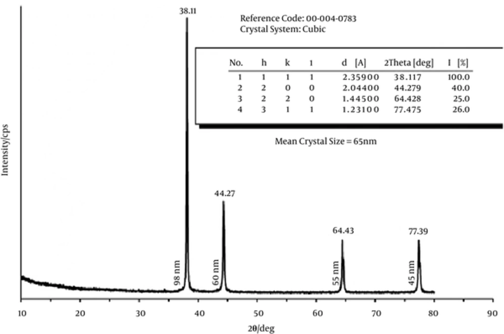
The silver nanoparticles (cubic crystals, 65 nm) used for the obtaining of Ag/RGO nanocomposite were prepared according to a known chemical reduction method (17, 18) by adding an excess of the reducing agent, NaBH4 to AgNO3, following the next chemical equation:
AgNO3 + NaBH4 → Ag + 1/2H2 + 1/2B2H6 + NaNO3
Briefly, 10 mL AgNO3 (1 mM) were added to 30 mL cooled NaBH4, (2 mM) drop wise (1 drop per second). The color of the solution converted from yellow at the beginning of the addition to yellowish-brown with the increase of the silver concentration. Finally, few drops of NaCl (1.5 M) were added to precipitate the colloid. The solution was settled for 2 hours before the filtration. The filtrate was washed many times with distilled water and dried at 80°C for 2 hours. The successful obtaining of silver nanoparticles (with size of about 65 nm) was confirmed by XRD analysis (Bruker D8 Advance, Cu Ka radiation) (Figure 1).
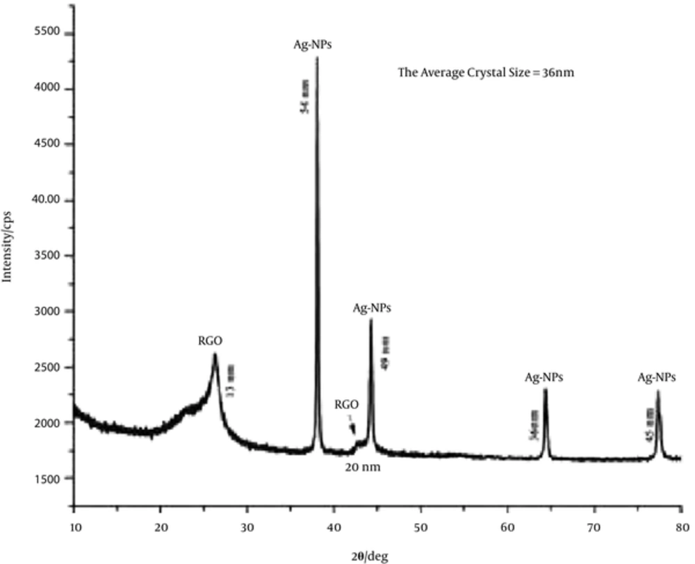
The RGO multi-layered sheets, used for preparation of Ag/RGO nanocomposites were the same that were employed in our former investigation; they were prepared by chemical exfoliation of purified natural graphite powder (99.9%, Alfa Aesar Co.) as it was already described (2). Ag/RGO nanocomposite was prepared by ultrasonic dispersion of AgNPs and RGO (1:2, weight ratio) in distilled water for 1 hour. The dispersion was then filtered and drayed at 80°C for 2 hours. The X-ray diffraction (Bruker D8 Advance, Cu Ka radiation) pattern of the AgNPs/ RGO (1:2, wt/wt) composition (Figure 2) shows the crystal phase of AgNPs and RGO that confirms the successful preparation of this nanocomposite. The average crystalline size of the obtained Ag/RGO nanocomposite, calculated from the diffraction line using Scherrer’s equation (19) was about 36 nm. The atomic emission spectral analysis (High Dispersion ICP-OES Prodigy spectrometer, Teledyne Leeman Labs, USA) of the Ag/RGO nanocomposite proved silver content of 15.63 wt%.
2.2. Preparation of Ag/RGO/SiO2 Nanocomposite
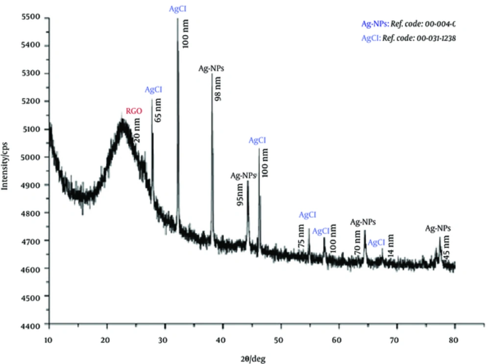
In trials, to decrease the aggregation of the Ag/RGO particles, Ag/RGO/SiO2 composite was prepared knowing that a silica matrix improves dispersion, reduces the agglomeration of nanoparticles, and acts as a carrier of other materials. The used in this investigation Ag/RGO/SiO2 composite was prepared by sol-gel technique, adding distilled water dispersions of both: AgNPs and of RGO, to a TEOS solution followed by gelation and drying to obtain Ag/RGO/SiO2 powder. Briefly, 0.38 g AgNPs were dispersed in 3.3 mL distilled water; 0.77 g RGO were dispersed in 3.3 mL distilled water; 10.1 mL TEOS were added to 7.9 mL ethanol. After, both the Ag NPs and RGO dispersions were added to the TEOS ethanol solution under stirring for 1 hour at 80°C. Few drops of HCl were added in order to obtain the gel, the last one dried at 80°C, for 2 hours, under vacuum to produce Ag/RGO/SiO2 powder. XRD patterns of the powder (Bruker D8 Advance, Cu Kα radiation) confirmed the obtaining of the Ag/RGO/SiO2 composite (Figure 3): the amorphous hollow at 2θ = 22.6° is connected to existing of amorphous silicate; there are different crystal phases such as RGO nanoparticles, AgNPs (Ref. code: 00-004-0783) and AgCl nanocrystals (Ref. code: 00-031-1238). By Scherrer’s equation (19), the average crystal size, calculated from the corresponding diffraction line, was of about 77 nm, 75 nm, and 20 nm for the AgNPS, AgCl, and RGO, respectively. The silver content in the nanocomposite was of 11.67 wt% (atomic emission spectral analysis, High Dispersion ICP-OES Prodigy spectrometer, Teledyne Leeman Labs Comp., USA).
2.3. Preparation of Collagen/(Ag/RGO) and Collagen/(Ag/RGO/SiO2) Composites
Type I fibrillar collagen gel with concentration of 2.64 wt% was extracted from calf hide using previously described technology (20). The concentration of the collagen gel was adjusted at 1% and pH at 7.3 (that of the physiological medium) using 1M sodium hydroxide and antimicrobial agent (powder of Ag/RGO or Ag/SiO2/RGO) in 2:1, 2:0.8, 2:0.6, 2:0.4, or 2:0.2 ratios (wt/wt) was added. As prepared collagen composites were cross-linked with 0.5% glutaraldehyde (to dry collagen) at 4°C for 24 hours and then lyophilized at -40°C to obtain porous (sponge) material using a Martin Christ freeze-dryer for 48 hours, as it was previously described (21, 22). Test samples with a diameter of 9 mm were prepared after that from each composite.
2.4. SEM Observations
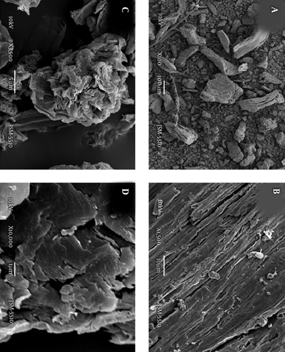
JEOL SEM, model JSM-5510, Japan apparatus was used to observe the morphological features of the studied antimicrobial agents and Porous Collagen Composites. The samples were gold-sputtered coated and viewed in the second electron mode with field emission gun.
2.5. Antimicrobial Activity Testing of Collagen/(Ag/RGO) and Collagen/(Ag/RGO/SiO2) Composites
The following microbial strains, 3 Gram negative bacteria: Salmonella enterica 2333, E. coli 264, P. putida 1090; 1 fungus, Candida lusitaniae 74 - 4, and 3 Gram positive bacteria: L. innocua, S. epidermidis 3486, and B. cereus were used in this investigation, all provided from the national bank of microorganisms and cell cultures (NBIMCC), Bulgaria. They were cultured in the most suitable for each one media. E. coli 264 and S. enterica 2333 were grown in nutrient broth (NB Conda, Spain) at 37°C and 180 rpm for 18 hour. B. cereus 1095 and S. epidermidis 3486 were propagated in nutrient broth and C. lusitaniae 74 - 4 in YGC, respectively at 30°C and 120 rpm. Pseudomonas putida 1090 (ATCC 12633) was cultivated in synthetic liquid medium (ISO10712) at 22 - 23°C and 180 rpm for 12 hour. Microbial density of 0.5 - 0.8 was determined according to McFarland. The aliquots of 100 µL microbial suspension was randomly spread on solid medium (Nutrient agar - NA and YGC agar) and discs of investigated material were put on them. The plates were left for 20 hours at 4 - 6°C to afford diffusion of the nanoparticles and after that cultivated for 24 hours at 37, 30 and 24°C, respectively. The formed sterile zones around the disks samples were measured in mm (± 0.5).
2.6. Antimicrobial Activity Testing of Ag/RGO and Ag/RGO/SiO2 Dispersions
The antimicrobial activity testing of Ag/RGO and Ag/RGO/SiO2 dispersions was included in this investigation since it is known that some GO and RGO in dispersions do not demonstrate antimicrobial activity. The antimicrobial activity of distilled water Ag/RGO and Ag/RGO/SiO2 dispersions with different concentrations, of 0.1 - 5.0 mg/mL, were tested against 5 bacteria: E. coli, B. cereus, S. holeresius, P. aeruginosa, and S. epidermidis as it is described above.
2.7. Cytotoxicity Evaluation
2.7.1. Eukaryotic Cells
Cytotoxicity was evaluated against 3 type eukaryotic cells: model osteoblast cells, MG-63; 3T3 fibroblasts, MDCK II kidney epithelial cell line, all provided from the NBIMCC, Bulgaria. The used eukaryotic cells were maintained at standard conditions in a humidified atmosphere with 5% СО2, at 37°C, in the corresponding medium F12 or DMEM (SIGMA), supplemented with 10% FBS (BioWhittaker TM) and 1% (v/v) antibiotic-antimitotic solution (penicillin 100 U/mL, streptomycin 100 ug/mL and amphotericin B 0.25 ug/mL BioWhittaker ТМ). For assessment of cytotoxicity, the materials were embedded in 96 wells-plates and hydrated with 200 μL culture medium for 12 - 24 hours. Then cells were seeded at a concentration of 1 × 10⁵ cells/mL. After 24 hours, the cytotoxicity was evaluated by crystal violet test and the cell morphology was observed by bright field microscopy.
2.8. Crystal Violet Staining and Microscopic Observation
Crystal violet staining was performed with some modifications (23), brifely: The residual cell monolayer was washed with phosphate-buffered saline (PBS) and fixed with 4% paraformaldehyde in PBS for 15 minutes. After that, plates were washed with water and 200 ul 0.1% crystal violet solutions were added to every well. After 20 minutes of incubation at room temperature, the plates were washed with water and the protein-bound dye (which is corresponding to the cell number) was extracted with 200 µL 10% acetic acid. The values of optical density were read on a micro plate reader (EPOCH UV/VIS Spectrometer) at 570 nm wavelength and the number of vital cells was calculated as a percentage from their total amount. Invert microscope pictures were taken out using supplied with a digital camera DV-130, XDS-2A microscope, China.
3. Results
3.1. SEM Images
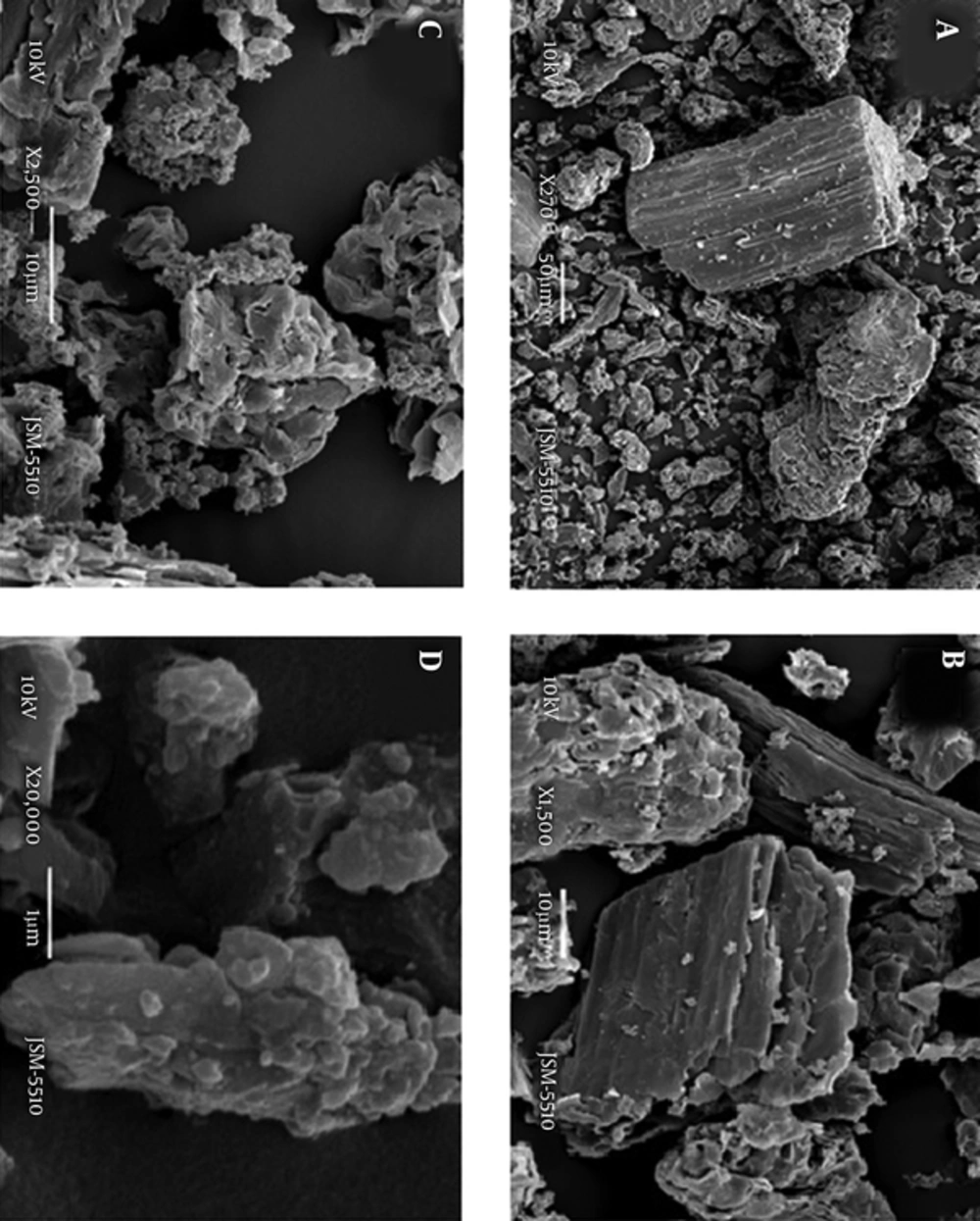
Figures 4 and 5 depict the morphology of the used as antimicrobial agents both, Ag doped RGO (Ag/RGO) and Ag/RGO/SiO2, respectively. Their aggregation as well as the relatively good dispersion of the AgNPs on the RGO sheets is clearly seen.
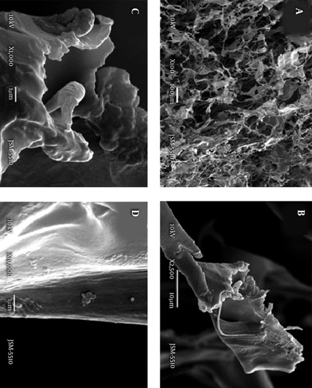
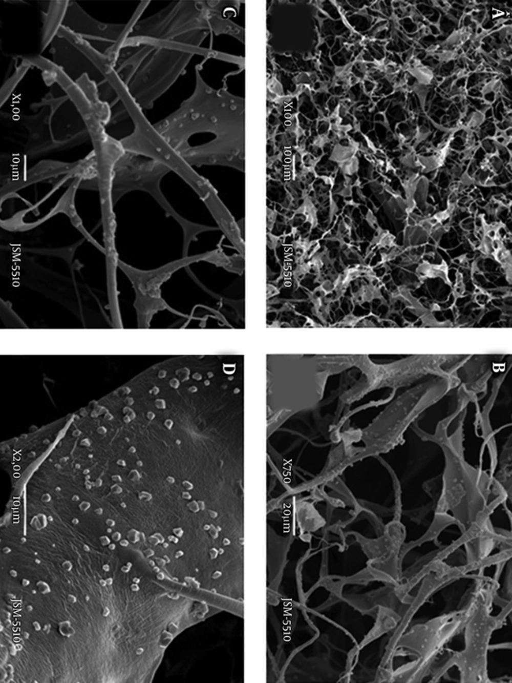
Figures 6 and 7 show the porous structure of Collagen/ (Ag/RGO) and Collagen/(Ag/RGO/SiO2) composites, respectively, in both cases at weight ratio collagen: antimicrobial agent of 2:1. The pictures of the Collagen/(Ag/RGO) and Collagen/(Ag/RGO/SiO2) composites at lower concentrations of the antimicrobial agent (Ag/RGO or Ag /RGO/SiO2), namely at 2:0.8, 2:0.6, and 2:0.4, wt/wt were similar and therefore they are not presented here. Figure 6A depicts an open and interconnected relatively homogeneous porous structure of the Collagen/(Ag/RGO) composite whereas the pictures at higher magnification (Figure 6B - D) show how the Ag/RGO aggregates are entrapped in the collagen matrix. It seems that a part of the Ag/RGO particles are coated by collagen. Figure 7A depicts the relatively homogeneous, open and interconnected pore structure of the Collagen/(Ag /RGO/SiO2) composite, similar to that of the Collagen/(Ag/RGO) composite. A comparison of the pictures in Figure 7B - D to those in Figure 6B- D are both taken out at higher magnification, indicates significant differences in the dispersion of Ag/RGO/SiO2 as compared to Ag/RGO particles in the collagen matrix: the Ag/RGO/SiO2 aggregates are smaller and do not coated by collagen. This confirms the expected improved dispersion due to added silane matrix in Ag/RGO/SiO2.
3.2. Antimicrobial Activity of Water Dispersions of Ag/RGO and Ag/RGO/SiO2
It is known that some GO and RGO dispersions do not demonstrate antimicrobial activity. Therefore the antimicrobial activity of Ag/RGO and Ag/RGO/SiO2 water dispersions was tested. The experimental results are presented in Table 1. It is evident that the water dispersions of Ag/RGO at concentrations of 0.1 - 1.0 mg/mL demonstrate insignificant antimicrobial activity, lack of activity against 3 Gram-negative (E. coli, P. aeruginosa and S. holeresius), and 1 Gram-positive (S. epidermidis bacteria and well expressed activity against B. cereus only at concentrations of 0.5 - 5.0 mg/mL. The water dispersions of Ag/RGO/SiO2 demonstrate concentration dependent, specific to the test bacteria antimicrobial activity: the highest and well expressed one against S. epidermidis in the whole concentration interval (0.1 - 5.0 mg/mL), with a sterile zone of 17.0 mm at concentration of 5.0 mg/mL. The Ag/RGO/SiO2 water dispersions, at concentrations above 2.5 mg/mL, demonstrate well expressed activity against all 5 test bacteria, Gram-negative and Gram-positive (the last 2 rows in Table 1). This indicates the higher antimicrobial activity of Ag/RGO/SiO2 as compared to RGO water dispersions (24) (Table 1), the last ones demonstrating significant antimicrobial activity at concentrations above 50 mg/mL.
| Antimicr agent. | Conc., mg/mL | E. coli | P. aerug. | S. holer. | S. epid. | B. cereus |
|---|---|---|---|---|---|---|
| Ag/RGO | 0.1 | 0 | 0 | 0 | 0 | 0 |
| 0.5 | 0 | 0 | 0 | 0 | 6.0 | |
| 1.0 | 0 | 0 | 0 | 0 | 8.5 | |
| 2.5 | 0 | 0 | 0 | 1.0 | 10.0 | |
| 5.0 | 0 | 0 | 0 | 2.5 | 11.5 | |
| Ag/RGO/SiO2 | 0.1 | 0 | 0 | 0 | 7.0 | 0 |
| 1.0 | 0 | 0 | 0 | 9.0 | 7.0 | |
| 2.5 | 9.5 | 9.0 | 13.0 | 10.0 | 11.5 | |
| 5.0 | 11.0 | 13.0 | 10.0 | 17.0 | 13.0 |
Antimicrobial Activity of Water Suspensions of Ag/RGO and Ag/RGO/SiO2 in the Concentration Range of 0.1 to 5.0 mg/mL
3.3. Antimicrobial Activity of Collagen/(Ag/RGO) and Collagen/(Ag/RGO/SiO2) Composites
The antimicrobial activity of the studied Collagen/(Ag/RGO) and Collagen/(Ag/RGO/SiO2) composites, tested against 3 Gram-negative and 2 Gram-positive bacteria, as well as against 1 fungus is presented in Table 2. It is evident that the doping of RGO with AgNPs increases the antimicrobial activity of the Collagen/(Ag/RGO) composites (Table 2, samples 1 - 4) as compared to the Collagen/RGO composites (24), (Table 2, samples 1 - 5): significant activity appears against E. coli and S. epidermidis (that lacks at Collagen/RGO composites) whereas the activity against the fungus C. lusitaniae is similar. In addition, the activity against B. cereus increases of about 3-folds. A comparison of samples 1 - 4 to samples 5 - 8 in Table 2 clearly demonstrates the additionally increased antimicrobial activity of the Collagen/(Ag/RGO/SiO2) composites compared to that of the Collagen/(Ag/RGO) composites: significant activity appears against two Gram-negative bacteria, S. enterica and P. aeruginosa; the activity against the Gram-negative bacterium, E. coli, as well as against the fungus C. lusitaniae and two Gram-positive bacteria, S. epiedermidis and B. cereus increases. This manifests a broad spectrum antimicrobial activity of the studied Collagen/(Ag/RGO/SiO2) composites, that is better expressed at the composites Collagen: Ag/RGO/SiO2 with weight ratios of 2:0.6 and above. The bactericidal effect of these Collagen/(Ag/RGO/SiO2) composites is comparable to that of the broad spectrum antibiotics and in addition, they possess antifungal activity.
| Sample, No. | Coll:AMA, wt:wt | Sterile Zoneb, mm | |||||
|---|---|---|---|---|---|---|---|
| S. enterica | P. aeruginosa | E. coli | C. lusitaniae | S. epidermidis | B. cereus | ||
| Collagen: Ag/RGO | |||||||
| 1 | 2:1.0 | 0 | 0 | 10.3 | 9.0 | 12.0 | 11.0 |
| 2 | 2:0.8 | 0 | 0 | 10.0 | 6.5 | 10.2 | 10.0 |
| 3 | 2:0.6 | 0 | 0 | 4.8 | 5.3 | 10.0 | 9.2 |
| 4 | 2:0.4 | 0 | 0 | 0 | 4.5 | ||
| 2.4 | 3.5 | ||||||
| Coll: Ag /RGO/SiO2 | |||||||
| 5 | 2:1.0 | 5.0 | 10.0 | 12.0 | 15.3 | 16.0 | 12.6 |
| 6 | 2:0.8 | 2.5 | 10.0 | 10.8 | 6.0 | 14.0 | 10.3 |
| 7 | 2:0.6 | 1.8 | 9.0 | 4.0 | 6.3 | 12.0 | 10.0 |
| 8 | 2:0.4 | 0.9 | 0 | 2.0 | 4.2 | 6.3 | 5.8 |
Antimicrobial Activity, Presented as a Sterile Zone, mm, of Collagen Sponges Loaded with Different Amounts of Antimicrobial Agents (AMA): Ag/RGO (Samples 1 - 4) or Ag/ RGO/SiO2 (Samples 5 - 8)a
3.4. Cell Viability
The large variety of possible potential applications of the studied new Collagen / (Ag/RGO) and Collagen/(Ag/RGO/SiO2) composites is the reason to investigate their cytotoxicity against 3 types of eukaryotic cells that are most often used in the tissue engineering, osteoblast, MG-63, fibroblast, 3T3, and kidney epithelial, MDCK II. The results of cytotoxicity evaluation are presented in Table 3.
| Eukaryotic Cells | Cell Viability, % | |||||
|---|---|---|---|---|---|---|
| Collagen/(Ag/RGO), wt/wt | Collagen/(Ag/RGO/SiO 2 ), wt/wt | |||||
| 2:1 | 2:0.8 | 2: 0.6 | 2:1 | 2:0.8 | 2:0.6 | |
| MG-63 | 39 ± 4 | 41 ± 4 | 50 ± 5 | 19 ± 1 | 17 ± 1 | 16 ± 3 |
| 3T3 | 51 ± 1 | 52 ± 1 | 56 ± 3 | 13 ± 1 | 19 ± 1 | 15 ± 2 |
| MDCK II | 44 ± 2 | 46 ± 3 | 64 ± 2 | 24 ± 2 | 20 ± 1 | 15 ± 2 |
Cristal Violet Assay of Eukaryotic Cells: Osteoblast, MG-63, Fibroblast, 3T3 and Kidney Epithelial, MDCK II on Collagen/ (Ag/RGO) and Collagen/ (Ag/RGO/SiO2) Composites
It is evident, that the viability of the used test eukaryotic cells is relatively good and dependent on the concentration of the antimicrobial agent, Ag doped RGO, in all cases it is the best at the its lowest concentration (Collagen: Ag/RGO = 2: 0.6, wt/wt). The viability of osteoblasts cells, MG-63 is good (living cells of above 50%) for Collagen/(Ag/RGO) composite, 2:0.6, wt/wt) and less for the composites containing higher amount Ag/RGO (Collagen: Ag/RGO = 2:0.8 and 2:1, wt/wt). The viability of the fibroblasts, 3T3 is better: the percentage of living cells is above 50% for all tested Collagen/(Ag/RGO) composites, including those with Collagen: Ag/RGO ratios above 2:0.6, respectively 2:0.8 and 2:1, wt/wt). Kidney epithelial cells, MDCK II also survive well on the tested Collagen/(Ag/RGO) composites. The living cells are above 50% for all samples with different Ag/RGO loading levels. Collagen: Ag/RGO of 2:0.6, 2:0.8 and 2:1, wt/wt. From Table 3, (the last 3columns of data), is evident that the viability of all test eukaryotic cells, osteoblast, fibroblast, and epithelial, on the Collagen/(Ag/RGO/SiO2) composites is significantly less, as compared to that on the above described Collagen/(Ag/RGO) composites at all concentrations of the antimicrobial agent (Collagen: Ag/RGO/SiO2 = 2:0.6, 2:0.8 and 2:1, wt/wt). The cell survival of 13% to 24% is far below IC 50 for all 3 types of eukaryotic cells that indicates a high cytotoxicity of these Collagen/(Ag/RGO/SiO2) composites.
3.5. Cell Morphology
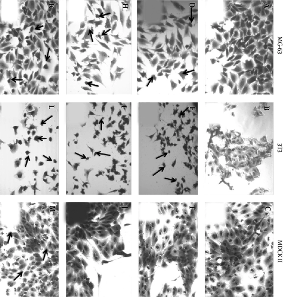
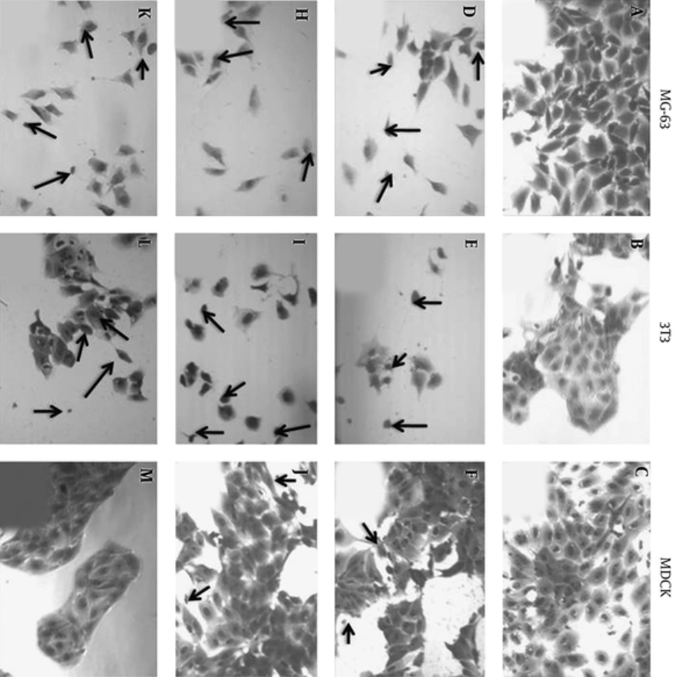
To understand more details about the cell viability, the morphology of the test eukaryotic cells, osteoblasts, fibroblasts, and epithelial cells were observed. The 3 types of control cells (MG 63, 3T3 and MDCK II, Figure 8A - C respectively), placed in the corresponding polystyrene plate wells, are well spread, tend to form monolayer, and have their typical morphology: spindle for the osteoblast, MG-63 (Figure 8A) and fibroblast, 3T3 (Figure 8B) and well expressed polygonal form for the epithelial, MDCK II cells (Figure 8C). Apoptotic cells or nuclei are not observed. The morphology of the osteoblasts, MG 63 on Collagen/(Ag/RGO) compounds with different Ag/RGO loading level is presented in Figure 8, Collagen: Ag/RGO = 2:1- (B), 2:0.8 - (H) and 2:0.6 - (K). No significant differences are observed in the MG 63 cell morphology on Collagen: Ag/RGO = 2:1 and 2:0.8 (Figure 8B and H respectively) but compared to the control MG 63 cells (Figure 8A), their number on the collagen samples is less and single cells are observed, a few of them are apoptotic. On the Collagen: Ag/RGO = 2:0.6, wt/wt sample, the morphology of MG 63 cells is similar to that of the control cells, excluding that some single cells are observed. A comparison of pictures (E), (I), and (L) to picture (B) in Figure 8 demonstrates that there is no significant alteration of the 3T3 cells morphology, but their number is less and some single and apoptotic cells are observed on the Collagen/(Ag/RGO) compounds. Contrary to the former 2 types, the epithelial cells form monolayer on all Collagen/(Ag/RGO) compounds (Figure 8F, J and M) even on those with the highest concentration of Ag/RGO (2:1 and 2:0.8, wt/wt) (Figure 8F and J, respectively), although some are slightly distant of each other. This observation of the cell’s morphology demonstrates that the 3 types test eukaryotic cells have a specific cell response to the studied antimicrobial Collagen/(Ag/RGO) composites.
Osteoblast, MG63 (A, D, H, K), fibroblast, 3T3 (B, E, I, L), and kidney epithelial, MDCK II (C, F, J, M); control (A, B, C, respectively) and on Collagen/(Ag/RGO) composites with weight ratios of Collagen: Ag/RGO at 2:1 (D, E, F, respectively), 2:0.8 (H, I, J, respectively) and 2:0.6 (K, L, M, respectively); black arrow - apoptotic cells; magnification 100x.
Collagen/(Ag/RGO) composites with different Collagen: Ag/RGO ratios, wt/wt: 2:1 (D, E, F, irrespectively), 2:0.8 (H, I, J, irrespectively) and 2:0.6 (K, L, M, irrespectively); black arrow -apoptotic cells, magnification 100x.
Similar comparative observation of the morphology of the same eukaryotic cells, MG 63, 3T3, and MDCK II on Collagen/(Ag/RGO/SiO2) composites demonstrated significant differences: the control cells present the typical morphology of osteoblast, fibroblast, and epithelial cells, respectively (Figure 9A - C); on the Collagen/(Ag/RGO/SiO2) samples, their morphology is altered, depending on the concentration of the antimicrobial agent, Ag/RGO/SiO2 (Figure 9D - M). A comparison of the pictures in Figure 9D, H and K to the picture in Figure 9A demonstrates the less number of MG 63 cells, among them some single and some apoptotic (marked by black arrows). The fibroblasts, 3T3, still form cell aggregates and monolayer on the Collagen/(Ag/RGO/SiO2) with lowest Ag/RGO/SiO2 loading level (Collagen: Ag/RGO/SiO2 = 2:0.6 wt/wt), Figure 9L whereas on the samples with higher Ag/RGO/SiO2 loading level (Collagen: Ag/RGO/SiO2= 2:0.8 and 2:1, wt/wt), Figure 9I and Figure 9E, respectively) single cells are observed, some apoptotic among them (marked by black arrows). The epithelial cells, MDCK II, form monolayer on all Collagen/(Ag/RGO/SiO2) compounds (Figure 9F, J and M), similar to those of the control epithelial cells (Figure 9C), in spite of some increase in the distance between the cells and appearance of few apoptotic cells. It is evident that the silane matrix in the Ag/RGO/SiO2 antimicrobial agent makes the studied Collagen/(Ag/RGO/SiO2) composites toxic to osteoblast, fibroblast, and epithelial cells as it was indicated also by crystal violet test.
Osteoblast, MG-63 (A, D, H, K), fibroblast, 3T3 (B, E, I, L), and kidney epithelial, MDCK II (C, F, J, M); control (A, B, C, respectively) and on Collagen/(Ag/RGO/SiO2) composites with different Collagen:Ag/RGO/SiO2 ratios, wt/wt: 2:1 (D, E, F, respectively), 2:0.8 (H, I, J, respectively) and 2:0.6 (K, L, M, respectively); black arrow - apoptotic cells; magnification 100x.
4. Discussion
4.1. Antimicrobial Activity of Ag/RGO and Ag/RGO/SiO2
The mechanism of antimicrobial action of RGO, Ag/RGO, and Ag/RGO/SiO2 is not fully understood. However, a relationship between RGO physicochemical properties (number of sheets and particles size) is known. The AgNPs size is critical for the antimicrobial activity. Their aggregation leads to a decrease of the antimicrobial efficiency. To avoid this problem, attempts are made for a decoration of RO and RGO sheets by AgNPs. However, the RGO sheets tend to aggregate themselves. In this study, the AgNPs are incrusted on micron size aggregates of RGO sheets, as it is seen from the SEM pictures in Figure 4. Similar is the distribution of the AgNPs particles in the Ag/RGO/SiO2 composites, which are observed as micron size aggregates in the SEM pictures (Figure 5). More fine dispersion in the water medium of the Ag/RGO/SiO2 particles compared to those of the Ag/RGO can be a reason for the better expressed activity in the first case.
4.2. Antimicrobial Activity of Collagen/(Ag/RGO) and Collagen/(Ag/RGO/SiO2) Composites
Both, Collagen/ (Ag/RGO) and Collagen/(Ag/RGO/SiO2) composites were prepared by sol-gel cryogen drying at -40°C. No chemical interactions under these conditions can be expected. It is possible that some AsNPs from the antimicrobial agents, both, Ag/RGO and Ag/RGO/SiO2, to be deliberated and to be dispersed in the collagen gel during the mixing before the cryogen drying. As demonstrated by SEM observations, Ag/RGO (Figure 6) and Ag/RGO/SiO2 (Figure 7) aggregates are relatively homogeneous dispersed and wrapped in the porous collagen matrix. Some Ag/RGO particles are included in the matrix collagen as submicron size particles (Figure 6C, D). As it was expected, the silane in the Ag/RGO/SiO2 composites improves its dispersion in the matrix collagen, as it is clearly seen in Figure 7C and D. This is a pre-requisite of a higher antimicrobial activity. The silver concentration of Ag/RGO of 15.63 wt% is higher than that of Ag/RGO/SiO2 of 11.76 wt%, (as proved by the elemental analysis) but the antimicrobial activity of Ag/RGO/SiO2 is higher than that of Ag/RGO. The antimicrobial activity of Collagen/(Ag/RGO/SiO2) composites is also higher than that of Collagen/(Ag/RGO). This indicates that the effect is connected to the silane matrix of the Ag/RGO/SiO2 for which is known to assist the dispersion. The better dispersion of Ag/RGO/SiO2 in distilled water is the reason for its higher antimicrobial activity (see Table 1). The higher antimicrobial activity of the Collagen/(Ag/RGO/SiO2) compared to that of Collagen/(Ag/RGO) composites, Table 2 is due to the better dispersion of the Ag/RGO/SiO2, as it is depicted in Figrue 7. These results prove the importance of the antimicrobial agent dispersion (24).
The 3 Gram-negative bacteria, S. enterica, P. aeruginosa, and E. coli have similar short rod body with flagella and similar cell envelope but only E. coli is sensitive to the Collagen/(Ag/RGO) composites. It could be suggested that both S. enterica, P. aeruginosa have some efflux mechanisms for silver but this mechanism could be blocked in the used E. coli strain. This is probably the explanation of the different sensitivity of these 3 Gram-negative test bacteria. The well-known differences of the cell wall structure at the different microbial strains can explain the different activity of the Collagen/(Ag/RGO/SiO2) against the used test microbial species (Gram-negative and Gram-positive bacteria and fungus).
4.3. Cytotoxicity
The Collagen/(Ag/RGO/SiO2) composites demonstrate higher toxicity to the test eukaryotic cells (osteoblast, fibroblast and epithelial) as compared to the Collagen/(Ag/RGO) composites due to the presence of silane in the Ag/RGO/SiO2. The morphology of the Collagen/(Ag/RGO/SiO2) composites clearly demonstrates a much finer dispersion of Ag/RGO/SiO2 (Figure 7) aggregates compared to those of Ag/RGO (Figure 6); in the first case they are smaller and the size of some of them go down to submicron (Figure 7). The toxic effect of SiO2 NPs and size-dependent cytotoxicity of mono-disperse silica nanoparticles to living cells are well known although its mechanism is insufficiently understood (25-27). A possible deliberation of some silica nanoparticles during the preparation of the Collagen/(Ag/RGO/SiO2) composites, together with the better dispersion of the Ag/RGO/SiO2 particles in the collagen matrix could be the explanation of their increased toxicity to the selected test eukaryotic cells, that are often used in the tissue engineering and originate from 3 different type tissues. The epithelial cells needs in cell lamina for their growth, whereas the osteoblasts and fibroblasts produce fibronectin themselves. The studied collagen composites mimic the natural lamina (their basic protein is collagen) that is probably the reason for the better adhesion and respectively higher viability of the epithelial cells on the studied collagen composites. As it was expected, the eukaryotic cells’ respond is specific and dependent on the composition of the studied collagen biomaterials.
4.4. Conclusions
New antimicrobial Collagen/(Ag/RGO) and Collagen/(Ag/RGO/SiO2) porous composites were developed with interesting antimicrobial activity. The last one is aided not only by the silver nanoparticles but also by the silane matrix.
The biological activity of the new collagen biomaterials is cell specific and dependent on the concentration of the antimicrobial agent, Ag/RGO or Ag/RGO/SiO2.
The broad spectrum antimicrobial activity of the Collagen/(Ag/RGO/SiO2) composites, well pronounced to Gram-negative, Gram-positive bacteria, and fungi is similar to that of the broad spectrum antibiotics.
Silver doping of RGO increases the biological activity of Collagen/(Ag/RGO) composites as compared to Collagen/RGO ones; the well pronounced activity against E. coli, S. epidermidis, B. cereus and fungus C. lusitinae, depending on the Ag/RGO concentration is combined with low cytotoxicity to eukaryotic cells at weight ratio Collagen:Ag/RGO of 2:0.6.
The silane matrix of the Ag/RGO/SiO2 increases sharply the antimicrobial activity of the Collagen/(Ag/RGO/SiO2) composites and cytotoxicity to osteoblasts, fibroblasts and epithelial cells, because of its better dispersion as well as due to a possible deliberation of silver and silica nanoparticles.
The novel Collagen/(Ag/RGO) and Collagen/(Ag/RGO/SiO2) composites with their specific and adjustable bioactivity are promising antimicrobial collagen biomaterials that widen the assortment of such for variety biomedical applications.
