1. Introduction
Kawasaki disease (KD), also known as the mucocutaneous lymph node syndrome, is an acute febrile illness of childhood and medium-sized vasculitis that particularly affects the coronary arteries. KD is the leading cause of acquired heart disease in children, and approximately 20% of untreated cases develop coronary artery aneurysms (1). The KD often diagnoses in children aged between six months to five years, and typically occurs with remitting fever lasting for at least five days, polymorphous exanthema, bilateral non-exudative conjunctival injection, non-suppurative cervical lymphadenopathy, erythema of the oral mucosa with strawberry tongue and red lips, extremity erythema, and edema (2). The most effective treatment for KD in its acute phase is 2 g/kg of intravenous immunoglobulin (IVIG) as a single infusion and high-dose aspirin. Although extreme irritability is a common manifestation among infants, neurological involvement, such as aseptic meningitis, hemiplegia, cerebral infarction, ataxia, seizures, cranial nerve palsies, and focal encephalopathy, are late symptoms and are uncommon. Neurological manifestations usually occur as a result of vasculitis or IVIG administration (3). In the current study, we discussed an atypical case of KD with unusual onset, which primarily occurred with aseptic meningitis.
2. Case Presentation
An eight-year-old girl was admitted to Besat Hospital complaining of fever since seven days ago. The patient had frequent vomiting and headache during the past three days. Past medical history was unremarkable. On physical examination, arterial blood pressure was 90/65 mmHg. The heart rate was 120 beat/min. The respiratory rate was 20/min, and the temperature (axillary) was 38°C. The heart, lungs, and abdominal examinations were normal. There was no cyanosis, mucosal erythema, tonsillar enlargement, neck rigidity, cervical lymphadenitis, edema, and limb movement limitation. Chest X-ray was routine, and the results of other initial laboratory investigations were as follow: urinalysis (U/A) was routine. Erythrocyte sedimentation rate (ESR) was 36 mm/h. C-reactive protein (CRP) was negative, and according to the complete blood count (CBC) hemoglobin, platelet, and white blood cells (WBC) were 13 g/dL, 263000/mm3, and 7100/mm3 (70% neutrophil), respectively.
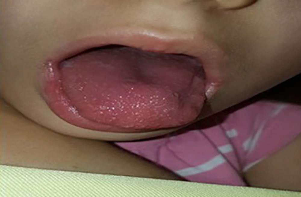
The fever, headache, and vomiting were indicating meningitis; therefore, lumbar puncture (LP) was performed. Besides, cerebral spinal fluid (CSF) analysis revealed routine glucose and protein levels, and elevated white blood cells, the results were as follow: white blood cells were 90/mm3 (41% PMN and 59 % Lymphocyte); glucose level was 67 mg/dL, and protein level was 60 mg/dL. To treat CSF pleocytosis, and elevated ESR, ceftriaxone, and vancomycin were administered. But, after 48 hours, no improvement was observed, and the CSF culture revealed a negative result. Hence, the patient considered as a case of aseptic meningitis, and antibiotics were discontinued. After two days, the patient developed a strawberry tongue as well as a bilateral mild conjunctival injection (Figure 1).
According to the atypical Kawasaki algorithm published by the American Heart Association (AHA), complementary laboratory tests and echocardiography were performed, because the patient had two clinical signs of KD (4). Results of the second laboratory investigation were as follow: routine U/A; ESR was 47 mm/h; negative CRP; HB = 12.7 g/d; PLT = 465000/mm3; WBC = 11600/mm3 (86% neutrophil). Also, serum albumin, aspartate aminotransferase (ALT), alanine aminotransferase (AST) levels were within their normal ranges. The echocardiography revealed a dilated left coronary artery, and the second echocardiography after two days indicated coronary artery aneurysm. Aspirin and IVIG were administered regarding atypical KD diagnosis according to the AHA guide (4). After 24 hours, the fever vanished, and typical desquamation of the fingertips developed.
3. Discussion
The criteria to diagnose KD are published by the AHA or the Japanese Kawasaki Disease Research Committee (5, 6). Although accurate criteria are available, the diagnosis of the KD is still challenging, particularly among children who do not fulfill the criteria and have atypical manifestations of KD. Atypical KD accounts for 15% - 20% of all KD cases, and occurs more among children aged > 5 years or < six months (7). Delayed diagnosis and treatment of patients increase the risk of coronary complications, particularly in the atypical form of KD, which makes the condition more challenging and more critical.
Extreme irritability is a common neurologic manifestation among infants. Other neurological symptoms of KD include aseptic meningitis, meningoencephalitis, subdural collection, ataxia, and sensorineural hearing loss. One percent of KD patients develop neurological complications, and five percent of them experience aseptic meningitis (8, 9). According to a study conducted by Sedighi et al. (10), neck stiffness occurs in less than five percent of Iranian children with KD. Ghandi et al. (11) reported that about four percent of Iranian patients develop aseptic meningitis. Aseptic meningitis mostly occurs during the acute phase of disease concurrent with other principal manifestation and usually is associated with increased intracranial pressure (ICP) (12). The case that is reported in the current study had a history of headache and vomiting, consistent with raised ICP. The patient’s response to antibiotics was not satisfying. The CSF culture was negative, hence, the diagnosis of aseptic meningitis became more pronounced.
The CSF profile of patients with acute KD can be misdiagnosed with that of patients with viral meningitis. The CSF profile of viral meningitis vary greatly, and commonly is manifested as a mononuclear cell predominant pleocytosis, routine CSF glucose, and mildly elevated CSF protein. Interestingly, the elevated levels of CSF protein are less common in patients with KD (13). The CSF profile of the patients diagnosed with KD commonly includes increased WBC (lymphocyte-predominant), routine glucose, and normal protein. Similarly, the CSF profile of our patient indicated elevated levels of WBC with a predominance of lymphocytes, routine glucose, and routine protein. Strawberry tongue and bilateral mild conjunctival injection shifted our attention to the atypical KD with aseptic meningitis. However, aseptic meningitis is very unusual in patients with KD, and only a few cases are reported so far, including the case reported by Salameh and the one reported by Attia (14, 15).
3.1. Conclusions
Aseptic meningitis is an uncommon manifestation of KD that develops late in the disease course, so that the diagnosis of KD is based on other clinical symptoms. Also, aseptic meningitis can occur as a complication of IVIG administration. Our case was primarily admitted due to aseptic meningitis, so the diagnosis of atypical KD was challenging. What makes this case interesting is aseptic meningitis as the first manifestation of the KD. Other symptoms, including strawberry tongue and coronary system ectasia, were developed following the treatment of the aseptic meningitis.
As the final conclusion in cases that patients with KD have patterns of aseptic meningitis in CSF analysis, but ESR or CRP are significantly high, and there is significant PMN dominant leukocytosis, clinicians should consider atypical KD with aseptic meningitis.

