1. Background
Biliary atresia (BA) is a severe cholestatic disease of infancy causing neonatal jaundice and is characterized by fibrous obliteration of biliary tree (1, 2). There is a variable incidence of 1 in 5000 to 1 in 19000 live births around the world with a slight female predominance. In untreated cases, it would be fatal by 2 years of age (1-3). Various etiologic mechanisms for BA have been postulated, including intrauterine or perinatal viral infection, genetic mutations, abnormal ductal plate remodeling, and immunologically mediated inflammation. However, the exact cause of BA remains unknown (3). As a neonatal cholestatic disorder, the cardinal signs and symptoms of BA are jaundice, clay - colored stool, and hepatomegaly (4-6). The liver becomes enlarged, firm, and green early in the course of BA. Microscopically, the biliary tracts contain inflammatory, and fibrous cells surrounding miniscule ducts (5). Later, the liver parenchyma becomes fibrotic and shows signs of cholestasis as many chronic liver disorders. Early management of the underlying etiology can postpone and even limit the progression of fibrosis, however, it cannot always prevent continuation to cirrhosis. Currently, liver biopsy is the gold standard for evaluation of the severity of liver fibrosis and cirrhosis. This procedure is invasive and can be associated with complications such as severe abdominal pain, bile leakage, hemobilia, and intraperitoneal hemorrhage, the last of which has an associated mortality rate of up to 0.5% (7). In addition, sampling error, inter- and intraobserver variability in interpretation, and inability to evaluate progression and regression of fibrosis limits the usefulness of this procedure (8-10). Furthermore, when repeated examination is needed, the acceptance of the procedure is limited by the patients. Less invasive techniques for determining the degree of liver fibrosis have been developed in adults (11). These methods have also been applied in pediatric patients with chronic liver diseases (12, 13). Among the serological indicators, the aspartate aminotransferase to platelet ratio index (APRI) is considered a good estimator of hepatic fibrosis and is derived from routine hematological and biochemical tests (12). This index has been proposed to assess BA patients during the last several years and potentially could be used to decrease the number of liver biopsies (14-16). APRI has been extensively studied in viral hepatitis B and C and its calculation is based on the formula by Wai et al., (17) [AST/upper limit of normal (ULN)]/platelet count (expressed as platelets × 10 9/L) × 100]. The laboratory results of AST and platelets count, which are performed within 1 week before the operation, are used for the analysis, and if more than one set of laboratory data is available, results closest to the date of operation are chosen (17-20). For the proper diagnosis and treatment of chronic hepatic disorders, the application of non - invasive procedures and avoidance of several liver sampling as much as possible are essential (21-25).
Few studies have been conducted on the relationship between biochemical and hematologic markers and indices of fibrosis in children with BA (26, 27). When assessing the value of APRI in liver fibrosis among BA patients, the results are controversial. We conducted the present study among children with neonatal cholestasis and preoperative diagnosis of BA, who underwent intraoperative wedge liver biopsy, to evaluate the role of aspartate aminotransferase to platelet ratio in diagnosis of liver fibrosis and its prognosis after surgery.
2. Methods
A retrospective, cohort study was conducted by studying the medical records of children with neonatal cholestasis and preoperative diagnosis of BA who underwent intraoperative liver biopsy in Mofid Children’s Hospital in a 10 year period from January 2008 to December 2016. In this study, an author - made checklist was used. This checklist consisted of 2 parts; the 1st part included the demographic characteristics (age, gender, age at diagnosis, and mother’s gestational age) and the 2nd part included the information related to performed tests such as hepatobiliary ultrasonography, nuclear scanning, intraoperative cholangiography, and serologic markers of aspartate aminotransferase (AST) and platelet. According to data available in the archives of Mofid Children’s Hospital, a total of 100 patients with preoperative diagnosis of BA entered the study. Patients aged less than 2 months were selected for the study, those patients who were simultaneously afflicted with heart disease, acute febrile illness, or skin rash were excluded from the study due to their possible impact on the final AST and platelet results.
The present study was approved by the Ethics Committee of Shahid Beheshti University of Medical Sciences. The obtained data were analyzed by SPSS software version 18 (SPSS Inc., Chicago, USA) using descriptive statistics (mean and standard deviation) and inferential statistics (Chi - square and ANOVA). Correlation was evaluated by the Spearman correlation coefficient. All of the possible cut - off values were associated with the probability of a true positive (sensitivity) and a true negative (specificity). P values less than 0.05 were regarded as statistically significant.
APRI for each patient was calculated using the test results and the formula suggested by Wai et al. (17). Upper normal limit for AST, based on the majority of previous studies, was set at 40 IU/L.
Liver sections of the enrolled patients were retrieved from the sample bank in the department of pathology and reviewed blindly by 2 experienced pediatric pathologists. Liver wedge tissue samples were obtained during surgical exploration or portoenterostomy. Sections were stained with hematoxylin and eosin (HE) as well as Masson’s trichrome stain. The severity of hepatic fibrosis was interpreted according to the meta - analysis of histological data in viral hepatitis (METAVIR) staging system, (24), which includes 5 stages of fibrosis: F0: no scarring (no fibrosis), F1: minimal scarring (mild fibrosis), F2: scarring has occurred and extends outside the areas in the liver that contains blood vessels (moderate or significant fibrosis), F3: bridging fibrosis is spreading and connecting to other area that contains fibrosis (severe fibrosis), and F4: cirrhosis or advanced scarring of the liver (cirrhosis). Scoring F0 - F1 was considered as no/mild fibrosis, F2 - F3 as significant fibrosis, and F4 as cirrhosis.
3. Results
A total of 100 patients were evaluated in this study according to the inclusion and exclusion criteria. There were 64 patients that were male (64%). A total of 50 cases had a BA, 10 cases total parenteral nutrition (TPN) - associated cholestasis, and 40 cases had other diagnosis.
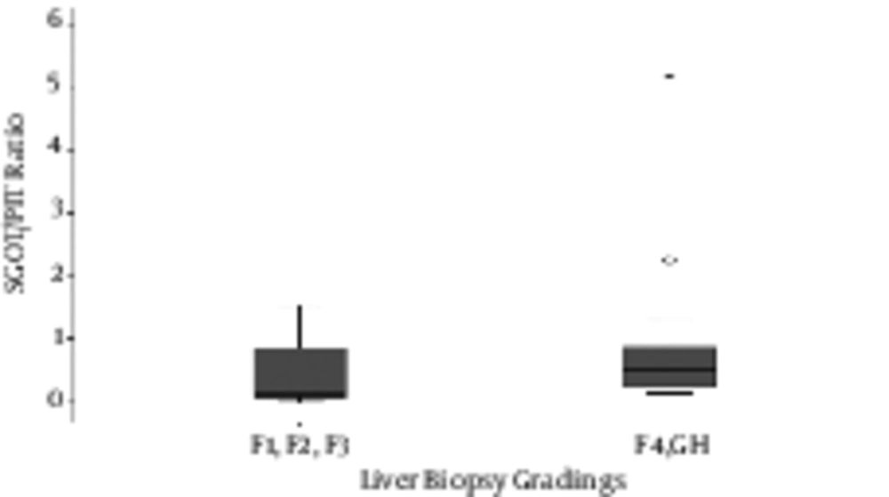
Patients’ delivery was at a minimum of 29 weeks and a maximum of 40 weeks. The average gestational age was 35.4 ± 5.84 weeks. The average weight of the patients was 2422.4 ± 937.3 gr. Based on analytical examinations, gestational age was significantly related to the final diagnosis of the disease (P = 0.011) (Table 1). Cholescintigraphy (HIDA) scan and the final diagnosis were also strongly correlated (p < 0.001) (Table 2). In addition, cholangiography findings showed significant relationship with the final diagnosis (p < 0.001) (Table 3). With a cut off value of aspartate aminotransferase to platelet ratio of 0.95, 25 patients (50%) with biliary atresia were in the group of significant fibrosis (F4, cirrhosis) and the others were in the non - significant fibrosis group (p value = 0.021) (Figure 1).
| Parameters | All Patients | Bailliary Atresia | ||||
|---|---|---|---|---|---|---|
| Mean ± SD | Pearson Correlation | P Value | Mean ± SD | Pearson Correlation | P Value | |
| Birth weight | 2430.4 ± 937.8 | r = - 0.91 | P = 0.402 | 2551.6 ± 842.6 | r = 0.157 | P = 0.287 |
| Gestational age | 35.5 ± 3.61 | r = - 0.195 | P = 0.072 | 36.1 ± 3.26 | r = 0.159 | P = 0. 280 |
| INR | 369.76 ± 1684.7 | r = - 0.004 | P = 0.976 | 2.20 ± 5.67 | r = - 0.106 | P = 0.520 |
| Parameters | All Patients | Billiary Atresia |
|---|---|---|
| Splenomegaly | (F 1 , 84 = 0.400 , P = 0.529) | (F 1 , 46 = 11.463 , P = 0.001) |
| Icter | (F 1 , 84 = 0.028 , P = 0.867) | (F 1 , 46 = 5.230 , P = 0.027) |
| Absent or constracted GB | (F 3 , 82 = 1.929 , P = 0.131) | (F 1 , 46 = 0.003 , P = 0.954) |
| Sonography | (F 1 , 84 = 3.731 , P = 0.057) | (F 1 , 46 = 9.716 , P = 0.003) |
| Ascite | (F 1 , 84 = 0.518 , P = 0.474) | (F 1 , 46 = 0.702 , P = 0.406) |
| Colangiography | (F 1 , 64 = 1.511 , P = 0.224) | There are fewer than two groups for dependent variable sgot.plt. No statistics are computed. |
| Hida scan | (F 1 , 84 = 0.400 , P = 0.529) | There are fewer than two groups for dependent variable sgot.plt. No statistics are computed. |
| Parameters | All Patients | Billiary Atresia | |||
|---|---|---|---|---|---|
| Splenomegaly | Positive, 12 (11.9%) | Negative, 88 (87.1%) | Positive, 2 (4%) | Negative, 48 (96%) | |
| Icter | Positive, 82 (81.2%) | Negative, 18 (17.8%) | Positive, 46 (92%) | Negative, 4 (8%) | |
| Absent or constracted GB | Positive, 20 (19.8%) | Negative, 80 (79.2%) | Positive, 12 (6%) | Negative, 44 (88%) | |
| Ascite | Positive, 16 (15.8%) | Negative, 84 (83.2%) | Positive, 10 (20%) | Negative, 40 (80%) | |
| Sonography | Normal, 36 (35.6%) | Abnormal, 64 (63.4%) | Normal, 6 (12%) | Abnormal, 44 (88%) | |
| Colangiography | Patent duct, 22 (21.8%) | Obliterated duct, 48 (47.5%) | Study not performed, 30 (29.7%) | Obliterated duct 48 (96%) | Patent duct, 2 (4%) |
4. Discussion
There are some serological markers that can indicate the current state of liver damage. These markers, in combination with various types of biochemical data e.g. Hepatoscore, King’s score, APRI (aspartate aminotransferase - to - platelet ratio index) and Fibro test, are known as class 2 markers and are used in hepatology to describe the level of liver damage (11-12, 14-15). The APRI, as a surrogate marker of liver fibrosis, has been used in some liver diseases to indicate the current state of liver damage. As an example of a non-invasive biochemical marker, APRI is presumed to reflect the degree of intrahepatic fibrosis (14). It was developed by Wai et al., in 2003 and introduced into adult practice to monitor and evaluate liver disease progression, typically hepatitis C, and to reduce dependence on serial liver biopsies (18). In the field of pediatric hepatology this index was used in few studies for predicting the liver fibrosis. The aim of the present study was to calculate APRI at a time of presentation and relate it to operative findings and the degree of hepatic fibrosis. In a study conducted by Grieve et al., in King’s College hospital, UK, a total of 260 patients with biliary atresia were evaluated. In their APRI correlated with age, spleen size, and bilirubin. The cut - off value of APRI in this study was about 1.22 and showed a sensitivity of 75% and a specificity of 84% for macroscopic cirrhosis (25). Many conditions can affect the level of liver enzymes and platelet counts (26).
In our study, patients who were suffering from acute diseases with fever or skin rash were excluded due to their effect on 2 main variables of AST and platelet. Cardiovascular patients were also left out due to the effect on the laboratory results. In the study by Kim et al., only patients with skin rash and acute diseases with fever were excluded from the study. However, Li - Yung Yong excluded cardiovascular patients as well as those with less than 15 days of age (27). In this study thirty - five patients with biliary atresia were enrolled. Clinical outcomes of patients were significantly different between cirrhotic and non - cirrhotic group based on APRI. Therefore, they considered APRI as a useful tool for assessing the liver fibrosis without additional risks in patients with BA during postoperative follow - up care (27). In a large study in China performed by Yang et al., (28) medical records of 153 infants with biliary atresia were reviewed and the efficacy of APRI for diagnosis of liver fibrosis was assessed. In their study, APRI was significantly correlated with METAVIR scores. The mean APRI value was 0.76 in no/mild fibrosis group, 1.29 in significant fibrosis group (F2 - F3), and 2.51 in cirrhosis group (F4) (p < 0.001). In addition, in their investigation the elevated preoperative APRI predicted the jaundice persistence after portoenterostomy (29). In our investigation we reached the same result; higher APRI levels were seen in patients with significant fibrosis and the cirrhotic group. Furthermore, other variables such as gestational age, birth weight, positive CPR, the need for respiratory support, liver biopsy categorizations, HIDA scans, and cholangiography (based on patency of bile duct) were all associated with the final diagnosis. Chrysanthos et al., showed that when using the APRI alone, the stage of fibrosis is incorrectly classified in 40-65% of patients (29). The diagnostic accuracy of APRI was improved in a study by Lok et al., after incorporation of ALT and the international normalized ratio (INR) (30). Furthermore, the APRI was also found to be of high diagnostic accuracy in assessing the progression of fibrosis in post liver transplant patients (31). In another investigation by Shin et al., the severity of fibrosis was checked in 47 patients with biliary atresia by comparing transient elastography and liver biopsy. They stated that it may help predict the outcomes among infants with biliary atresia before surgery or invasive liver biopsy (32).
4.1. Conclusion
In our study, aspartate aminotransferase to platelet ratio was calculated and showed significant relationship to the final diagnosis based on liver biopsy categorizations among Infants with Biliary Atresia after Portoenterostomy.

