


1. Background
Portal hypertension (PH), a serious complication of liver disease, refers to a clinical syndrome characterized by an elevation in the pressure gradient between the portal and hepatic veins (1). Given that the normal portal pressure is less than or equal to 5 mmHg, a pressure gradient of 6 mmHg or more is defined as PH (2). The PH results primarily from increased blood flow resistance and obstruction in the portal vein, which often occurs in the liver. This condition can also occur outside the liver. Various etiological factors with different clinical presentations are involved in the pathogenesis of PH. Although cirrhosis is the most frequent cause of the disease, it can also be categorized as non-cirrhotic PH, which is due to intrahepatic or extrahepatic causes. Of note, after cirrhosis, extrahepatic PH has been reported as the most common cause of the disease (3, 4).
Accumulating evidence suggests that PH is the leading cause of hospitalization, death, and the need for liver transplantation in cirrhotic patients (5). Most patients with PH experience no significant symptoms until the progression of the disease and its complications; however, it is mainly presented by splanchnic vasodilatation and variceal bleeding (6). The importance of PH is determined by the complication’s severity and frequency. Some of these complications are the direct results of PH, such as gastrointestinal (GI) bleeding from gastroesophageal varices, and in other complications, PH plays a critical role (7). However, the prevention of bleeding in patients with PH is the main purpose.
The mortality rate for the first variceal bleeding in patients with PH and cirrhosis has been estimated at 30 - 50% (8). Esophageal varices are responsible for the major complication of PH, upper gastrointestinal bleeding (UGIB) (9). This type of varices, with an annual incidence of 5 - 10%, is considered one of the most disease presentations (10-12). Other complications of PH include splenomegaly and thrombocytopenia, increased intestinal permeability, hepatic encephalopathy, ascites, hepatopulmonary syndrome, growth failure, biliopathy, vascular coagulation, and poor overall quality of life (4).
Since PH with multiple organ involvement is a life-threatening complication, its proper management and diagnosis are challenging. There are different approaches to PH diagnosis; nevertheless, the early identification of patients based on the effective diagnostic-therapeutic protocol will prevent disease-related complications.
2. Objectives
This study aimed to investigate the etiology, clinical manifestations, and endoscopic findings in patients with PH referred to Mofid children’s hospital in Tehran, Iran.
3. Methods
3.1. Study Design and Participants
This cross-sectional study was performed at the specialized pediatric clinics of Mofid Children’s Hospital within 2014 - 2020. A total of 102 admitted cases of PH were enrolled in this study after obtaining their consent. In this study, the census method was used without any sampling. All children (< 18 years) with a definite diagnosis of PH were included in the study. The patients were excluded if the information in their medical records were insufficient. The demographic details of the patients were collected from their medical records. Clinical manifestations, age at diagnosis, family history, and endoscopic findings were also recorded. The patients were divided into two main groups, extrahepatic and intrahepatic PH, regarding disease etiology.
3.2. Data Analysis
All statistical analyses were conducted using IBM SPSS software (version 23; IBM. Armonk, NY, USA). Descriptive statistics of quantitative variables using mean with standard deviations and qualitative variables with frequency and percentage were reported. The chi - square test was used to investigate the relationship between clinical manifestations and endoscopic findings with the cause of the disease. In addition, the significance level was set at < 0.05.
3.3. Ethical Considerations
The current study was approved by the Research Ethics Committee of the Shahid Beheshti University of Medical Sciences, Tehran, Iran (ethics code: IR.SBMU.MSP.REC.1398.522).
4. Results
Table 1 shows the demographic and clinical characteristics of the patients with PH. During the study period, 102 patients were enrolled, 57 (56.0%) and 45 (44.0%) of whom were male and female, respectively. The mean age and the mean age at diagnosis of the study population were 6.81 ± 4.46 and 5.09 ± 3.77 years, respectively. The extrahepatic obstruction of the portal vein was the cause of PH in 50 (49.0%) cases; nevertheless, intrahepatic causes were observed in 46 patients (45.0%). Idiopathic PH was reported only in 6 patients (6.0%). Significantly, patients with the extrahepatic cause of PH were older than the intrahepatic group (P = 0.009). There were no differences in gender and body mass index between the patients with intrahepatic and extrahepatic causes of PH.
| Parameters | Extrahepatic (N = 50) | Intrahepatic (N = 46) | P-Value |
|---|---|---|---|
| Age (y) | 7.99 ± 3.95 | 5.59 ± 4.83 | 0.009 |
| Gender | 0.44 | ||
| Male | 31 (62.0) | 25 (54.3) | |
| Female | 19 (38.0) | 21 (45.7) | |
| BMI (kg/m2) | |||
| < 5 (y) | -0.44 ± 1.78 | -0.81 ± 1.95 | 0.34 |
| ≥ 5 (y) | 16.64 ± 3.29 | 15.56 ± 2.46 | 0.14 |
| Consanguinity | 6.0 (12.0) | 17.0 (37.0) | 0.004 |
| Family history | 3.0 (6.0) | 2.0 (4.3) | 0.71 |
| Clinical manifestations | |||
| Jaundice | 1.0 (2.0) | 23.0 (50) | < 0.001 |
| Hepatomegaly | 3.0 (6.0) | 12 (26.1) | 0.007 |
| Splenomegaly | 18.0 (36.0) | 13.0 (28.3) | 0.41 |
| Ascites | 1.0 (2.0) | 6.0 (13.0) | 0.03 |
| UGIB | 34.0 (68.0) | 13.0 (28.3) | < 0.001 |
| LGIB | 11.0 (22.0) | 7.0 (15.2) | 0.39 |
| Endoscopic findings | |||
| Esophageal varices | 48.0 (96.0) | 36.0 (90.0) | 0.02 |
| Gastric varices | 12.0 (25.0) | 10.0 (25.0) | 1.00 |
| Hypertensive gastropathy | 25.0 (52.1) | 25.0 (62.5) | 0.32 |
Demographics and Clinical Characteristics of Patients with Portal Hypertension
As shown in Table 1, five patients (6.9%) had a family history of liver disease, and 24 patients (23.5%) were born to consanguineous parents. Clinically, UGIB was the most frequent presenting symptom (47.1%), followed by splenomegaly (36.3%), jaundice (23.5%), lower gastrointestinal bleeding (LGIB) (17.6%), hepatomegaly (17.6%), abdominal pain (13.7%), malaise (8.8%), and ascites (7.8%). No significant difference was observed in clinical presentations, including splenomegaly and LGIB, between the two groups (P > 0.05); nonetheless, there was an association among splenomegaly, jaundice, and ascites between the two groups. Based on the endoscopic analysis, esophageal varices were the most common findings observed in patients with PH (95.8%).
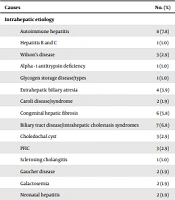
Furthermore, the obtained results revealed that autoimmune hepatitis and portal vein thrombosis (PVT) were the most common etiology of intrahepatic and extrahepatic PH, respectively (Table 2). In the current study, PVT was reported in 48 patients, among whom 21 patients with unknown etiology (43.7%) were observed. Table 3 shows the underlying causes of PVT in the remaining patients. Based on the results, thrombophilia and malignancy were the causes of PVT in 7.8% of patients; however, portal vein injuries, including trauma, abdominal surgery, and umbilical vein catheterization, were the causes of PVT in 18.6% of cases. In addition, 44.46% of patients with known PVT had a history of umbilical cord catheterization in infancy.
| Causes | No. (%) |
|---|---|
| Intrahepatic etiology | |
| Autoimmune hepatitis | 8 (7.8) |
| Hepatitis B and C | 1 (1.0) |
| Wilson’s disease | 3 (2.9) |
| Alpha - 1 antitrypsin deficiency | 1 (1.0) |
| Glycogen storage disease/types | 1 (1.0) |
| Extrahepatic biliary atresia | 4 (3.9) |
| Caroli disease/syndrome | 2 (1.9) |
| Congenital hepatic fibrosis | 6 (5.8) |
| Biliary tract disease/intrahepatic cholestasis syndromes | 7 (6.8) |
| Choledochal cyst | 3 (2.9) |
| PFIC | 3 (2.9) |
| Sclerosing cholangitis | 1 (1.0) |
| Gaucher disease | 2 (1.9) |
| Galactosemia | 2 (1.9) |
| Neonatal hepatitis | 2 (1.9) |
| Extrahepatic etiology | |
| PVT/hematologic | 8 (7.8) |
| PVT/portal vein injury | 19 (18.6) |
| PVT/idiopathic | 21 (20.6) |
| Chronic congestive heart failure | 2 (2.0) |
Main Underlying Causes of Extrahepatic and Intrahepatic Portal Hypertension in Study Population
| Parameters | No. (%) |
|---|---|
| PVT/hematologic | |
| Factor V Leiden mutation | 1 (3.7) |
| Malignancy (AML) | 1 (3.7) |
| Proteins C, S & antithrombin III deficiency | 6 (22.2) |
| PVT/portal vein injury | |
| Trauma | 1 (3.7) |
| Abdominal surgery | 6 (22.2) |
| Umbilical vein catheterization or sepsis | 12 (44.4) |
Underlying Causes of Portal Vein Thrombosis in Patients
5. Discussion
The PH refers to portal vein obstruction, which, based on its site, is categorized into prehepatic, intrahepatic, and post hepatic (13). According to PH types, various etiological factors are considered to be related to disease susceptibility (14). Focus on the assessment and management of PH has the potential to prevent the development of variceal hemorrhage or other related complications. Due to the location of the portal venous system, direct measurement is invasive and not routine.
The diagnosis of PH considering splenomegaly, ascites, and anatomy of intrahepatic and extrahepatic portal veins can be reliably made by ultrasound measurements (15). A large number of studies were conducted on PH in adults and children. The PH in children is frequently due to extrahepatic portal vein obstruction (EHPVO); nevertheless, the intrahepatic pattern is the main cause in adults (16-18). Additionally, EHPVO is rarely observed in western countries. The EHPVO in children can be idiopathic or due to congenital anomalies, hypercoagulable state, local inflammatory and systemic autoimmune disorders, vasculitis, and portal vein injury or occur after liver transplant (19).
Based on the obtained results of the current study, extrahepatic diseases were the most prevalent cause of PH in nearly half of the study population. Extrahepatic diseases were the most common etiology of PH in the studied patients. A study conducted in South India showed similar results to the present study (20). However, the findings of the current study are in contrast with the results of studies that reported the presence of PH in pediatrics due to intrahepatic diseases (21). Based on the current study’s results, there was a significant relationship between age and PH. The PH can occur at different ages; however, Sooraj et al., in a study, reported that age > 8 years is significantly related to PH (22). Based on the evidence, the frequency of various causes in PH patients can vary due to different age groups (20). On the other hand, the etiology of PH varies at different ages. In children, the primary cause is EHPVO (23); nonetheless, cirrhosis is the main cause in adults (7).
In the case of PH, patients might have no symptoms; however, increased pressure within the portal vein can lead to several related symptoms, including splenomegaly, GI bleeding, and ascites (24). It should be noted that PH itself does not cause signs and symptoms; nevertheless, some of its consequences can lead to a variety of symptoms. The GI bleeding is one of the first noticeable symptoms of PH. The current study’s findings are in agreement with previous studies reporting GI bleeding, jaundice, hepatomegaly, and ascites as the most common clinical presentations in patients with PH. Although UGIB was significantly higher in our patients with extrahepatic etiology than in those with intrahepatic etiology, patients with cirrhosis, due to a variety of lesions, including lesions associated with PH, might develop UGIB (25).
Based on the evidence, EHPVO is responsible for most pediatric UGIB (68 - 84%) (26). Grama et al., in a study, reported that UGIB (49.21%) was the first symptom in children with PH, followed by splenomegaly (34.92%) (27). Esophageal variceal bleeding is the major cause of morbidity and mortality associated with PH in patients (28). The results of this study showed that esophageal varices were the most common endoscopic finding in the patients. However, this finding was significantly more common in patients with the extrahepatic type of PH than in those with the intrahepatic type.
Regarding the results of previous studies, alcoholic, metabolic, and autoimmune liver diseases can be considered the etiology of PH in children (29). Although different underlying diseases might be involved, autoimmune hepatitis was the most common cause of intrahepatic PH in the current study. Other but less frequent causes included biliary tract disease and congenital hepatic fibrosis. The results also indicated the notable role of PVT in extrahepatic PH cases. In accordance with the present study’s results, Sooraj et al. also reported PVT as the most frequent etiology (63%), followed by liver cirrhosis (19%) and biliary atresia (18%) (22). In a study by Imanieh et al., cryptogenic cirrhosis (26.6%), biliary atresia (24.4%), Wilson’s disease (17.7%), and autoimmune hepatitis (6.6%) were the most prevalent causes of intrahepatic PH in patients (21).
The PVT, the most frequent cause of PH and specifically one of the causes of EHPVO, has been studied for many years (30). PVT is a multifactorial disease that risk factors such as, malignancy; liver diseases, inflammatory diseases, systemic diseases, and inherited thrombophilia, are considered its underlying causes, although, in 30 - 40% of cases, the etiology remains unknown (31, 32). In the present study, no possible cause of PVT was recognized for 20.6% of the patients. In line with previous studies, inherited thrombophilia, myeloblastic leukemia, and portal vein injuries, including umbilical cord catheterization, were the causes of PVT in patients. In several studies, umbilical vein catheterization has been reported as the most prevalent risk factor for developing PVT in pediatrics (33).
Since all studies have limitations as a natural occurrence, there are some limitations for the current study that might influence the outcomes of this study. The insufficient sample size for statistical measurements was one of the significant limitations of the current study. In addition, given the invasive approaches to measuring PH, there is a relatively small number of well - documented pediatric experiences with PH.
5.1. Conclusions
Given that the pathogenesis of PH is complex, the accurate identification of the cause of port hypertension is essential to select the appropriate treatment. However, in children, PH might be caused by a broad spectrum of etiologies. This study revealed that the extrahepatic type of disease was the most common etiology of PH in studied children referred to the Pediatric Medical Center at Mofid Children’s Hospital. However, the cause of the disease was unknown in several patients. An ultrasound assessment of PH in children with risk factors for EHPVT should be considered for timely diagnosis and treatment.
