1. Introduction
Central venous catheterization (CVC) is an imperative tool in critically ill children to administer fluids, medications, parenteral nutrition, and for monitoring the central venous pressure. The Seldinger technique is the most commonly used method for central venous catheter insertion and is considered quite safe when properly performed (1). However, it can lead to various complications such as arterial puncture, hemothorax, pneumothorax, nerve injury, or air embolism. A rare iatrogenic complication of CVC is guidewire retention, which can be recognized loss of guidewire during procedure, or unrecognized failure to remove guidewire. The retention can lead to dysrhythmia, vascular damage, thrombosis, embolism, infection, cardiac perforation, and tamponade (2-4). Although most of these cases are reported from adult patients, this can complicate the CVC in children also. We report a case of inadvertent guidewire retention following femoral venous catheterization and its successful management.
2. Case Presentation
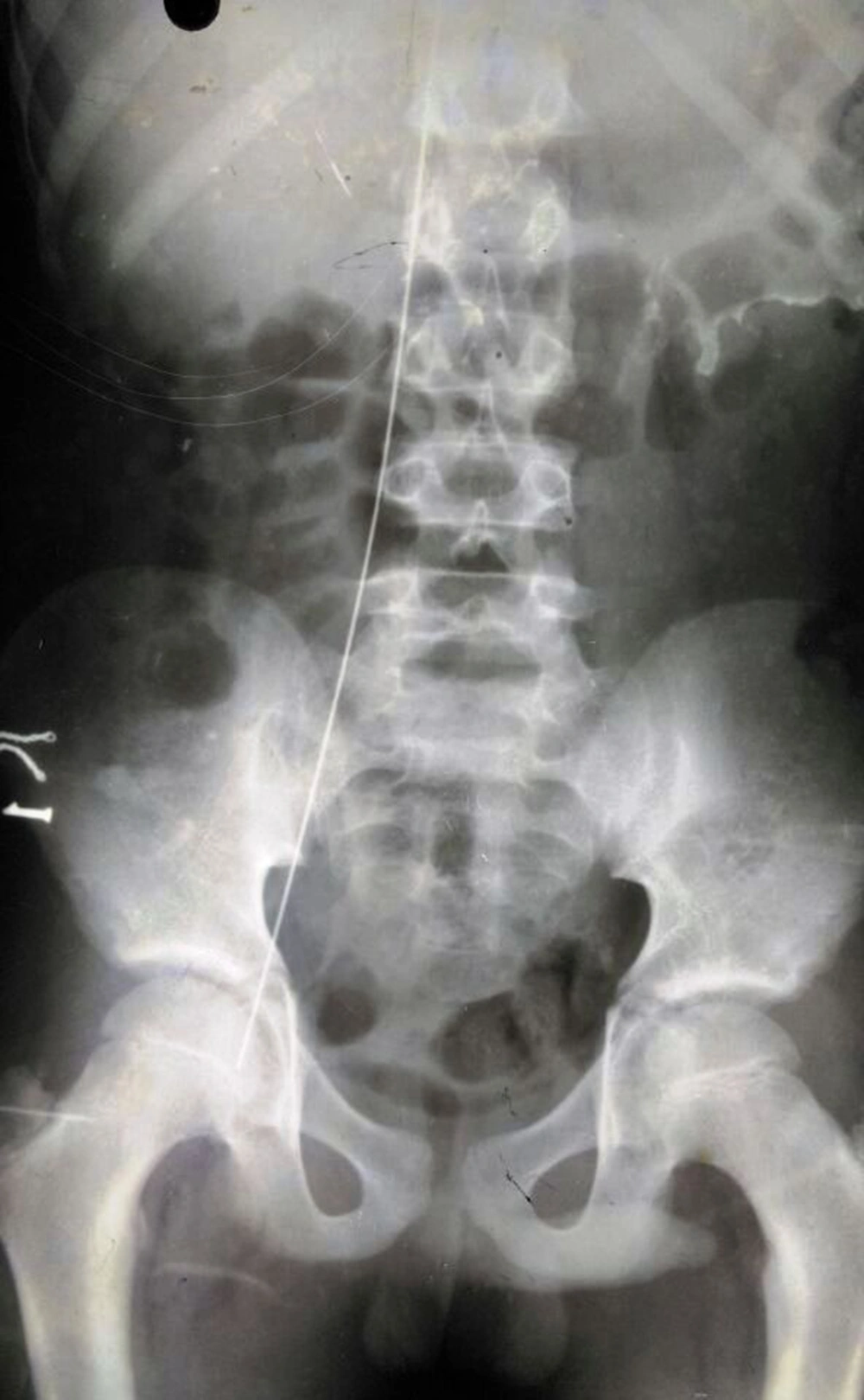
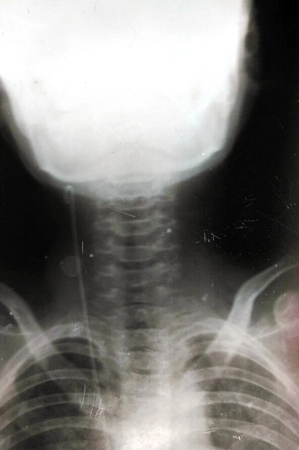
An 11-year-old male child was admitted to the pediatric intensive care unit (PICU) with a provisional diagnosis of traumatic tetanus in view of history of trauma and his unvaccinated state and supported by presence of spasms, trismus and rigidity. The treatment was started as per protocol to manage tetanus; however as the frequency of spasm were increased, child was mechanically ventilated and midazolam infusion was started along with intermittent boluses of vecuronium. Central venous catheterization was planned for fluid management and drug administration. After taking written consent from the parents, the procedure was assigned to a second-year resident under supervision of a faculty. Under all aseptic precautions, right femoral vein was cannulated using an introducer needle (18G, 70 mm) after local infiltration. A J-tipped guidewire (0.035 mm, 50 cm) was introduced smoothly through the needle after positive aspiration of venous blood. Then, a dilator (6.0 Fr) was advanced over guidewire to create a track followed by insertion of catheter on the guidewire (5.0 Fr, 20 cm, double lumen, Romson’s Centro, India). During this procedure, guidewire was inadvertently pushed further ahead into the vein and after realizing this, catheter was removed but the guidewire was missing. Urgent x-rays of the kidneys, ureters, bladder region, upper abdomen and chest were done, in which the guidewire was clearly visible inside the vein and seems up to the right side of the neck (Figures 1 and 2). Ultrasonography (USG) of the abdomen and chest also confirmed that the guidewire was inside the vein. Low-molecular weight heparin was started as per the advice of cardiothoracic surgeon at a dose of 0.5 mg/kg/dose twice a day given subcutaneously. The guidewire was then removed by a pediatric surgery team from the right posterior triangle of the neck through incision in an internal jugular vein followed by ligation. Whole procedure from central venous catheter insertion to guidewire removal was uneventful and no arrhythmia was recorded and subsequent echocardiogram also did not reveal any evidence of cardiac dysfunction.
Gradually, the patient started improving with cessation of spasms and extubated after 15 days of mechanical ventilation. He was shifted out of PICU after 22 days and admission and discharged after another 7 days of hospital stay.
3. Discussion
The Seldinger’s technique of placement of a central venous catheter involves insertion of introducer needle into the vein, advancing the guidewire through needle, removing the needle, and then advancing the catheter over guidewire. Once catheter is in place, guidewire is removed. However, a rare but potentially hazardous, iatrogenic complication of this procedure is the retention of a guidewire which can lead to dysrhythmia, vascular damage, thrombo-embolism, infection, cardiac perforation, and tamponade. However, it is usually asymptomatic as in our case. It can be found incidentally, even several months after the procedure, on X-ray done for other reason (4). The exact incidence of guidewire retention is difficult to track; however, it has been estimated at one per few thousand placements of central venous catheter. According to New York state health department report, retained catheters and guidewires were the most commonly reported nonsurgically retained objects with 80 cases reported between 2008 and 2009 (5). Essentially introduction of an excessive length of a guidewire during CVC, due to many factors, leads to its retention. Primary reason is the design of guidewire (absence of marks and straight proximal end) leading to difficulty in ensuring that this end has been secured. Fear of losing vascular access, use of circular advancer, and concerns over contamination of proximal end also contribute to excessive introduction of guidewire (6). Operators’ inexperience, fatigue, inattention, and inadequate supervision of trainees were suggested as other predisposing factors (6-8). In our case, inattention and not lack of care caused this complication. The resident, overwhelmed with various dimensions of the patient’s care, unknowingly pushed the guidewire while inserting a central venous catheter. The inadvertent intravascular insertion of entire guidewire is an avoidable complication and every effort should be made to prevent such an incident as it can be life-threatening also (9). Holding on to the proximal tip of the wire at all times is fundamental to prevent this mistake. A small, lightweight retention device to be attached to the proximal end of the wire, a bright or different colour of proximal wire tip and longer guidewires may possibly decrease the risk of guidewire loss (6-8). Retention should be suspected if the wire or a piece of wire is missing after completion of procedure, if there is resistance to injection or poor backflow from distal lumen, or if imaging shows a guidewire (7, 8). Interventional radiology methods are preferred for retrieving a retained guidewire preferably by catching the wire with a gooseneck snare; other techniques being 2-wire technique, endovascular forceps or Dormier basket (10). Surgical venotomy and median sternotomy may be required in some cases (3). An anticoagulant, usually heparin, should be given before and during wire removal. Retention of guidewire is a completely avoidable complication of CVC and is an important issue from the patient’s safety point of view. Therefore, measures to prevent and early diagnosis of this complication should be promulgated among those who perform central venous catheter insertion procedure.

