


1. Background
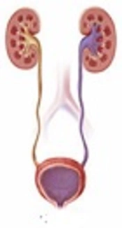
Vesicourethral reflux (VUR) is defiened as retrograde passage of urine from the bladder into kidney and upper urinary tract (1, 2). This condition can increase transporting bacteria from the bladder to the kidney and then predisposes patients to acute pyelonephritis (3, 4). Pyelonephritis as an acute and morbid event requires acute care in children and is a possible indication for hospitalization in infants. Also, loss of renal parenchyma (renal scarring) can occure by urinary tract infection (UTI) (5).
International classification of reflux study classified it I to V intensity grades of reflux nephropathy based on voiding cystourethrography (VCUG). Rating of reflux nephropathy mentioned below:
- Grade I: reflux limited to the ureter.
- Grade II: reflux up to the renal pelvis.
- Grade III: mild dilatation of ureter and pelvicalyceal system.
- Grade IV: tortuous ureter with moderate dilatation and blunting of fornices but preserved papillary impressions.
- Grade V: tortuous ureter with severe dilatation of ureter and pelvicalyceal system and loss of fornices and papillary impressions (6, 7).
In the diagnosis of this condition, VCUG is a method of choice. However, this is an invasive procedure that has irradiation risks on patients and their parents (8). For VUR diagnosis, it is very important to choose a procedure that is safe, noninvasive, and accepted by the patient and the parents. For urinary tract system evaluation, technetium 99 m-labeled dimercaptosuccinic acid scan (DMSA) are used for abnormalities urological system and renal parenchymal scars (9). Efficacy of DMSA scan was predicted in some studies and ruled out by others (1, 10, 11).
For the DMSA scan, dimercaptosuccinic acid is injected intravenously and arrives to the kidney through the bloodstream (12). In addition, the kidneys are scanned by special cameras, that show the structure of the kidney (10). For this, test preparation is not necessary, eating and drinking for the individual is permitted. Except for infants who do not have necessary cooperation, sedation (sedative or hypnotic) or anesthesia have to be used (11, 13). The scan has 2 phases: injecting radioactive substances as a first step and taking scans of children as a second step. Between these two stages, there are about 2 to 4 hour intervals (3). Injected radioactive substance has been excreted through the kidneys. Hydration of children (getting enough fluids) can help fast disposal of radioactive materials. In addition, washing the urogenital area is an important act after urination (9, 14). Since VCUG can induce and increase children radiation, DMSA scan has suggested for evaluation of different grades of VUR.
2. Objectives
In our study, we consider the DMSA as an approach to evaluate prediction of VUR and its grade in children and to find a better diagnostic approach for VUR.
3. Methods
3.1. Study Setting
It was a hospital-based study that was conducted in the pediatric clinic of Amir-Kabir hospital.
3.2. Study Population
Study populations were all male and female children lower than 6 years of age by indication for VCUG (first UTI) and pediatrician confirmation of VUR in six months in 2016. With these criteria, others in following, samples (101 patients) were randomly chosen and enrolled in the study for study group.
3.3. Measurements
A VCUG and then DMSA renal scan were done for all patients. VUR has detected by VCUG and approved by pediatric nephrologists and were divided into I and VI, according to the VCUG test and based on the international reflux study classification. Medical, demographic, and epidemiological information was taken from the patients by questionnaire.
In order to evaluate renal parenchymal, we carry out the DMSA scan for children. The scan was done 3 - 4 hours after injection of technetium-99m and renal parenchymal is evaluated based on the technetium uptake.
3.4. Ethical Considerations
Ethical issues were completely observed by the authors. The study group adheres to the principles of medical ethics introduced by the health ministry and the declaration of Helsinki and legislation in the Medical ethics committee of Arak University of Medical Sciences. In addition, the ethical committee of Arak University of Medical Sciences approved the protocol of the study.
3.5. Statistical Analysis
The data were analyzed by the SPSS program and P < 0.05 was considered as a significant value. We consider the t-test for quantitative variables and X2 test for qualitative variables.
3.6. Inclusion and Exclusion Criteria
Each child that was under the age of 6, by diagnosis of VUR, were recruited based on the Nelson book and medical supervision of pediatrics nephrologists. The children should not have any mental and physical illness. Parents and children who did not cooperate in entering to study, presence of any chronic kidney disease or other urinary tract problems, and chronic use of any drug were considered as the exclusion criteria.
4. Results
In total, 101 patients were examined, 88 cases were male (87.1%) and 13 cases were female (12.9%). In addition, children under the age of 6 and other characteristics of children with VUR have been presented as No. (%) (Table 1).
| Variables | Children with Vesicoureteral Reflux |
|---|---|
| Gender | |
| Male | 13 (12.9) |
| Female | 88 (87.1) |
| Age, yb | |
| 1 and lower | 44 (43.6) |
| 1 - 5 | 42 (41.6) |
| 5 and higher | 15 (14.9) |
| Birth weightc | |
| VLBW | 18 (17.8) |
| LBW | 69 (68.4) |
| NBW | 8 (7.9) |
| HBW | 6 (5.9) |
| Household incomesd | |
| Low | 32 (31.6) |
| Moderate | 65 (64.3) |
| High | 2 (2) |
| Paternal education | |
| University | 13 (12.9) |
| High school | 85 (84.2) |
| Elementary school | 3 (2.9) |
| Maternal education | |
| University | 31 (30.8) |
| High school | 70 (69.2) |
| Elementary school | 0 (0) |
| Consanguineous marriage | |
| Yes | 10 (9.9) |
| No | 91 (90.1) |
| Gestational age, week | |
| Full term (= 37) | 98 (97.1) |
| Premature (< 37) | 3 (2.9) |
| Post-term (> 40) | 0 (0) |
| Patient with siblings | |
| Yes | 73 (72.3) |
| No | 28 (27.7) |
| Location | |
| City | 37 (36.6) |
| Village | 64 (63.4) |
| Maternal age, y | |
| < 20 | 62 (61.4) |
| 20 - 30 | 36 (35.6) |
| > 31 | 3 (3) |
| Paternal age, y | |
| < 20 | 0 (0) |
| 20 - 30 | 81 (80.2) |
| > 31 | 20 (19.8) |
aThe values are presented as No. (%)
bBased on the years.
cVLBW, > 1500; LBW, 1500 - 2500; NBW, 2500 - 4000; HBW, > 4000; based on the gr scale.
dLow, family monthly income < 5 million Rials; Moderate, family monthly income of 5 million to 10 million Rials; High, family monthly income > 10 million Rials.
As shown in Table 2, the mean of VUR in the renal scaring (P = 0.0001) and pyelonephritis in the DMSA scan (P = 0.0001) were significantly different between the two groups. In addition, the age (P = 0.24) and gender (P = 0.4) of children have no significant difference in the two groups (Table 3).
| Variables | Mean of Vesicoureteric Reflux Grade | P Value | |
|---|---|---|---|
| LK | RK | ||
| Renal scar | 0.0001 | ||
| Yes | 2.7 ± 1.06 | 1.9 ± 1.52 | |
| No | 1.78 ± 1.34 | 1.6 ± 1.3 | |
| Pyelonephritis | 0.0001 | ||
| Yes | 2 ± 1.7 | 2.6 ± 0.57 | |
| No | 1.9 ± 1.34 | 1.58 ± 1.3 | |
Abbreviations: LK, Left Kidney; RK, Right Kidney.
aThe Values are presented mean ± SD
| Variables | Grade of Vesicoureteric Reflux | P Value | |||||||||||
|---|---|---|---|---|---|---|---|---|---|---|---|---|---|
| 0 | 1 | 2 | 3 | 4 | 5 | ||||||||
| LK | RK | LK | RK | LK | RK | LK | RK | LK | RK | LK | RK | ||
| Ageb, y | 0.24 | ||||||||||||
| 1 and lower | 12 (11.8) | 9 (8.9) | 1 (0.9) | 2 (1.9) | 11 (10.9) | 18 (17.7) | 15 (14.8) | 10 (9.9) | 2 (1.9) | 5 (4.9) | 3 (2.8) | 0 (0) | |
| 1 - 5 | 9 (8.9) | 17 (16.8) | 1 (0.9) | 3 (2.9) | 18 (17.7) | 10 (9.9) | 11 (10.9) | 10 (9.9) | 3 (2.9) | 2 (1.9) | 0 (0) | 0 (0) | |
| 5 and higher | 4 (3.9) | 7 (6.9) | 3 (2.9) | 1 (0.9) | 4 (3.9) | 4 (3.9) | 4 (3.9) | 3 (2.9) | 0 (0) | 0 (0) | 0 (0) | 0 (0) | |
| Gender | 0.4 | ||||||||||||
| Male | 4 (3.9) | 2 (1.9) | 1 (0.9) | 2 (1.9) | 2 (1.9) | 3 (2.9) | 2 (1.9) | 2 (1.9) | 1 (0.9) | 3 (2.9) | 2 (1.9) | 0 (0) | |
| Female | 21 (20.5) | 26 (25.4) | 4 (3.9) | 4 (3.9) | 27 (26.4) | 26 (25.4) | 21 (20.5) | 18 (17.6) | 3 (2.9) | 2 (1.9) | 0 (0) | 0 (0) | |
Abbreviations: LK, Left Kidney; RK, Right Kidney.
aThe values are presented as No. (%)
bBased on the years
5. Discussion
Our results showed that DMSA scan in pyelonephritis and renal scar is a diagnostic approach, and according to DMSA scan, high graded VUR can be predicted.
In a study by Tseng et al., specificity and sensitivity of DMSA scan for prediction of VUR, respectively, were 71%, 58%, 44%, and 88% (15). Camacho et al., reported PPV and NPV respectively as 88% and 48% for prediction of VUR by DMSA scan and concluded that DMSA scan is normal during acute UTI and renal damage risk is low (16). In Alshamsam and Mahant studies, sensitivity, specificity, PPV, and NVP of ultrasonography was reported respectively as 40%, 76%, 32%, and 82%, for prediction of VUR (17). Moorthy et al., in a study, reported that 16% of children with VUR had a abnormal kidney. In addition, they concluded that ultrasonography examination for predication of VUR is not recommended and VCUG is preferred to ultrasonography (18). Kass et al., mentioned that the ability of DMSA or ultrasonography for prediction of VUR is not recommended (19). Based on our study, accuracy of DMSA scan in high graded VUR was approved. In a study by Ajdinovic et al., the specificity and sensitivity of ultrasonography for prediction of high grade VUR (III and higher) were 84% (20). Sorkhi et al., evaluated prediction of VUR and DMSA scan and concluded that DMSA scan or ultrasonography cannot predict VUR (especially low grade VUR), however, based on NPV, absence of VUR can be predicted with these tests (8). However, with the reasons that few clinical studies have been carried out in regards to this test, further studies are needed before the verification accuracy of DMSA scan as a better diagnostic approach.
The limitation was parental noncompliance due to the use of the DMSA scan as a non-routine diagnostic approach for VUR. However, when we explained to parents that DMSA scan is a cheap and safe test in compare to VCUG scan, they were convinced. In addition, Due to a low number of clinical studies, we recommend further studies with larger sample sizes.
5.1. Conclusion
Our funding showed that DMSA scan, as an alternative dignostic approach, can be used in the diagnosis of high graded VUR, renal scar, and pyolonephrit, however, not in low garded VUR. Since VCUG is golden standard in VUR, DMSA scan is better in a high grade of VUR due to the fact that VCUG is invasive, which causes anxiety in patients and their parents. In addition, DMSA has lower complications and is better for children.
