1. Background
Tetralogy of Fallot (TOF) is one of the most prevalent cyanotic heart lesions. Total surgical repair has proven to be a definite cure since 1954 (1). About 80% of these patients have pulmonary valve stenosis (2); thus, they need widening of the pulmonary annulus with tran annular patch (TAP) (3). Pulmonary regurgitation (PR) is an inevitable long-term complication of the surgery that can lead to right ventricular systolic and diastolic dysfunction. Evaluating the Tei index is a non-invasive method for assessing the right ventricular function. This index can be obtained by pulsed Doppler (PW) or tissue Doppler imaging (TDI).
2. Objectives
This study aimed to investigate the two methods in calculating the Tei index in TOF patients with PR following surgery.
3. Methods
3.1. Study Population
This cross-sectional study was conducted between March 2017 and March 2018, with 21 subjects as cases and 21 as controls. The inclusion criteria were defined as the TOF patients who undergone total correction surgery with TAP without any history of previous palliative surgery that had at least moderate pulmonary valve regurgitation. The exclusion criteria were defined as the existence of an arrhythmia and residual pulmonary valve stenosis with a pressure gradient of more than 40 mmHg. The control group was chosen from healthy children with no history of cardiac involvement following a full examination. Ethical approval for the study was obtained from the institutional review board.
3.2. Echocardiographic Evaluations
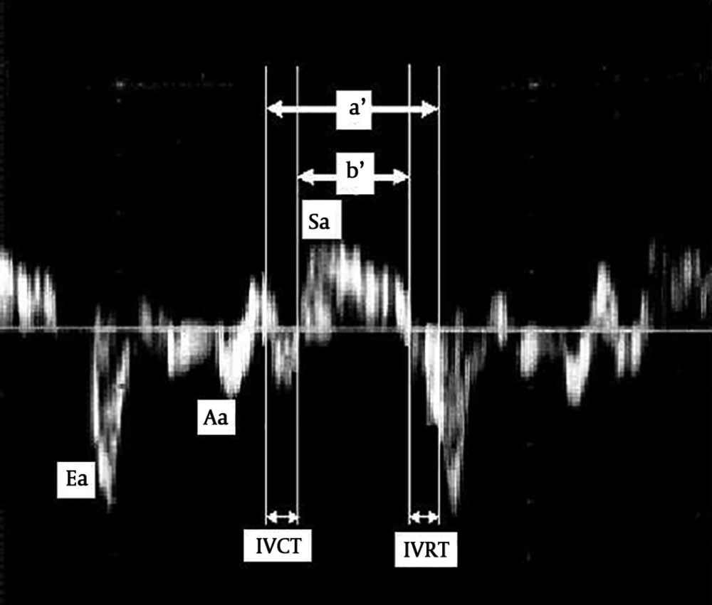
The PW two-dimensional echocardiography was performed using Mylab 60 Echocardiography device with 3.5 and 5 MHz transducers. The tissue Doppler imaging was performed by activating the TDI in the same device. Positioning the Doppler sample volume at the tips of tricuspid leaflets, the tricuspid inflow velocity patterns were recorded from the four-chamber view. Angle correction was not performed. In the same apical four-chamber view, a 2-mm sample volume was located on the external side of the Tricuspid valve annulus with a sweep speed of 100 mm/s for acquiring peak myocardial velocities during systole (Sa), early diastole (Ea), and late diastole (Aa). Two carefully placed vertical cursors were used for recording the intervals (Figure 1).
Using the pulsed Doppler recordings, the tricuspid closing-to-opening time (a) was measured as the interval from the end to the onset of the tricuspid inflow velocity pattern. The RV ejection time (b) was measured from the onset to the end of the RV outflow velocity pattern. The Tei index was calculated as a-b/b. For measuring the Tei index from TDI, the time interval from the end to the onset of the tricuspid annular velocity pattern during diastole was determined (a′). The duration of the S wave (b′) was measured from the onset to the end of the S wave. The Tei index was calculated as a′-b′/b′. The TAPSE (Tricuspid annular plane systolic excursion) was recorded by locating M-mode sampling line on the external side of the tricuspid annulus with the speed of 100 mm/s and the distance between the peak and the bottom of the annulus movement was recorded.
All data were analyzed by SPPS version 17. Statistical significance was considered at P value = 0.05.
4. Results
The study recruited 42 subjects, including 21 TOF patients as cases and 21 healthy children as controls. Neither age (3.09 ± 1.81 vs. 3.04 ± 2.38; P = 0.936) nor heart rate (102.6 ± 12.1 vs. 100.1 ± 7.0; P = 0.422) differed between the cases and controls.
4.1. Echocardiographic Results for the Controls
Obtained by PW and TDI, the values of a and a′ (359.5 ± 14.6 vs. 358.7 ± 15.8; P = 0.851), b and b′ (266.7 ± 15.8 vs. 265.1 ± 18.1; P = 0.768), and the Tei index (0.35 ± 0.06 vs. 0.36 ± 0.06; P = 0.666) did not differ statistically in the control group (Table 1).
| a, msec | a', msec | b, msec | b', msec | MPI | MPI' | |
|---|---|---|---|---|---|---|
| Normal | 359.5 ± 14.6 | 358.7 ± 15.8 | 266.7 ± 15.8 | 265.1 ± 18.1 | 0.35 ± 0.06 | 0.36 ± 0.06 |
| TOF | 393.5 ± 16.4 | 397.3 ± 18.2 | 306.7 ± 28.0* | 266.8 ± 28.0 | 0.30 ± 0.10† | 0.50 ± 0.10 |
aValues are expressed as mean ± SD.
4.2. Echocardiographic Results for the Cases
The values of a and a′ had no difference in the patient group (393.5 ± 16.4 vs. 397.3 ± 18.2; P = 0.479), but the b value was significantly lower than the b′ value (306.7 ± 28.0 vs. 266.8 ± 28.0; P < 0.001). Consequently, the Tei index obtained by the TDI method (MPI′) was higher than the Tei index obtained by PW (MPI) (0.50 ± 0.10 vs. 0.30 ± 0.10; P = 0.001) (Table 1).
4.3. Comparing the Cases and Controls
The groups had no difference concerning the Tei index obtained by PW (0.35 ± 0.06 vs. 0.30 ± 0.10; P = 0.059). However, the TDI method showed a higher Tei index in cases than in controls (0.50 ± 0.10 vs. 0.36 ± 0.06; P < 0.001) (Table 2).
| TOF (n = 21) | Normal (n = 21) | P Value | |
|---|---|---|---|
| Tei index calculated by PW | 0.30 ± 0.10 | 0.35 ± 0.06 | 0.059 |
| Tei index calculated by TDI | 0.50 ± 0.10 | 0.36 ± 0.06 | < 0.001 |
aValues are expressed as mean ± SD.
5. Discussion
Tetralogy of Fallot (TOF) is one of the most prevalent cyanotic congenital heart diseases. Pulmonary valve stenosis is found in 80% of these patients (2). In 1954, the complete repair of the defect was performed, including ventricular septal defect closure and pulmonary stenosis resolving, which has been preferred for almost every patient since then (1). Most of the patients have small pulmonary annulus that necessitates the surgeons to use the transannular patch (TAP) for widening the pulmonary annulus (3). Pulmonary valve regurgitation (PR) is known as an important long-term complication of the TOF total repair, which additionally leads to right ventricle systolic and diastolic dysfunction, dilation, and arrhythmia (4-6). Sudden death may occur as a result of pulmonary valve regurgitation plus impaired right ventricle function (2). Furthermore, based on our study, the impaired RV and PR following TOF total repair caused longer contraction time and isovolumic loosening, showing systolic and diastolic ventricle function, as well as diastolic velocity (Ea and Aa) and systolic myocardial (Sa) decrement. The IVCT and IVRT were longer and the values of Ea, Aa, Sa, and TAPSE were significantly lower in TOF patients than in the control group.
The Tei index, first described by Tei et al., is a Doppler index for the assessment of both systolic and diastolic functions of the left ventricle (7). Moreover, it has been found useful for assessing the right ventricle function in the fetus (8, 9) and children (10-12). However, this index is not sensitive enough in patients with significant PR who had undergone TOF total repair (13). The Tei index obtained by tissue Doppler imaging (TDI) has proven to have a high correlation with that obtained by pulsed Doppler (PW) (14, 15).
In our study, similar to previous studies (13, 16), the Tei index values obtained by PW were not significantly different between TOF patients and controls (0.30 ± 0.10 vs. 0.35 ± 0.06; P = 0.059). However, the Tei index obtained by TDI was higher in TOF patients than in the control group (0.50 ± 0.10 vs. 0.36 ± 0.06; P < 0.001). Our findings are congruent with those reported by Yasuoka et al. (16).
We found no difference in the values of a and a′ obtained by PW and TDI between TOF patients and controls. However, in TOF patients, b′ measured by TDI was shorter than b measured by PW. Thus, it can be concluded that IVRT is longer in TOF patients than in the control group (43.53 ± 8.82 vs. 25.77 ± 10.14; P < 0.001) while IVCT is longer in TOF patients (87.73 ± 10.91 vs. 68.36 ± 8.96; P < 0.001). These will result in the decrement of b′ and the increment of the Tei index obtained by TDI in TOF patients.
PR brings about the augmentation of the ejection fraction of the right ventricle, which results in no changes in the b time obtained by PW. In conclusion, the Tei index will be lower than the real amount in these patients. In other words, the Tei index obtained by PW will be in the normal range falsely. Based on our findings, calculating the Tei index by TDI is more accurate than by DW for evaluating the overall performance of the right ventricle in PR patients after TOF reconstruction.

