


1. Background
The thyroid gland is a vital hormonal gland located superficially in the anterior and lower part of the neck and consists of two lobes joined together by isthmus and is asymmetrical in a way that the right lobe is larger (1). The thyroid gland is responsible for production, storage, and secretion of thyroxine (T4) and triiodothyronine (T3) (2). Thyroid hormones are essential for many physiological functions of the body, including the regulation of metabolism and the development of the body and proper mental development (3). Approximately, 200 million people suffer from some types of thyroid disease worldwide (4) and women are 5 - 20 times more at risk for thyroid problems than men (5). The World Health Organization (WHO) assessed that the number of elder people will increase from 900 million to two billion between 2015 and 2050 (6).
The size and shape of the thyroid gland is very different in normal people and is enlarged in tall and oval in short people, respectively (7, 8).
Many factors affect the size of the thyroid gland, such as age, sex, weight, height, Body Mass Index (BMI), and body surface area (BSA), diet iodine, smoking, race, genetics, and geographic area (9-11). Thyroid gland enlargement is referred to as thyromegaly, which is also known as stroma and goiter (12). The most common cause of endemic goiter is iodine deficiency worldwide (13). However, in countries with adequate iodine intake, such as the United States, thyromegaly is usually caused by autoimmune thyroid disorders (14), the most common of which are Hashimoto's thyroiditis, Graves' disease, and thyroid nodules (15). Other causes of thyromegaly include the impaired intrinsic production of thyroid hormone, as well as the use of various drugs, including aminosalicylic acid, lithium, and even excessive iodine intake (16).
The thyroid gland is easily palpable considering its superficial location, and a thyroid examination is the first stage in assessing thyroid disease (17). It is important to estimate the thyroid volume in a variety of pathological conditions (18), and skilled physicians can detect thyromegaly through touch (19). Although, this estimated size differs from ultrasound estimates in one-third of cases, and overall physical examination has little sensitivity or specificity (20). In cases such as planning for surgery, calculating iodine 131 required for the thyrotoxicosis treatment (21), and monitoring the effectiveness of various thyroid treatments, it is necessary to determine the exact volume of the thyroid gland, which is not possible by clinical evaluation. Recently, accurate estimation of thyroid volume has received much more attention with the introduction of minimally invasive surgery because the most important limiting factor is thyroid volume greater than 20 mL (22).
There are other methods for measuring thyroid volume, such as ultrasonography, radionuclide scanning, computed tomography (CT), and magnetic resonance imaging (MRI) (20, 23, 24). Among these methods, ultrasound is the preferred and standard for thyroid evaluation and a proven useful and practical method considering its availability, low cost, lack of radiation risk due to ionizing radiation, and ease of use (20, 25).
The American College of Radiology (ACR), the WHO and most relevant organizations use the ellipsoid formula to calculate thyroid volume, and measure
longitudinal (L) and anterior-posterior or depth (AP), and width (W) diameter of each lobe and calculate the volume of each lobe and report its total as thyroid volume using AP×W×L×π/6 formula and the isthmus volume is not calculated (26). Besides, AP dimeter of isthmus is calculated (8), which is generally a time-consuming process because careful attention should be placed (27). Moreover, it has not still been determined that an increase in which of these dimensions or the combination of two or three dimensions can be a symptom of thyromegaly. The thyroid dimensions, like other organs, vary from population to population mainly due to age, sex, anthropometric parameters, and genetic factors, and the relationship between thyroid volume and anthropometric characteristics is controversial (8, 9, 28-32).
2. Objectives
Therefore, the aim of the present study was to find the most important ultrasound measurement of thyroid to detect thyromegaly and also to find the relationship between these measurements and some anthropometric characteristics in the study population. The results of the current study could be helpful in increasing the rate of detection of thyromegaly and other thyroid diseases.
3. Methods
3.1. Samples and Data Collection
This case-control study was carried out at Imam Reza Hospital in Kermanshah, Iran after being approved by the Ethics Committee of Kermanshah University of Medical Sciences (code of ethics IR.KUMS.REC.1398.1013).
The sample size formula was calculated by statistics specialist. An endocrinologist request for ultrasound imaging on 131 individuals referred to the Ultrasound Department of Imam Reza Hospital of Kermanshah for diagnostic examination of thyromegaly during the years 2017 and 2019. Exclusion criteria included the following in addition to participants’ dissatisfaction:
- History of thyroid surgery
- Ambiguity in the margin or each dimension of the thyroid and retrosternal thyroid
- The presence of a large mass (nodule) or a very large cyst
Eligible individuals entered the research after completing the conscious consent form and receiving full explanations about the research objective from the responsible radiologist. Demographic information was collected through a researcher-made questionnaire. Anthropometric factors, including height (in centimeters) and weight (an accuracy of 100 g) were measured and BMI was measured based on the following formula: BMI = weight (kg)/height² (m²). Then, thyroid dimensions, including L, AP, and W diameters of both sides, and AP diameter of isthmus were measured in the supine position using the Samsung WS80 ultrasound device and a 16Mhz multi-frequency surface probe. The volume of each lobe was calculated based on the formula (AP×W×L×π/6) and the total volume of the both lobes was recorded as the total thyroid volume (10 parameters in total). The isthmus volume was not calculated while calculating the thyroid volume. People with thyroid volume greater than 11.1 for women and 11.6 for men were considered as the case group (n = 69) and the rest as the control group (n = 62) by three radiologists who were unaware of the research.
Data analysis was carried out by statistics specialist on parameters measured in the two groups (Table 1), along with the demographic information of the studied individuals, based on the main objectives, the most important ultrasound measurement parameters of thyroid to detect thyromegaly and also find the relationship between these measurements and some anthropometric characteristics in SPSS ver. 26.
Table 1.Demographic and Anthropometric Information of Case and Control Groups a
| Variables | Case | Control | P-Value |
|---|---|---|---|
| Gender (female/male) | 11.58 | 5.57 | - |
| Age | 41.99 ± 12.25 | 37.6 ± 13.32 | 0.065 |
| Height | 165.86 ± 6.97 | 164.13 ± 7.016 | 0.207 |
| Weight | 73.14 ± 12.51 | 65.65 ± 10.12 | 0.000 |
| BMI | 26.65 ± 3.96 | 24.39 ± 3.66 | 0.000 |
Abbreviation: BMI, Body Mass Index.
a Values are expressed as mean ± SD.
3.2. Statistical Analysis
Mann-Whitney and chi-square tests, receiver operating characteristic (ROC) curve, and logistic regression tests were analysis by statistics specialist in the present study. To select the best cut-off point based on the sensitivity and specificity values in the ROC curve method, the criterion was expressed as in the Unal (33).
Besides, the conditional forward method in logistic regression was used to determine the effective variables in thyromegaly and the enter method was used to investigate the effect of weight on thyromegaly in this model.
4. Results
According to the results of the Mann–Whitney U test, there was no significant difference between the case and control groups in terms of sex, age, and height, (P > 0.05), while the weight and BMI were significantly higher in the case group than the control group.
In addition, Mann–Whitney U nonparametric test was used to compare the average width of the right lobe, depth of the right lobe, length of the right lobe, volume of the right lobe, width of the left lobe, depth of the left lobe, length of the left lobe, volume of the left lobe, total volume, and isthmus volume in both the case and control groups and the results showed that the mean of all these factors in the case group was significantly higher than the control group (P < 0.05).
Also, the results of this test showed that the mean depth and length of the left and right lobes did not differ significantly in the case group (P > 0.05), while the mean width of the left and right lobes is significantly different (P < 0.05) (the average width of the right lobe is significantly larger than the left lobe). The opposite is true in the control group. That is, the mean width of the left and right lobes is not significantly different (P > 0.05), while the mean length and depth of the left and right lobes are significantly different (P < 0.05) (the average length and depth of the right lobe is significantly larger than the left lobe).
Besides, the results of the ROC curve on the width of the right lobe, depth of the right lobe, length of the right lobe, volume of the right lobe, width of the left lobe, depth of the left lobe, length of the left lobe, volume of the left lobe, total volume and isthmus volume, are shown in Figure 1.
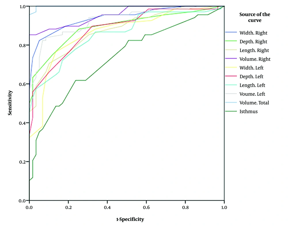
Figure 1.
Receiver operating characteristic (ROC) curve results for all variables measured.
Cut-off point values, according to the criterion expressed in (34), for the width of right lobe, depth of the right lobe, length of the right lobe, volume of the right lobe, width of the left lobe, depth of the left lobe, length of the left lobe, volume of the left lobe, total volume, and isthmus volume were 16.5, 15.5, 47.5, 6.45, 15.5, 14.5, 44.5, 5.55, 11.15, and 2.45, respectively. According to this method, if the desired variable in a person is less than these numbers, the thyroid is normal and otherwise the person has a thyromegaly.
Besides, forward conditional method in logistic regression was used to determine the most important factors in detecting thyromegaly. The width of the right lobe, the depth of the right lobe, the length of the right lobe, the width of the left lobe, the depth of the left lobe, the length of the left lobe, and the isthmus volume were taken as independent variables and the individual's health status (thyromegal or healthy) was considered as the dependent variable. The results are shown in Table 2.
Table 2.Summary of Significant Step to Include Variables in the Regression Equation
| Variables | B | Wald | Sig. |
|---|---|---|---|
| Width.right | -2.042 | 10.963 | 0.001 |
| Depth.right | -0.678 | 4.616 | 0.032 |
| Length.right | -1.180 | 6.097 | 0.014 |
| Constant | 61.234 | 9.763 | 0.002 |
In fact, this method yielded significant results only until step 3 and it was not significant in the next steps with the entry of these 3 variables in the model. Therefore, the three factors of width of the right lobe, depth of the right lobe, and length of the right lobe, respectively, were selected as the most important factors in detection of thyromegaly.
Also, according to the results of chi-square test (P-value > 0.05), sex has no effect on thyromegaly, and the results of using logistic regression with Enter method showed that the odds ratio of thyromegaly increases by 0.941 with increasing weight.
5. Discussion
The results of the present study showed that the mean of all ultrasound parameters measured in the case group is higher than the control group. In the case group, width of the right lobe was greater than in the left lobe, and the right lobe length and depth were greater than in the left lobe in the control group. The cut-off points values for width of the right lobe, depth of the right lobe, length of the right lobe, volume of the right lobe, width of the left lobe, depth of the left lobe, length of the left lobe, volume of the left lobe, total volume, and isthmus volume were 16.5, 15.5, 47.5, 6.45, 15.5, 14.5, 44.5, 5.55, 11.15, and 2.45 respectively. According to this method, if the desired variable is less than these numbers, the thyroid is normal and otherwise the person suffers from thyromegaly. Accordingly, normal thyroid volume is considered < 11.6 mL in men, < 11.1 mL in women, and < 11.15 mL in general. Also, the three factors of width of the right lobe, depth of the right lobe, and length of the right lobe, respectively, were selected as the most important factors in detection of thyromegaly.
Viduetsky and Herrejon (35) stated that none of L, W, and AP dimensions could be clearly used to determine the maximum thyroid size, and it is unclear whether all three dimensions, a combination of the two dimensions, or just one of these dimensions, must be higher than normal range in order to detect thyromegaly. Other studies have not referred to the superiority of any these parameters in the detection of thyromegaly and the conventional ellipsoid formula was used to calculate thyroid volume (8, 9, 28, 29, 32, 35-38). Normal thyroid volumes vary in different geographical areas and based on body conditions, age, and sex (35). Currently, many countries determine the reference value of their normal thyroid volume because there is a great deal of variation based on demographic and anthropometric, genetic and environmental factors. For example, the mean thyroid volume was 6.26 ± 2.96 mL (8) and 9.14 ± 2.97 mL in another study (29) in the Pakistani population, 6.44 ± 2.34 in Sudan (32), 6.6 ± 2.5 mL in Nepal (36). In two studies in Iran (28), this value was 9.53 ± 3.68 and 8.34 ± 2.37 mL (37), respectively.
Accordingly, there is a significant difference between studies in other countries and the present studies in terms of cut-off point values for normal thyroid volume (< 11.15 mL), but it is consistent with other studies conducted in Iran (28, 37) and in Pakistan (29).
There was no significant difference between the case and control groups in terms of sex, age and height (P > 0.05), while weight and BMI of individuals in the case group were significantly higher than people in the control group, therefore, there was no significant relationship between thyroid volume with age, sex, and height, but it was directly related to weight and BMI.
According to previous studies, there was a different relationship and sometimes inconsistent between thyroid volume with these anthropometric factors, and here we mention some of them:
In a study in Murakami et al. (39), thyroid volume was related to height and then BSA.
Adibi et al. (28) showed in a study in Isfahan that thyroid volume is related to sex and it was higher in men. Thyroid volume is related with age and weight, and it increases with increasing weight (28). One study showed that higher thyroid volume in men than women is mainly due to weight and muscle mass differences (9). A large difference in the thyroid volume in both males and females is only due to the body weight differences (9).
Kayastha et al. (36) reported in a study in Nepal a significant and positive relationship between thyroid volume with age, height, weight, BMI, and BSA but, not a significant relationship with sex.
Yousef et al. (32) also showed in a study in Sudan that there is a significant relationship between thyroid volume and sex, that is it was higher in men. The right lobe is larger in both sexes, which is consistent with previous studies in the Caucasus and China (32).
In a study in Pakistan, Memon (29) found a relationship between thyroid volume and sex and was higher in men, the right lobe was larger, however, they reported no relationship between thyroid volume and BMI.
In another study in Pakistan, Kamran et al. (8) found that thyroid volume was significantly related with height, weight, and BSA, but not with BMI.
In a study conducted in Iran, Nafisi Moghadam et al. (37) showed a significant relationship between thyroid volume and sex and was higher in men (higher weight and muscle mass). It was directly related with weight and BSA but not with age.
Similarly, in another study in Iran, Adibi et al. (28) stated that thyroid volume was related to sex and it was higher in men. They also found a direct relationship between thyroid volume with age, height, and BSA, but not with BMI. Thyroid volume values greater than 10.14 mL is indicative of thyromegaly, which is almost equal to the amount obtained in the present study (28).
In a study in Nepal, Lamichhane found a relationship between thyroid volume and age, and the right lobe was larger in both sexes (38).
Considering the great discrepancy in the evidence regarding the relationship between thyroid volume and anthropometric factors in various studies and the lack of studies on the most important ultrasound parameters in detecting thyromegaly, it is recommended to carry out a more comprehensive multi-center studies in different geographical areas (coastal, mountainous and desert) with a larger sample sizes to answer these ambiguities.
5.1. Conclusions
Considering the increasing use of ultrasound in the detection of thyroid disease and its acceptable accuracy in determining the thyroid volume, the radiologists need to first determine the normal thyroid volume and be well aware of the effect of physiological variables (such as age, sex, body weight, and BMI) on thyroid volume in their community so that they can distinguish pathological cases from normal ones. A total cut-off point of 11.15 was obtained for thyromegaly and the thyroid dimensions, including L, AP, and W diameters of the both lobes and isthmus were determined. Also, the three factors of width of the right lobe, depth of the right lobe, and length of the right lobe, respectively, were selected as the most important ultrasound parameters in detection of thyromegaly. There was no significant relationship between thyroid volume with sex, age, and height, but was directly related to weight and BMI.
