1. Background
Midlife is associated with the development of various cardiovascular diseases such as coronary artery disease, hypertension, and heart failure (1). Physical inactivity during midlife leads to weight gain in these individuals (2). The weight gain can result in the development of a variety of cardiovascular diseases, thereby increasing the risk of developing cardiovascular disease in adults (3). In this regard, it has been shown that every decade of midlife affects the integrity of the cardiovascular system, even in the absence of pathologic factors. These changes in the cardiovascular physiology due to midlife are different from the pathological changes and rises to its maximum level in old age (4, 5). The existing documents suggest that the aging process significantly affects the structure and function of the cardiovascular system, as aging is associated with molecular changes in the heart muscle. The variations in the function of cardiomyocytes are pivotal factors in the aging-dependent changes since they play an important role in hemodynamics of the cardiovascular system, and aging affects the function of cardiomyocytes at different levels. Midlife has a direct impact on calcium homeostasis, cardiac muscle contraction, paired stimulation of contractile elements, and cellular integrity of cardiomyocyte organelles (6, 7). These factors are essential in the neurohumoral regulation of cardiomyocyte function through adrenergic and renin-angiotensin systems (8-12). Additionally, midlife also causes changes in the components and quality of extracellular matrices, which affect not only the structures of the cardiomyocytes but also the cardiac function (13, 14). One of the most important changes in the heart structure during midlife is the cardiac hypertrophy caused by various underlying factors (15). Obesity, aging, high blood pressure, and oxidative pressure are among the most important (16-18). Also, the heart is capable of responding to environmental conditions, with the ability to grow or shrink. The heart size can increase, which depends on the strength and duration of stimulation, and can be categorized into pathological and physiological hypertrophy. Physiological hypertrophy is characterized by normal or incremental levels of increase in contractile function and the normal organization of the heart structure (19). Pathological hypertrophy is associated with increased cell death, fibrosis, and remodeling, and is characterized by decreased systolic and diastolic function, which often leads to heart failure. The stimuli are responsible for various cellular responses, including gene expression, protein synthesis, accumulation of sarcomeres, and cell metabolism, as well as the development of cardiac hypertrophy (20-22).
On the other hand, cardiac hypertrophy based on heart geometry can be divided into eccentric and concentric. Eccentric hypertrophy develops due to volume overload, and non-pathological eccentric hypertrophy is characterized by an increase in the ventricular volume and wall and septal thickness. Pathological eccentric hypertrophy usually develops due to heart diseases, such as myocardial infarction and dilated cardiomyopathy, leading to dilatation of the ventricles and elongation of cardiomyocytes. Concentric hypertrophy is associated with an increase in the wall and septal thickness and a decrease in the left ventricular dimensions (23-25). Generally, sedentary adults or elderly suffer from pathological hypertrophy. The important issue is the geometric type of hypertrophy that develops during adulthood and the effect of weight gain on it. In the review by Cuspidi et al. (2014), it has been pointed out that eccentric hypertrophy is more prevalent among the obese than concentric hypertrophy (26). Also, research has shown that midlife if accompanied by low mobility, leads to an increase in oxidative stress (27). There are also verified evidence of the effect of oxidative stress in pathological cardiac hypertrophy 16. However, there is still no clear view on how the heart dimensions change due to midlife and the effects of resulting weight gain and changes in oxidative pressure. Therefore, the purpose of this study was to compare the heart dimensions in 4-month-old (young) and 15-month-old (adults) rats, as well as to investigate the statistical correlation between weight gain and oxidative stress with dimensions and weight of the heart.
2. Methods
This experimental study was conducted using young and adult male rats. Twenty rats including 10 young rats (4 months of age) and 10 adult rats (11 - 14 months of age) were procured from one of the research centers in Iran. The rats were kept for 10 days under normal housing conditions with 12:12 h light-dark cycle at about 22°C in polycarbonate boxes with free access to food and water. Then, the rats were slaughtered under anesthetic conditions after measuring the body weight (The GF6100 scale, Japan). The laboratory stages of this research were conducted at the Tabriz University of Medical Sciences.
2.1. Measurement of Left Ventricular Wall Thickness and Internal Diameter
After sacrificing the rats, the heart weight was measured (The GF6100 scale, Japan), and then, the heart was dissected diagonally into two parts. One part was stored immediately at -80°C to measure immunoblotting. The other part was placed in 10% formalin, and then, embedded in paraffin and transected into 5 µm thick sections for hematoxylin and eosin staining and Masson’s trichrome staining. The heart dimensions were also measured using the ImageJ software (USA) and nanometer calibration.
2.2. Measurement of H2O2
The amount of H2O2 in the heart tissue was measured using the specific kit (Sigma-Aldrich, USA).
2.3. Statistical Analysis
The statistical tests used included Kolmogorov-Smirnov test to verify the normality of the distribution of the data, independent t-test to compare the research variables of the two groups, and linear regression to investigate the relationship between the research variables. All statistical analyses were performed using the SPSS software (Chicago, USA) version 23.
3. Results
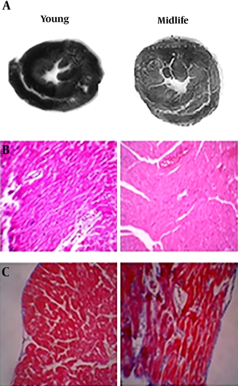
In this study, we examined some variables in young and middle-aged rats and also looked at the relationship of the variables with each other. Accordingly, the findings of this study showed that midlife causes an increase in heart weight (P = 0.001), body weight gain (P = 0.001), and pathological cardiac hypertrophy; consequently, a significant increase was seen in the ventricular wall thickness (P = 0.001) and ventricular internal diameter (P = 0.001). There was a significant correlation between weight gain due to midlife and increase in ventricular wall thickness (r =0.959, P = 0.001), ventricular internal diameter (r = 0.771. P = 0.001) and heart weight (r = 955, P = 0.001). Also, there was a significant relationship between the amount of H2O2 and ventricular wall thickness (r = 0.886, P = 0.001), ventricular internal diameter (r = 0.761, P = 0.001) and heart weight (r = 0.891, P = 0.001). There was a significant correlation between body weight and H2O2 (r = 0.952, P = 0.001). In the young rats, there was no significant relationship between these variables (Tables 1 - 3 and Figure 1).
| Variables | Midlife | Young | P Value |
|---|---|---|---|
| Internal diameter, nm | 1482.2 ± 128.47 | 464.5 ± 29.25 | 0.001 |
| Ventricular wall thickness, nm | 7978.8 ± 581.4 | 6455.5 ± 622.25 | 0.001 |
| Heart weight, g | 1.29 ± 0.14 | 0.633 ± 0.1 | 0.001 |
| Body weight, g | 646.4 ± 33.65 | 183.78 ± 14.96 | 0.001 |
| H2O2, intensity of DCF/mgP | 11.2 ± 0.85 | 7.43 ± 0.85 | 0.001 |
| Variables/Age | Response | |||||
|---|---|---|---|---|---|---|
| Internal Diameter | Ventricular Wall Thickness | Heart Weight | ||||
| r | P Value | r | P Value | r | P Value | |
| Body weight | ||||||
| Young | 0.609 | 0.1 | 0.02 | 0.94 | 0.309 | 0.456 |
| Midlife | 0.959 | 0.001 | 0.771 | 0.001 | 0.955 | 0.001 |
| H2O2 | ||||||
| Young | 0.275 | 0.51 | 0.156 | 0.71 | 0.368 | 0.39 |
| Midlife | 0.886 | 0.001 | 0.761 | 0.001 | 0.891 | 0.001 |
| Variables/Age | Response | |
|---|---|---|
| H2O2 | ||
| r | P Value | |
| Body weight | ||
| Young | 0.07 | 0.86 |
| Midlife | 0.952 | 0.001 |
4. Discussion
The findings of this study showed that midlife causes weight gain and increased heart weight. The midlife was associated with the development of pathological concentric cardiac hypertrophy. Also, weight gain resulted in an increase in the left ventricular wall thickness, ventricular internal diameter, and heart weight. In line with the findings of this study, Avelar et al. (2007) demonstrated that weight gain leads to cardiac hypertrophy (28). Cuspidi et al. (2014) also showed that midlife causes cardiac hypertrophy (26). It seems that midlife, through various mechanisms, can cause weight gain and cardiac hypertrophy (26). Previous studies have revealed that weight gain is inevitable if aging is associated with physical inactivity (2) because different tissues undergo hypertrophy and fibrosis (29), as well as an increase in the body fat (2). The heart is one of the tissues that is affected significantly by aging and physical inactivity which inevitably result in high oxidative stress (30). Various studies have also shown that aging can lead to increased oxidative stress (30). Our findings also indicated that the adult rats experienced a significant increase in H2O2 levels, and a significant relationship was seen between the elevated oxidative stress and ventricular internal diameter, ventricular wall thickness, and heart weight. Other studies also found that sedentary adult men suffered from an increase in oxidative stress (12, 31). Increasing oxidative stress is due to mitochondrial impairment and damage; thus, physical inactivity can effectively cause these effects (30). Bailey-Downs et al. (2012) reported that aging exacerbates obesity-induced oxidative stress and inflammation in perivascular adipose tissue in mice (32). Our findings also showed a significant relationship between weight gain and high levels of H2O2. The mitochondrial damage and impairment of bioenergetics caused by aging along with physical inactivity may reduce the antioxidant capacity to cope with reactive oxygen species, and this process can lead to the development of oxidative stress (30). Angiotensin-2 is one of the factors known to increase oxidative stress and cause cardiac hypertrophy, and midlife causes an increase in angiotensin-2 activity (11). The above changes can lead to fibrosis in the cardiac tissue. Imaging of heart tissue in adult rats in this study also showed fibrosis.
In this regard, our findings showed that the sedentary adult rats are confronted with pathological concentric cardiac hypertrophy. Murdolo et al. (2015) in a meta-analysis reported similar results (33). The left ventricular concentric hypertrophy is caused by diseases such as hypertension, which increases the risk of adverse cardiovascular prognosis with a high risk of death from cardiovascular disease (34). Existing evidence suggests that some degree of stress in the ventricular end-diastolic wall acts as a signal regulating hypertrophy. This can lead to more induction of left ventricular concentric hypertrophy in patients with hypertension with a small elevation of ventricular end-diastolic wall pressure, which is characterized by a significant increase in the relative wall thickness. Cardiomyocytes in myocardial concentric remodeling that have excessive thickness are indicative of increased diameters, but there is no significant increase in length. Therefore, the mean length by width (L/W) ratio is significantly reduced (35). This process is due to the orientation of the contractile sarcomere units added to cardiomyocytes (36). It seems that concentric cardiac hypertrophy appearing in adult rats may be associated with impaired blood supply to other tissues and reduce cardiac efficiency as well as cardiac and respiratory endurance.
4.1. Conclusions
To conclude, this study suggests that midlife is associated with pathological concentric cardiac hypertrophy in rats. Concomitant with weight gain, an increase is seen in the heart weight, left ventricular wall thickness, and left ventricular internal diameter. Therefore, midlife accompanied by physical inactivity causes cardiac fibrosis and impaired cardiac function.

