Abstract
Background:
Cisplatin (CPN) is widely used for the management of various malignant tumors.Objectives:
This study investigated the effects of Thymoquinone (TQN) on the expression of the p62 gene in CPN-induced testicular damage in mice.Methods:
Histomorphometry, testis injury scores, expression of p62, and protein levels of LC3-II were assessed.Results:
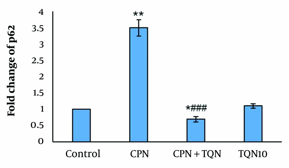
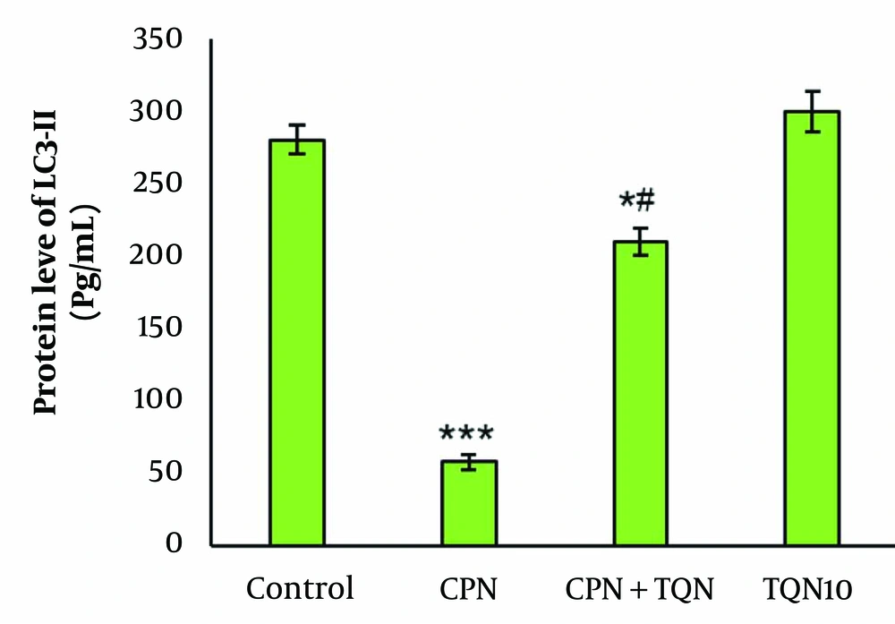
Cisplatin induced histological changes, increased p62 expression (P < 0.01), and reduced LC3-II levels (P < 0.001). Thymoquinone pretreatment decreased p62 expression while increasing LC3-II protein levels. Thymoquinone significantly reversed the testicular injury scores and improved histomorphometric parameters.Conclusions:
The results indicate that TQN enhances autophagy and improves testicular tissue in CPN-intoxicated mice.Keywords
1. Background
Cisplatin (CPN) is widely used for the management of various malignant tumors. However, CPN impairs the male reproductive system and can cause infertility in men undergoing chemotherapy (1-3). Cisplatin leads to impaired steroidogenesis, germ cell apoptosis, and changes in testicular histology (1, 4, 5). Previous studies have shown that CPN disrupts autophagy in the testes (6, 7).
Autophagy-related genes are highly expressed in spermatozoa, suggesting that autophagy regulates sperm quality. LC3, a biomarker of autophagy, is activated during sperm capacitation and the acrosome reaction (8-10).
P62 is an autophagy receptor that suppresses autophagy. This multifunctional protein integrates death and survival signals, modulating apoptosis and autophagy to maintain cellular homeostasis (11).
Black seed (Nigella sativa) is widely used in traditional medicine in Iran. Thymoquinone (TQN), derived from Nigella sativa, has shown numerous pharmacological and biological effects (12-15).
Thymoquinone has been found to enhance spermatogenesis and improve testicular damage caused by ischemia-reperfusion injury (16) and testicular torsion (17). Moreover, TQN protects against testicular damage induced by bleomycin (18), cyclophosphamide, and methotrexate through anti-inflammatory, anti-apoptotic, and antioxidant mechanisms (18-20).
2. Objectives
Given the important role of autophagy in spermatogenesis and spermiogenesis, this study aimed to investigate the effects of TQN and CPN on autophagy in mouse testicular tissue by evaluating the expression of p62 and LC3-II.
3. Methods
3.1. Animals
Thirty-two NMRI mice (6 - 8 weeks old, 25 - 30 g) were kept under standard conditions (12-hour dark/light cycles, 20 - 25°C). The animals were divided into the following groups:
- Control: Intraperitoneal (i.p.) injection of normal saline for 35 days.
- CPN: i.p. injection of CPN (7 mg/kg) on days 30 - 35 (21).
- TQN10: i.p. injection of TQN (10 mg/kg) for 35 days (22).
- TQN + CPN: i.p. injection of TQN for 35 days and CPN on days 30 - 35.
At the end of the experiment, the right testicles were preserved in Bouin's fixative for histomorphometric assessment, and the left testicles were stored at -70°C for evaluating autophagy.
3.2. Histology
After tissue processing, the slides were prepared and stained with Hematoxylin and Eosin. Two researchers, blinded to group allocation, independently analyzed the slides. Seminiferous tubule diameters and germinal epithelium height were calculated using Motic software. One hundred tubules per animal were evaluated.
Testicular injury was scored using the Cosentino grading system (23), which grades testicular damage from I to IV:
- Grade I: Normal testis tissue.
- Grade II: Closely packed seminiferous tubules with non-cohesive germinal cells.
- Grade III: Sloughing and shrinking of the seminiferous epithelium.
- Grade IV: Closely packed seminiferous tubules with coagulated necrosis.
2.3. Real-time PCR
The RNA from the testicles was isolated using a RNeasy Mini kit (Qiagen) and converted to cDNA. A PCR reaction (25 μL) containing cDNA, primers (Table 1), DEPC water, and SYBR Green (Qiagen) was prepared. The relative expression of p62 was normalized to GAPDH using REST software.
Primer Sequences
Genes | Forward | Reverse |
---|---|---|
p62 | GCTCAGGAGGAGACGATGAC | AGAAACCCATGGACAGCATC |
GAPDH | GCTGGACATTGGACTTCCTC | ACCACTGTGACCTGCTCCA |
2.4. Protein Levels of LC3-II
The levels of LC3-II were measured in the culture supernatants using a commercially available ELISA kit (Abcam, USA) according to the manufacturer's instructions. Both primary and secondary antibodies were added to the tissue lysates. The protein levels were quantified using an ELISA reader (BioTek, USA) by measuring the absorbance at 450 nm.
2.5. Statistical Analysis
A one-way analysis of variance (ANOVA) was performed using SPSS (version 21.0), followed by a post hoc test (LSD or Kruskal-Wallis). A P-value of < 0.05 was considered statistically significant.
4. Results
4.1. Histology
The mean Cosentino score in the CPN-treated mice was significantly reduced compared to the control group. Thymoquinone treatment was able to reverse the mean Cosentino score (Table 2).
Histological Assessment and Mean Cosentino Score of Right Testicles a
Groups | Grade I | Grade II | Grade III | Grade IV | Mean ± SD |
---|---|---|---|---|---|
Control (n = 8) | 8 | - | - | - | 1.0 ± 0.0 |
CPN (n = 8) | - | - | 5 | 3 | 3. 8 ± 0.4** |
CPN+TQN (n = 8) | 1 | 3 | 2 | 2 | 1.6 ± 0.2*# |
TQN10 (n = 8) | 8 | - | - | - | 1.0 ± 0.0 |
There were no significant changes in the morphometric parameters in the TQN10 group. However, in the CPN group, morphometric parameters were significantly decreased compared to the control (P < 0.01). In the TQN + CPN group, morphometric parameters were significantly higher than those in the CPN group (P < 0.05) (Figure 1).
Morphmetric parameters (mean ± SD; n = 8). * and # indicate a comparison to the control and cisplatin (CPN).
4.2. Autophagy Assessments
There were no significant changes in p62 gene expression or LC3-II protein levels in the TQN10 group. In the CPN group, p62 expression was significantly upregulated, while LC3-II protein levels were downregulated (P < 0.01). In the TQN + CPN group, LC3-II levels were markedly increased compared to the CPN-treated mice, and p62 expression was significantly reduced compared to the CPN group (Figures 2 and 3).
Expression of p62 gene (mean ± SD; n = 4). * and # indicate a comparison to the control and cisplatin (CPN).
Protein level of LC3-II (mean ± SD; n = 4). * and # indicate a comparison to the control and cisplatin (CPN).
5. Discussion
In this study, CPN induced impaired testicular tissue, increased expression of the p62 gene, and reduced expression of the LC3-II protein. The administration of TQN significantly reversed these effects.
The decrease in germinal epithelium height and seminiferous tubule diameter indicates the toxic impact of CPN. The increased Cosentino score further confirms the testicular toxicity caused by CPN.
The decline in morphometric parameters and the rise in the Cosentino score may suggest an apoptotic effect of CPN. A previous study showed that CPN reduced sperm quality, decreased the diameter of seminiferous tubules, and induced apoptosis in rats (24). In our earlier research, CPN increased Bax (a pro-apoptotic factor) in the mouse testis (25). The observed improvement in histological changes suggests that TQN protects against CPN-induced testicular damage. In another study, TQN ameliorated histological changes in testicular tissue following ischemia (17).
The improvement in histomorphometric parameters and reduction in the Cosentino score in this study may indicate the anti-apoptotic effects of TQN. Anti-apoptotic effects of TQN on damaged testes have been reported previously (26-28). Thymoquinone was shown to reduce apoptosis in the testicles of doxorubicin-intoxicated animals (29). In another study, TQN significantly decreased apoptosis in varicocele-induced rats (26).
In this study, the increased fold change of the p62 gene and the reduced protein level of LC3-II suggest that CPN suppresses autophagy. Therefore, CPN may impair spermatogenesis by inhibiting autophagy.
In our study, TQN pretreatment elevated LC3-II protein levels while decreasing the expression of p62 in the CPN group. These findings suggest that TQN promotes autophagy in CPN-induced testicular damage. Liao et al. showed that CPN activated autophagy in renal damaged tissue through a marked increase in p62 (30).
Interestingly, we found that CPN-induced testicular damage was associated with autophagy inhibition. In contrast, TQN decreased p62 expression, increased LC3-II protein levels, and improved testicular histology.
Liu et al. demonstrated that TQN reduces doxorubicin-induced apoptosis in H9c2 cardiomyocytes by inducing autophagy (31). In another study, TQN induced autophagy in renal cancer cells (32). Additionally, TQN attenuates lipid accumulation by activating autophagy in nonalcoholic fatty liver disease (33).
5.1. Conclusions
Our results demonstrated that TQN activated autophagy by suppressing p62 expression. These data suggest that TQN could be a promising adjunct therapy against CPN-induced testicular toxicity in future anticancer clinical practice.
References
-
1.
Park HJ, Kim JS, Lee R, Song H. Cisplatin Induces Apoptosis in Mouse Neonatal Testes Organ Culture. Int J Mol Sci. 2022;23(21). [PubMed ID: 36362147]. [PubMed Central ID: PMC9658841]. https://doi.org/10.3390/ijms232113360.
-
2.
Abdel-Latif R, Fathy M, Anwar HA, Naseem M, Dandekar T, Othman EM. Cisplatin-Induced Reproductive Toxicity and Oxidative Stress: Ameliorative Effect of Kinetin. Antioxidants (Basel). 2022;11(5). [PubMed ID: 35624727]. [PubMed Central ID: PMC9137797]. https://doi.org/10.3390/antiox11050863.
-
3.
Akbal C, Turker P, Ozyurek M, Erkanli G, Simsek F, Turkeri L. A new cause of male infertility after cisplatin exposure: the effect of cisplatin on Y chromosomes. Urol. 2009;73(5):1145-9. [PubMed ID: 18407334]. https://doi.org/10.1016/j.urology.2008.02.028.
-
4.
Garcia MM, Acquier A, Suarez G, Gomez NV, Gorostizaga A, Mendez CF, et al. Cisplatin inhibits testosterone synthesis by a mechanism that includes the action of reactive oxygen species (ROS) at the level of P450scc. Chem Biol Interact. 2012;199(3):185-91. [PubMed ID: 22940207]. https://doi.org/10.1016/j.cbi.2012.08.012.
-
5.
Yadav YC. Effect of cisplatin on pancreas and testies in Wistar rats: biochemical parameters and histology. Heliyon. 2019;5(8). e02247. [PubMed ID: 31453403]. [PubMed Central ID: PMC6700420]. https://doi.org/10.1016/j.heliyon.2019.e02247.
-
6.
Ceylan T, Karabulut D, ÖZtÜRk E, AkİN AT, Kaymak E, Yakan B. Histological evaluation of the effects of rapamycin and 3-methyladenine on cisplatin-induced epididymal injury in rats. Cukurova Med J. 2021;46(3):1184-90. https://doi.org/10.17826/cumj.924352.
-
7.
Yuan M, Yao Y, Wu D, Zhu C, Dong S, Tong X. Pannexin1 inhibits autophagy of cisplatin-resistant testicular cancer cells by mediating ATP release. Cell Cycle. 2022;21(15):1651-61. [PubMed ID: 35373707]. [PubMed Central ID: PMC9291690]. https://doi.org/10.1080/15384101.2022.2060655.
-
8.
Aparicio IM, Espino J, Bejarano I, Gallardo-Soler A, Campo ML, Salido GM, et al. Autophagy-related proteins are functionally active in human spermatozoa and may be involved in the regulation of cell survival and motility. Sci Rep. 2016;6:33647. [PubMed ID: 27633131]. [PubMed Central ID: PMC5025659]. https://doi.org/10.1038/srep33647.
-
9.
Guo Y, Ma Y, Zhang J, Jiang S, Yuan G, Cheng J, et al. Alteration in autophagy gene expression profile correlates with low sperm quality. Reprod Biol. 2021;21(4):100546. [PubMed ID: 34428669]. https://doi.org/10.1016/j.repbio.2021.100546.
-
10.
Aparicio IM, Rojo-Dominguez P, Castillejo-Rufo A, Pena FJ, Tapia JA. The Autophagy Marker LC3 Is Processed during the Sperm Capacitation and the Acrosome Reaction and Translocates to the Acrosome Where It Colocalizes with the Acrosomal Membranes in Horse Spermatozoa. Int J Mol Sci. 2023;24(2). [PubMed ID: 36674454]. [PubMed Central ID: PMC9862423]. https://doi.org/10.3390/ijms24020937.
-
11.
Yan XY, Qu XZ, Xu L, Yu SH, Tian R, Zhong XR, et al. Insight into the role of p62 in the cisplatin resistant mechanisms of ovarian cancer. Cancer Cell Int. 2020;20:128. [PubMed ID: 32322174]. [PubMed Central ID: PMC7164250]. https://doi.org/10.1186/s12935-020-01196-w.
-
12.
Kazemi R, Yazdanpanah E, Esmaeili SA, Yousefi B, Baharlou R, Haghmorad D. Thymoquinone improves experimental autoimmune encephalomyelitis by regulating both pro-inflammatory and anti-inflammatory cytokines. Mol Biol Rep. 2024;51(1):256. [PubMed ID: 38302802]. https://doi.org/10.1007/s11033-023-09148-z.
-
13.
Isaev NK, Genrikhs EE, Stelmashook EV. Antioxidant Thymoquinone and Its Potential in the Treatment of Neurological Diseases. Antioxidants (Basel). 2023;12(2). [PubMed ID: 36829993]. [PubMed Central ID: PMC9952318]. https://doi.org/10.3390/antiox12020433.
-
14.
Shabani H, Karami MH, Kolour J, Sayyahi Z, Parvin MA, Soghala S, et al. Anticancer activity of thymoquinone against breast cancer cells: Mechanisms of action and delivery approaches. Biomed Pharmacother. 2023;165:114972. [PubMed ID: 37481931]. https://doi.org/10.1016/j.biopha.2023.114972.
-
15.
Hofni A, Ali FEM, Ibrahim ARN, Aboubaker EM. Renoprotective Effect of Thymoquinone against Streptozotocin-Induced Diabetic Nephropathy: Role of NOX2 and Nrf2 Signals. Curr Mol Pharmacol. 2023;16(8):905-14. [PubMed ID: 36698232]. https://doi.org/10.2174/1874467216666230125150112.
-
16.
Erol B, Sari U, Amasyali AS, Ozkanli S, Sogut S, Hanci V, et al. Comparison of combined antioxidants and thymoquinone in the prevention of testis ischemia - reperfusion injury. Androl. 2017;5(1):119-24. [PubMed ID: 27748062]. https://doi.org/10.1111/andr.12268.
-
17.
Ayan M, Tas U, Sogut E, Cayli S, Kaya H, Esen M, et al. Protective effect of thymoquinone against testicular torsion induced oxidative injury. Andrologia. 2016;48(2):143-51. [PubMed ID: 25906970]. https://doi.org/10.1111/and.12424.
-
18.
Yaghutian Nezhad L, Mohseni Kouchesfahani H, Alaee S, Bakhtari A. Thymoquinone ameliorates bleomycin-induced reproductive toxicity in male Balb/c mice. Hum Exp Toxicol. 2021;40(12_suppl):S611-21. [PubMed ID: 34818114]. https://doi.org/10.1177/09603271211048184.
-
19.
Savran M, Ascı H, Armagan İ, Erzurumlu Y, Azırak S, Kaya Ozer M, et al. Thymoquinone could be protective against valproic acid‐induced testicular toxicity by antioxidant and anti‐inflammatory mechanisms. Andrologia. 2020;52(7). https://doi.org/10.1111/and.13623.
-
20.
Adana MY, Imam A, Bello AA, Sunmonu OE, Alege EP, Onigbolabi OG, et al. Oral thymoquinone modulates cyclophosphamide‐induced testicular toxicity in adolescent Wistar rats. Andrologia. 2022;54(4). https://doi.org/10.1111/and.14368.
-
21.
Aldemir M, Okulu E, Kosemehmetoglu K, Ener K, Topal F, Evirgen O, et al. Evaluation of the protective effect of quercetin against cisplatin-induced renal and testis tissue damage and sperm parameters in rats. Andrologia. 2014;46(10):1089-97. [PubMed ID: 24266675]. https://doi.org/10.1111/and.12197.
-
22.
Hassan E, El-Neweshy M, Hassan M, Noreldin A. Thymoquinone attenuates testicular and spermotoxicity following subchronic lead exposure in male rats: Possible mechanisms are involved. Life Sci. 2019;230:132-40. https://doi.org/10.1016/j.lfs.2019.05.067.
-
23.
Cosentino MJ, Nishida M, Rabinowitz R, Cockett AT. Histopathology of prepubertal rat testes subjected to various durations of spermatic cord torsion. J Androl. 1986;7(1):23-31. [PubMed ID: 3944017]. https://doi.org/10.1002/j.1939-4640.1986.tb00862.x.
-
24.
Favareto AP, de Toledo FC, Kempinas Wde G. Paternal treatment with cisplatin impairs reproduction of adult male offspring in rats. Reprod Toxicol. 2011;32(4):425-33. [PubMed ID: 22019602]. https://doi.org/10.1016/j.reprotox.2011.10.003.
-
25.
Shojaedini M, Hemadi M, Saki G, Fakhredini F, Khodayar MJ, Khorsandi L. Thymoquinone effects on autophagy, apoptosis, and oxidative stress in cisplatin-induced testicular damage in mice. J Assist Reprod Genet. 2024;41(7):1881-91. [PubMed ID: 38568464]. [PubMed Central ID: PMC11263301]. https://doi.org/10.1007/s10815-024-03097-7.
-
26.
Gur FM, Timurkaan S, Taskin E, Guven C, Gur HE, Senturk M, et al. Thymoquinone improves testicular damage and sperm quality in experimentally varicocele-induced adolescent rats. Andrologia. 2021;53(5). e14033. [PubMed ID: 33660882]. https://doi.org/10.1111/and.14033.
-
27.
Erboga M, Aktas C, Kurt O, Uygur R, Caglar V, Turan BC, et al. Protective effects of thymoquinone on experimental testicular ischaemia-reperfusion injury: an apoptotic, proliferative and biochemical study. Andrologia. 2016;48(2):222-30. https://doi.org/10.1111/and.12436.
-
28.
Attari SS, Mohammadi S, Ebrahimzadeh A, Hosseinzadeh H, Soukhtanloo M, Rajabzadeh A. Effects of Thymoquinone on Sperm Parameters, Apoptosis, Testosterone Level, and Oxidative Stress in a Mouse Model of D-Galactose-Induced Aging. Pharmaceutical Sci. 2018;24(3):180-6. https://doi.org/10.15171/ps.2018.26.
-
29.
Öztürk E, Kaymak E, Akin AT, Karabulut D, Ünsal H, Yakan B. Thymoquinone is a protective agent that reduces the negative effects of doxorubicin in rat testis. Hum Experimental Toxicol. 2020;39(10):1364-73. https://doi.org/10.1177/0960327120924108.
-
30.
Liao W, Wang Z, Fu Z, Ma H, Jiang M, Xu A, et al. p62/SQSTM1 protects against cisplatin-induced oxidative stress in kidneys by mediating the cross talk between autophagy and the Keap1-Nrf2 signalling pathway. Free Radic Res. 2019;53(7):800-14. [PubMed ID: 31223046]. https://doi.org/10.1080/10715762.2019.1635251.
-
31.
Liu D, Zhao L. Thymoquinone‑induced autophagy mitigates doxorubicin‑induced H9c2 cell apoptosis. Exp Ther Med. 2022;24(5):694. [PubMed ID: 36277157]. [PubMed Central ID: PMC9535332]. https://doi.org/10.3892/etm.2022.11630.
-
32.
Zhang Y, Fan Y, Huang S, Wang G, Han R, Lei F, et al. Thymoquinone inhibits the metastasis of renal cell cancer cells by inducing autophagy via AMPK/mTOR signaling pathway. Cancer Sci. 2018;109(12):3865-73. [PubMed ID: 30259603]. [PubMed Central ID: PMC6272120]. https://doi.org/10.1111/cas.13808.
-
33.
Zhang D, Zhang Y, Wang Z, Lei L. Thymoquinone attenuates hepatic lipid accumulation by inducing autophagy via AMPK/mTOR/ULK1-dependent pathway in nonalcoholic fatty liver disease. Phytother Res. 2023;37(3):781-97. [PubMed ID: 36479746]. https://doi.org/10.1002/ptr.7662.