


1. Background
Cervical cancer is the fourth most frequent malignancy that affects women. The World Health Organization (WHO) estimates that 530,000 new cases of cervical cancer are diagnosed worldwide that lead to 275,000 death each year (1). Approximately 90% of deaths are reported from low- and middle-income countries. The age-standardized rate (ASR) for cervical cancer in Iran is estimated to be 2.8, and its mortality is 1.2 per 100,000 patients. Cervical cancer is a preventable malignancy because it has long precancerous conditions, screening programs, and effective treatments for precancerous lesions (2).
Human Papillomavirus (HPV) has more than 100 genotypes, at least 12 of which are high-risk and associated with several cancers (3). Persistent infection with high-risk HPV types causes cellular abnormalities that can be an important precursor for carcinogenesis. The detection of HPV is a strong predictor of the development of invasive carcinoma. Human papillomavirus 16 and HPV18 are the most common genotypes linked to genital cancers. These two high-risk types cause about 70% of cervical carcinomas (4).
Cytology testing is commonly used to screen for cervical cancer, although its sensitivity for detecting disease stages or intraepithelial lesions is not very high. False-negative results in previous studies have ranged from 15 to 65% (5). Colposcopy is another method for the examination of the cervix. It has high sensitivity (94%) for detecting the stages of cervical carcinoma. Colposcopy can detect high-grade cervical intraepithelial neoplasia cases, but its specificity and reproducibility in women with minor cytological cervical lesions are not high (less than 50%) (6). Previous studies indicated that the genome of HPV is present in more than 90% of cervical carcinoma cases (7). Several studies have shown that HPV detection is better than cytology for the detection of severe cervical lesions. In a study conducted in Italy with a large sample size, screening with HPV testing was more effective than common cytology testing in the prevention of invasive cervical carcinoma (8).
Human papillomavirus testing can help detect pre-cancerous changes that are not clinically visible or detectable by cytology examination. Several studies demonstrated that combined cytology and HPV testing were not more effective than sole HPV testing for cervical cancer screening. Therefore, molecular testing for the detection and genotyping of HPV in cervical samples can improve the efficacy of cancer screening (9). The US Preventive Services Task Force (USPSTF) recommends screening every three years with cervical cytology alone, every five years with HPV testing alone, or in combination with cytology testing in women aged 30 to 65 years (10). It is noticed that the cytology and colposcopy examination may be cheaper than molecular tests in some countries; therefore, it is more used for cervical cancer screening (11).
2. Objectives
Data on the prevalence of high-risk HPV genotypes among women are not yet available for the total regions of Iran. The present study aimed to determine the prevalence of high-risk human papillomavirus types among women screened for cervical carcinoma in Yazd and compare the cytology, histology, and colposcopy examinations for the detection of high-grade cervical lesions.
3. Methods
3.1. Patients
This cross-sectional study enrolled 402 women referring to Gynecology Clinics of Shahid Sadoughi University of Medical Sciences, Yazd, Iran, between March 2016 and February 2017. Women with a history of abnormal Pap smear results, cervical colposcopy/biopsy, malignancy, and high-risk behaviors were excluded from the study. Demographic data, including age, number of pregnancies, age at first intercourse, and duration of marriage, were collected by a questionnaire. Women were examined by a gynecologist for genital lesions.
3.2. Human Papillomavirus Detection
Samples of all 402 women were examined by polymerase chain reaction (PCR) for detecting the HPV genome. Viral genomic DNA was isolated from samples using the AmpliSens® RIBO-prep Nucleic acid extraction kit (Russia) according to the manufacturer’s recommendation and was stored at -80°C. The HPV-DNA was detected and genotyped by the MolecuTech REBA HPV-ID kit (Molecules and Diagnostics, Wonju, Korea), which is a PCR-based reverse blot hybridization assay. This kit could identify 18 high-risk HPV genotypes (16, 18, 26, 31, 33, 35, 39, 45, 51, 52, 53, 56, 58, 59, 66, 68, 69, and 73), one probable high-risk HPV (34), and 13 low-risk HPV genotypes (6, 11, 32, 40, 42, 43, 44, 54, 70, 72, 81, 84, and 87). The nested PCR was performed with MY11 and MY9 primers in step 1, followed by the GP5/GP6 primer pair in step 2, using the Labcycler PCR system (SensoQuest, Germany). Then, PCR products were denatured with a denaturation solution and added into the membrane strip labeled with HPV genotype-specific probes. After washing unhybridized PCR products, the alkaline-phosphatase enzyme reaction was performed, and chromogen was added to develop detection signals. Finally, the strips were scanned using REBASCAN (YD Diagnostics Corp).
3.3. Cytology Examination
Cervical cytology specimens were collected using a cytobrush. The cells obtained were spread on two glass slides, and a common cytological examination with Papanicolaou staining was carried out. The Smears were seen by an experienced cytopathologist, and the findings were classified according to the Bethesda classification system (12).
3.4. Colposcopy
Colposcopy examinations were performed on women diagnosed with high-risk HPV by a gynecologic oncologist. The colposcopic findings were graded according to the Reids Colposcopic index (RCI) (13). Biopsies were taken from women for histological examinations.
3.5. Histological Examination
Biopsy samples were stained with hematoxylin-eosin and evaluated by a pathologist. The tissue specimens were classified according to the cervical intraepithelial neoplasia (CIN) classification system as normal (without dysplasia), low-grade dysplasia (CIN I), moderate dysplasia (CIN II), high-grade dysplasia (CIN III) (14). The pathologist had no information about the results of other tests, including HPV PCR, cytology, and colposcopy.
3.6. Statistical Analysis
The characteristics of the study participants were presented as numbers and proportions. Statistical analysis was carried out by SPSS version 18.0 software. The sensitivity, specificity, positive predictive value (PPV), negative predictive value (NPV), and accuracy were determined for colposcopy and Pap smear with the biopsy result as the gold standard. The chi-square test was used for comparing the number of pregnancies between women with and without HPV. Student’s t-test was performed for comparing the mean age, age at first intercourse, and duration of marriage between HPV-positive and HPV-negative women. P-values of less than 0.05 were considered significant.
4. Results
A total of 402 women aged 21 to 39 years with a mean of 30.82 ± 4.05 years were screened by cytology examination and HPV typing. The characteristics of the study population and the results of HPV testing are summarized in Table 1. There was no significant difference in the mean age, the number of pregnancies, and duration of marriage between HPV-positive and HPV-negative women (P-value > 0.05). The HPV-positive women had a lower mean age at first intercourse than HPV-negative people (P-value < 0.0001).
| Variable | HPV-Positive | HPV-Negative | Total | P-Value |
|---|---|---|---|---|
| Number of pregnancies | 0.441 | |||
| 0 | 10 | 76 | 86 | |
| 1 | 13 | 133 | 146 | |
| 2 | 6 | 11 | 17 | |
| 3 | 3 | 44 | 47 | |
| 4 | 0 | 7 | 7 | |
| Age | 31.03 ± 4.95 | 30. 8 ± 3.97 | 30.82 ± 4.05 | 0.760 |
| Age at first intercourse | 19.6 ± 2.44 | 22.1 ± 3.19 | 21.9 ± 3.21 | < 0.0001 |
| Duration of marriage, y | 10.03 ± 5.77 | 8.6 ± 4.18 | 8.74 ± 4.34 | 0.080 |
aValues are expressed as mean ± SD.
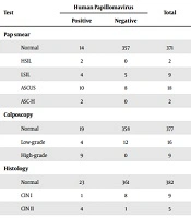
Thirty-two (7.97%) women were positive for high-risk HPV types (Table 2), of whom 11 (34.4%) patients were in the range of 20 - 30 years and 21 patients aged more than 30 years. Besides, HPV16 and HPV18 were the most frequent genotypes detected in participants. One woman was co-infected with subtypes 16 and 18. The results of Pap smear, colposcopy, and histology examinations in women infected with high-risk HPV types are summarized in Table 3. The results of Pap smear were abnormal in 56.2% of HPV-positive women, and ASCUS was the most common finding (31.2%). Among 32 women with high-risk HPV, 13 people had positive colposcopy results. The histology examination of eight HPV-positive patients showed CIN II and CIN III, while one woman had CIN I and 23 subjects had normal tissues. Physical examinations revealed genital warts in two HPV-positive women and cervical wounds in four patients, while 26 women had no visible cervical lesions.
| HPV Type | Values |
|---|---|
| 16 | 5 (15.6) |
| 16 and 18 | 1 (3.1) |
| 16 and others | 7 (21.9) |
| 18 and others | 2 (6.3) |
| Others | 17 (53.1) |
aValues are expressed as No. (%).
| Test | Human Papillomavirus | Total | |
|---|---|---|---|
| Positive | Negative | ||
| Pap smear | |||
| Normal | 14 | 357 | 371 |
| HSIL | 2 | 0 | 2 |
| LSIL | 4 | 5 | 9 |
| ASCUS | 10 | 8 | 18 |
| ASC-H | 2 | 0 | 2 |
| Colposcopy | |||
| Normal | 19 | 358 | 377 |
| Low-grade | 4 | 12 | 16 |
| High-grade | 9 | 0 | 9 |
| Histology | |||
| Normal | 23 | 361 | 382 |
| CIN I | 1 | 8 | 9 |
| CIN II | 4 | 1 | 5 |
| CIN III | 4 | 0 | 4 |
| Total | 32 | 370 | 402 |
Twenty-four women diagnosed with high-risk HPV types had anal intercourse. Ten women with CIN I or normal histology examination and eight patients with CIN II had abnormal Pap smear. Pap smear testing for the detection of women infected with high-risk types of HPV and cancerous lesions had 100% sensitivity and 58.3% specificity, while these values for colposcopy were 75% and 87.5%, respectively (Table 4). Combined colposcopy/Pap smear examination did not increase the sensitivity and specificity of detecting cancerous lesions in high-risk HPV-positive women compared to the Pap test.
| Test | Sensitivity, % | Specificity, % | PPV | NPV | Accuracy |
|---|---|---|---|---|---|
| Pap smear | 100 | 58.3 | 44.4 | 100 | 68.7 |
| Colposcopy | 75 | 87.5 | 66.7 | 91.3 | 84.3 |
| Pap smear and colposcopy | 100 | 58.3 | 44.4 | 100 | 100 |
Abbreviations: NPV, negative predictive value; PPV, positive predictive value.
5. Discussion
Human papillomavirus is a sexually transmitted infectious agent that is known as an important risk factor for developing cervical carcinoma. In the present study, the prevalence of high-risk HPV types among women living in Yazd, Iran, was about 8%. Besides, HPV16 and HPV18 were found in 40.6% of women infected with high-risk types. Data on the prevalence of high-risk HPV types among women are not yet available for the total regions of Iran. The rate of high-risk HPV types in most studies conducted in Iran has been reported higher than in the present study. In the study of Yousefzadeh et al. (15), 30.3% of women in Tehran were infected with high-risk types of HPV, 10.1% of which were subtypes 16 and 18. High-risk HPV types were prevalent among 61.3% of HPV-positive women in Kermanshah, and types 16 and 18 were detected in 26% of them (16). The prevalence of HPV genotypes among populations is varied because of the difference in laboratory tests for the detection of the virus, such as the used primers in PCR, the region and geographic locality, age categories, and type of population (2).
In the current study, Pap smear tests were abnormal in 56.2% of high-risk HPV-positive patients, and 71.9% of HPV-infected patients had normal histology in biopsy examinations. These results indicate that a significant number of women were infected with high-risk types of HPV, but they had normal cytology. They are at risk of developing cancer in the coming years. Hopman et al. (17) followed 68 women with normal cervical cytology who were infected with high-risk HPV types. Seventeen patients developed abnormal cytology within four years after virus detection, and high-grade dysplasia was seen in eight women (17). The HPV screening of women can help decrease the occurrence of precancerous and cancerous events in the cervix. The results of the current study highlight the importance of HPV screening relative to Pap smear for detecting predictable future cancer events.
In the present study, normal colposcopic results were obtained in 71.9% of high-risk HPV-positive women. The colposcopy had 75% sensitivity and 87.5% specificity for the detection of high-grade cervical lesions. Generally, the diagnostic sensitivity of CIN 2 or worse lesions by colposcopy is reported to be 30 - 70% (18). In a study by Zandvakili et al. (19) conducted in Iran, the sensitivity and specificity of colposcopy were 69.23% and 73.46%, respectively that were lower than our results. Zamani et al. (20) also reported a sensitivity of 60% and specificity of 64% for colposcopy. The specificity of colposcopy was higher in the current study than in studies conducted by Zivadinovic et al. (57%) (21), Adamopoulou et al. (50%) (9), Kohi et al. (57%) (22), and Ramesh et al. (46%) (23), but its sensitivity was lower than in the studies mentioned above.
Incompatible with the findings of the present study, several studies have reported that the sensitivity of colposcopy is higher than that of Pap test for the detection of high-grade cervical lesions (1, 22, 24). In a study conducted in Yazd, Iran, the sensitivity of Pap smear and colposcopy was 50% and 80%, respectively, which is in contrast to our results (25). However, a lower number of high-grade lesions in the current study than in previous studies may be a possible reason for the conflicting results. In addition, the difference in the quality of performing colposcopy and biopsy may affect the results of studies.
5.1. Conclusions
The prevalence of high-risk HPV types was relatively low among women living in Yazd than in those from other provinces of Iran. A significant percentage of high-risk HPV-infected patients had normal cervical cytology and histology. Therefore, the HPV screening test is recommended to decrease the development of high-grade cervical lesions and carcinoma. Colposcopy has an acceptable sensitivity and specificity for the detection of precancerous and cancerous cervical lesions.
