Establishment and Application of a Model for SARS-CoV-2 Infection of Human Nasal Mucosa Organoid

authors:

avatar Yan Yu 1 , avatar Zexin Chen 2 , avatar Junyuan Cao 3 , avatar Minmin Zhou 3 , avatar Jinyan Wei 1 , avatar Hairui Zheng 1 , avatar Junlang Li 4 , avatar Wei Wang 3 , avatar Gang Li 1 , *

Nanfang Hospital, Southern Medical University, Guangdong, China
Guangdong Research Center of Organoid Engineering and Technology, Guangdong, China
Wuhan Institute of Virology, Chinese Academy of Sciences, Wuhan, China
Guangzhou No.3 High School, Guangzhou, China

How To Cite Yu Y, Chen Z, Cao J, Zhou M, Wei J, et al. Establishment and Application of a Model for SARS-CoV-2 Infection of Human Nasal Mucosa Organoid. Jundishapur J Microbiol. 2024;17(10):e155640. https://doi.org/10.5812/jjm-155640.

Abstract

Background:

The COVID-19 pandemic underscores the need for effective models to study SARS-CoV-2 infection and evaluate antiviral therapies.

Objectives:

This study aimed to develop a human nasal organoid model to assess susceptibility to SARS-CoV-2 and its variants and to evaluate the antiviral effects of compounds such as camostat, remdesivir, and bergamottin.

Methods:

Nasal organoids were infected with SARS-CoV-2 and HCoV-OC43 pseudoviruses, followed by wild-type and variant strains in a BSL-3 laboratory. Viral content was measured at 2, 24, and 48 hours post-infection using qPCR, and infected cells were identified via immunofluorescence.

Results:

The organoids demonstrated susceptibility to SARS-CoV-2 and HCoV-OC43 pseudoviruses (P < 0.001) and were effectively infected with SARS-CoV-2 variants. Remdesivir, hesperidin, and bergamottin exhibited dose-dependent antiviral effects (P < 0.0001).

Conclusions:

Human nasal organoids represent a valuable model for studying SARS-CoV-2 infection and evaluating antiviral drugs, particularly for nasal applications in vitro.

1. Background

The SARS-CoV-2 outbreak has emphasized the need for effective treatment and control strategies. Traditional research models face limitations, such as the lack of physiological relevance in 2D cultured cell lines (1, 2) and discrepancies in animal models (3). Understanding the infection mechanisms of SARS-CoV-2, particularly in the nasal cavity—the primary target for respiratory viruses—is crucial (4). However, limited information is available regarding its infection routes and replication in the nasal mucosal epithelium, necessitating a model that accurately reflects human nasal cavity infection to advance early detection and antiviral evaluations (5).

Organoids, three-dimensional cultures derived from stem cells, mimic the structures and functions of human organs (6-8). They are valuable tools for studying tissue biology, disease modeling, and drug discovery (9-11). While research on respiratory organoids in the context of SARS-CoV-2 infection exists (12-15), studies on nasal mucosal organoids (NMOs) are scarce. This research focuses on NMOs, cultivated from human nasal mucosal basal cells, which include ciliated and secretory cells (16). Nasal mucosal organoids exhibit characteristics of typical nasal epithelium and express ACE2 and TMPRSS2, both essential for SARS-CoV-2 infection.

2. Objectives

This study aims to develop an in vitro viral infection model using NMOs to accurately simulate human nasal mucosa. This model, combined with pseudo-viral tools, offers a more precise representation of the infection process than traditional models. By evaluating the efficacy of antiviral drugs, we aim to support the development of effective nasal treatments for SARS-CoV-2, contributing to epidemic prevention and clinical care.

3. Methods

3.1. Tissue Samples

Human NMOs were generated from tissues obtained from Nanfang Hospital, in compliance with ethical guidelines.

3.2. Viruses

The study utilized several SARS-CoV-2 virus and pseudovirus strains (Appendix 1), with all experiments conducted in a biosafety level 3 (BSL-3) laboratory.

3.3. Nasal Mucosal Organoid Culture

Nasal tissues were processed according to the method described for nasal mucosal tissue preparation (6), digested with enzymes (collagenase IV and dispase), and cultured in Matrigel within trans-well plates (Corning, USA). The culture medium (Accuroid, China) was replaced every 2 - 3 days.

3.4. SARS-CoV-2 Infection

Nasal mucosal organoids were seeded at a density of 2.0 × 10⁴ cells per well in 100 μL and incubated at 37°C for 3 days. After the addition of the virus inoculum for 2 hours, the inoculum was replaced with culture medium for an additional 24 hours. Infection was assessed using fluorescence microscopy and a luciferase assay.

3.5. qRT-PCR

Total RNA was extracted from the organoids using TRIzol. The RNA was then converted to cDNA, and qRT-PCR was performed using custom primers (Appendix 2).

3.6. Flow Cytometry

Flow cytometry of NMOs was performed using an Attune CytPix flow cytometer (ThermoFisher, USA) with various cell marker antibodies detailed in Appendix 3.

3.7. Immunofluorescence

Post-infection, organoid samples were fixed, embedded, and processed for immunofluorescence analysis using primary and secondary antibodies to identify viral proteins.

3.8. Drug Treatment

Organoids were treated with camostat, remdesivir, and bergamottin (Appendix 4) for 1 hour prior to viral infection. Viral RNA was then measured by qPCR at 24 hours post-infection. To evaluate the impact of bergamottin, a CellTiter-Glo (Promega, USA) cell viability assay was conducted.

3.9. Statistics

Data were analyzed using three or more replicates, presented as mean ± standard deviation, and statistical significance was assessed using t-tests with a threshold of P < 0.05.

4. Results

4.1. Long-Term Culture and Validation of Nasal Mucosal Organoids

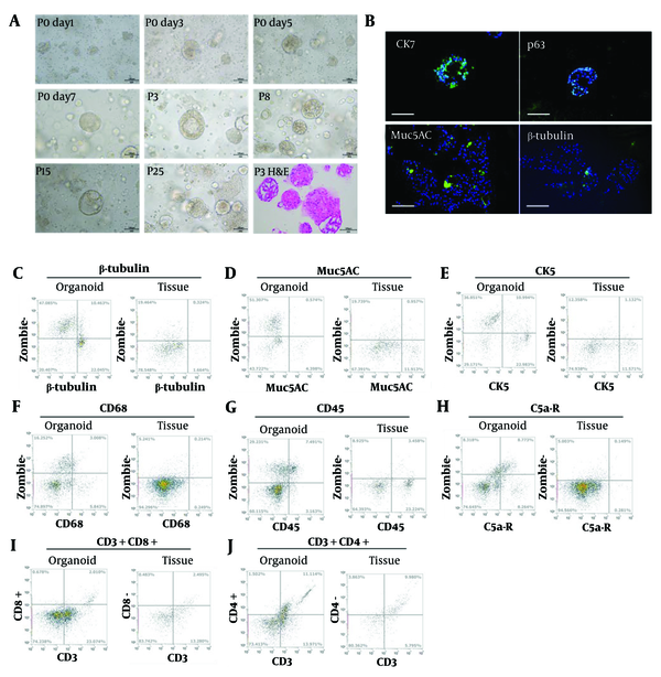
After seeding in Matrigel, NMOs grew from 20 μm to 200 μm within 3 days, forming solid or hollow structures (Figure 1A). Enzymatic and mechanical dissociation were utilized for passaging every 7 days, allowing at least 6 months of efficient expansion. Histological analysis using H&E staining (Figure 1A) confirmed that the NMOs closely resembled native tissue. Immunofluorescence staining identified key cell types (17), including epithelial cells (Cytokeratin 7) and ciliated cells (β-tubulin) (Figure 1B).

Growth dynamics of nasal mucosal organoids (NMOs). A, bright field image of NMO at day 1, 3, 5, 7 of passage 0; passage 3, 8, 15, and 25; and the H&E staining (Scale bar = 100 μm); B, immunofluorescence staining of Cytokeratin 7 (CK7), p63, Muc5AC, and β-tubulin. The flow-cytometry results of β-tubulin; C, Muc5AC; D, CK5; E, CD68; F, CD45; G, C5a-R; H, CD3 + CD8 +; I, CD3 + CD4 +; J of NMO and original tissue; K. Quantified results of the expression of cellular markers based on flow cytometry.

Flow cytometry revealed a higher percentage of CK7-positive epithelial cells and a lower percentage of CD45-positive lymphocytes (Figure 1C-J), along with an increase in β-tubulin-positive ciliated cells, which are essential for nasal mucosal function (18). Additionally, the NMOs supported immune cells, providing a foundation for further studies on the immune microenvironment.

4.2. Nasal Mucosal Organoids Susceptible to SARS-CoV-2 Pseudovirus and HCoV-OC43 Pseudovirus

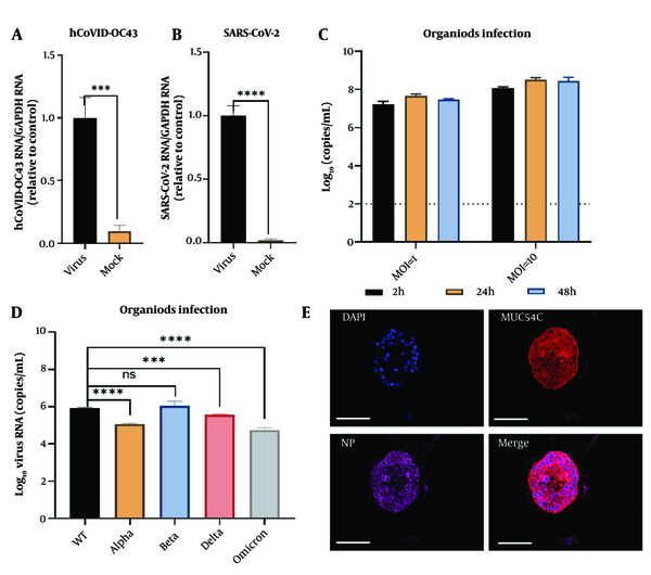
SARS-CoV-2 and HCoV-OC43 pseudoviruses were added to a 96-well plate (30 μL per well) and incubated at 37°C for 2 hours. The supernatant was then replaced with 100 μL of organoid culture medium and incubated for an additional 24 hours. After discarding the medium, the organoids were collected for intracellular viral RNA detection. Viral mRNA expression was significantly higher in the HCoV-OC43 group (P < 0.001) (Figure 2A) and even more elevated in the SARS-CoV-2 group (P < 0.0001) (Figure 2B), confirming the successful establishment of infection models for both viruses in human nasal mucosal epithelial organoids.

Modeling of SARS-CoV-2 infection in nasal mucosal organoids (NMOs). Pseudovirus virus HCoV-OC43 (A) and SARS-CoV2 (B) infection of nasal mucosal organoid; C, virus content in the supernatant at different times of infection for MOI = 1 and MOI = 10; D, detection of viral content in the supernatant under different types of virus of infection; E, immunofluorescence detection of goblet cells infected by SARS-CoV-2. nuclei (DAPI), goblet cells (Muc5AC), viral nucleoprotein (NP), and Merge (Scale bar = 100 μm).

4.3. Assessing Susceptibility of Nasal Mucosal Organoids to SARS-CoV-2 Live Virus

The virus was introduced to NMOs at multiplicities of infection (MOIs) of 1 and 10, followed by a 24-hour incubation. Detection of viral RNA revealed that the MOI = 10 group exhibited higher SARS-CoV-2 copy numbers compared to the MOI = 1 group (Figure 2C), suggesting a correlation between viral exposure and nasal cell susceptibility. Testing various SARS-CoV-2 strains demonstrated similar susceptibility and replication in NMOs (Figure 2D), with viral nucleocapsid primarily expressed in goblet cells, indicating potential sites for viral persistence (Figure 2E).

4.4. Antiviral Drugs Inhibit SARS-CoV-2 Infection in Nasal Mucosal Organoids

The antiviral drug testing capability of the NMO model was evaluated using three antiviral agents: Camostat, remdesivir, and bergamottin. Camostat and remdesivir have established antiviral efficacy, blocking TMPRSS2 and inhibiting RNA-dependent RNA polymerase, respectively (19, 20). Therefore, drug treatments were administered prior to viral introduction to evaluate their preventive efficacy.

Since the adverse effects of bergamottin are unclear, a safety test was conducted first. An ATP assay indicated no significant difference in NMO survival at bergamottin concentrations below 125 μM compared to the DMSO control, consistent with morphological assessments (Figure 3A). H&E staining confirmed the absence of pathological changes or apoptosis at these concentrations (Figure 3C).

Cell validation evaluation of bergamottin against nasal mucosal organoids (NMOs). A, morphological observation of NMOs treated with 125μM bergamottin scale bar = 1000 μm; B, H&E staining of NMOs under the influence of bergamottin; C, cell viability of NMOs under the influence of different concentrations of bergamottin at 24, 48, and 72 hours.

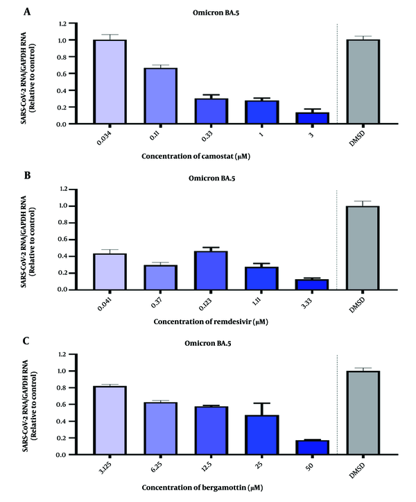
Following the infection of NMOs with SARS-CoV-2 Omicron BA.5 at MOI = 1 for 2 hours, camostat (3 μM) demonstrated dose-dependent inhibition (Figure 4A), while remdesivir reduced viral RNA levels by approximately 40% at 0.041 μM (Figure 4B). Bergamottin showed comparable inhibitory effects, particularly at 50 μM (Figure 4C).

Detection of the inhibitory effect of drugs on SARS-CoV-2 in human nasal mucosal organoids (NMOs). A, camostat at a concentration of 3 μM shows a dose-dependent inhibition of SARS-CoV-2 infection of human NMOs; B, remdesivir at a concentration of 3.33 μM shows a dose-dependent inhibition of SARS-CoV-2 infection of human NMOs. (Compare with DMSO control, **** P < 0.0001); C, bergamottin at a concentration of 50 μM shows a dose-dependent inhibition of SARS-CoV-2 infection of human NMOs. (Compare with DMSO control, **** P < 0.0001).

5. Discussion

Traditional virus research models, such as 2D-cultured immortalized nasal mucosal epithelial cell lines, are limited by their lack of tissue structure and physiological function, as they are derived from monoclonal cell lines. Consequently, they fail to accurately replicate infection processes and pathophysiological changes within the organ. These cell lines also exhibit a uniform genetic background, making them unsuitable for capturing inter-individual variations, thereby restricting their utility in population studies and high-throughput analyses (2, 21-24).

In contrast, coronavirus animal models, including rhesus macaques, transgenic mice, and ferrets, present significant physiological disparities compared to humans and are associated with high costs and biosafety risks (3, 25). The nasal cavity, as the primary barrier and initial target organ for respiratory viruses, plays a critical role in viral invasion, replication, and transmission. The nasal mucosa serves as a reservoir for viral replication and a trigger point for immune responses in COVID-19 infection (5). This makes NMOs an ideal model for investigating the mechanisms of COVID-19.

Pseudoviruses, engineered to mimic wild-type viruses, are non-replicating and undergo only a single round of infection, enhancing biosafety while allowing for detection and analysis via reporter genes (26). Both HCoV-OC43 and SARS-CoV-2, classified as β-coronaviruses, can cause a range of diseases in humans and animals (27, 28). Initial experiments using NMOs infected with HCoV-OC43 and SARS-CoV-2 pseudoviruses confirmed the susceptibility of NMOs to coronaviruses.

The nasal mucosa, composed of ciliated and goblet cells, exhibits high expression of ACE2 and TMPRSS2, making it the primary site of SARS-CoV-2 infection (29). The virus spreads through goblet cell secretions and ciliary movement (30). Our experiments demonstrated a positive correlation between higher viral exposure doses and increased infection rates and replication. Immunofluorescence analysis revealed that goblet cells are particularly susceptible to SARS-CoV-2, consistent with previous findings (30).

Post-infection, we observed a continuous increase in viral gene copy numbers in NMOs, indicating effective infection and replication. This confirms that NMOs can replicate the complete lifecycle of the virus. The nasal mucosa's large surface area and rich blood supply facilitate efficient drug delivery while minimizing systemic side effects (31-34). Following the COVID-19 outbreak, various nasal-administered antiviral drugs have been developed, yet the lack of robust preclinical models remains a significant bottleneck. The establishment of our human nasal mucosa model for SARS-CoV-2 provides a cost-effective, high-throughput platform for drug evaluation.

The primary limitations of this study include a small sample size and the absence of analysis on the variability of viral infection across different populations. Additionally, the antiviral mechanism of bergamottin was not addressed in this study and warrants further investigation.

5.1. Conclusions

Our research demonstrates that camostat, remdesivir, and bergamottin exhibit dose-dependent antiviral properties against SARS-CoV-2 in NMOs, confirming the model’s efficacy as a drug evaluation platform. In conclusion, we have developed a stable in vitro model of SARS-CoV-2 infection using NMOs, which can be utilized for studying infection mechanisms and screening antiviral drugs, offering valuable insights into nasal drug administration for COVID-19 prevention and treatment.
References

  • 1.

    Ballard DH, Boyer CJ, Alexander JS. Organoids—preclinical models of human disease. New England J Med. 2019;380(20):1981-2. https://doi.org/10.1056/NEJMc1903253.

  • 2.

    Kondo J, Inoue M. Application of cancer organoid model for drug screening and personalized therapy. Cells. 2019;8(5). [PubMed ID: 31108870]. [PubMed Central ID: PMC6562517]. https://doi.org/10.3390/cells8050470.

  • 3.

    Cleary SJ, Magnen M, Looney MR, Page CP. Update on animal models for COVID‐19 research. British J Pharmacol. 2020;177(24):5679-81. https://doi.org/10.1111/bph.15266.

  • 4.

    Li C, Chiu MC, Yu Y, Liu X, Xiao D, Huang J, et al. Establishing human lung organoids and proximal differentiation to generate mature airway organoids. 1940-087X; 2022, [cited 181]. Available from: https://app.jove.com/63684.

  • 5.

    Zou L, Ruan F, Huang M, Liang L, Huang H, Hong Z, et al. SARS-CoV-2 viral load in upper respiratory specimens of infected patients. N Engl J Med. 2020;382(12):1177-9. [PubMed ID: 32074444]. [PubMed Central ID: PMC7121626]. https://doi.org/10.1056/NEJMc2001737.

  • 6.

    Wang XW, Xia TL, Tang HC, Liu X, Han R, Zou X, et al. Establishment of a patient-derived organoid model and living biobank for nasopharyngeal carcinoma. Ann Transl Med. 2022;10(9):526. [PubMed ID: 35928731]. [PubMed Central ID: PMC9347035]. https://doi.org/10.21037/atm-22-1076.

  • 7.

    Liu Y, Zhou Y, Chen P. Lung cancer organoids: Models for preclinical research and precision medicine. Front Oncol. 2023;13:1293441. [PubMed ID: 37941550]. [PubMed Central ID: PMC10628480]. https://doi.org/10.3389/fonc.2023.1293441.

  • 8.

    Schutgens F, Clevers H. Human organoids: Tools for understanding biology and treating diseases. Annu Rev Pathol. 2020;15:211-34. [PubMed ID: 31550983]. https://doi.org/10.1146/annurev-pathmechdis-012419-032611.

  • 9.

    Tsang JO, Zhou J, Zhao X, Li C, Zou Z, Yin F, et al. Development of three-dimensional human intestinal organoids as a physiologically relevant model for characterizing the viral replication kinetics and antiviral susceptibility of enteroviruses. Biomedicines. 2021;9(1). [PubMed ID: 33477611]. [PubMed Central ID: PMC7831294]. https://doi.org/10.3390/biomedicines9010088.

  • 10.

    Bredenoord AL, Clevers H, Knoblich JA. Human tissues in a dish: The research and ethical implications of organoid technology. Science. 2017;355(6322). [PubMed ID: 28104841]. https://doi.org/10.1126/science.aaf9414.

  • 11.

    Li M, Izpisua Belmonte JC. Organoids—preclinical models of human disease. New England J Med. 2019;380(20):1981-2. https://doi.org/10.1056/NEJMc1903253.

  • 12.

    Shapira T, Monreal IA, Dion SP, Buchholz DW, Imbiakha B, Olmstead AD, et al. A TMPRSS2 inhibitor acts as a pan-SARS-CoV-2 prophylactic and therapeutic. Nature. 2022;605(7909):340-8. [PubMed ID: 35344983]. [PubMed Central ID: PMC9095466]. https://doi.org/10.1038/s41586-022-04661-w.

  • 13.

    Elbadawy M, Kato Y, Saito N, Hayashi K, Abugomaa A, Kobayashi M, et al. Establishment of intestinal organoid from rousettus leschenaultii and the susceptibility to bat-associated viruses, SARS-CoV-2 and pteropine orthoreovirus. Int J Mol Sci. 2021;22(19). [PubMed ID: 34639103]. [PubMed Central ID: PMC8509532]. https://doi.org/10.3390/ijms221910763.

  • 14.

    Lamers MM, Beumer J, van der Vaart J, Knoops K, Puschhof J, Breugem TI, et al. SARS-CoV-2 productively infects human gut enterocytes. Science. 2020;369(6499):50-4. [PubMed ID: 32358202]. [PubMed Central ID: PMC7199907]. https://doi.org/10.1126/science.abc1669.

  • 15.

    Han Y, Duan X, Yang L, Nilsson-Payant BE, Wang P, Duan F, et al. Identification of SARS-CoV-2 inhibitors using lung and colonic organoids. Nature. 2021;589(7841):270-5. [PubMed ID: 33116299]. [PubMed Central ID: PMC8034380]. https://doi.org/10.1038/s41586-020-2901-9.

  • 16.

    Chiu MC, Li C, Liu X, Yu Y, Huang J, Wan Z, et al. A bipotential organoid model of respiratory epithelium recapitulates high infectivity of SARS-CoV-2 Omicron variant. Cell Discov. 2022;8(1):57. [PubMed ID: 35710786]. [PubMed Central ID: PMC9203776]. https://doi.org/10.1038/s41421-022-00422-1.

  • 17.

    Ualiyeva S, Lemire E, Wong C, Perniss A, Boyd AA, Aviles EC, et al. A nasal cell atlas reveals heterogeneity of tuft cells and their role in directing olfactory stem cell proliferation. Sci Immunol. 2024;9(92):eabq4341. [PubMed ID: 38306414]. [PubMed Central ID: PMC11127180]. https://doi.org/10.1126/sciimmunol.abq4341.

  • 18.

    Davis JD, Wypych TP. Cellular and functional heterogeneity of the airway epithelium. Mucosal Immunol. 2021;14(5):978-90. [PubMed ID: 33608655]. [PubMed Central ID: PMC7893625]. https://doi.org/10.1038/s41385-020-00370-7.

  • 19.

    Hoffmann M, Hofmann-Winkler H, Smith JC, Kruger N, Arora P, Sorensen LK, et al. Camostat mesylate inhibits SARS-CoV-2 activation by TMPRSS2-related proteases and its metabolite GBPA exerts antiviral activity. E Bio Med. 2021;65:103255. [PubMed ID: 33676899]. [PubMed Central ID: PMC7930809]. https://doi.org/10.1016/j.ebiom.2021.103255.

  • 20.

    Gordon DM, Neifer KL, Hamoud AA, Hawk CF, Nestor-Kalinoski AL, Miruzzi SA, et al. Bilirubin remodels murine white adipose tissue by reshaping mitochondrial activity and the coregulator profile of peroxisome proliferator–activated receptor α. J Biological Chem. 2020;295(29):9804-22. https://doi.org/10.1074/jbc.RA120.013700.

  • 21.

    Papazian D, Wurtzen PA, Hansen SW. Polarized airway epithelial models for immunological co-culture studies. Int Arch Allergy Immunol. 2016;170(1):1-21. [PubMed ID: 27240620]. https://doi.org/10.1159/000445833.

  • 22.

    Qin X, Sufi J, Vlckova P, Kyriakidou P, Acton SE, Li VSW, et al. Cell-type-specific signaling networks in heterocellular organoids. Nat Methods. 2020;17(3):335-42. [PubMed ID: 32066960]. [PubMed Central ID: PMC7060080]. https://doi.org/10.1038/s41592-020-0737-8.

  • 23.

    Charles DD, Fisher JR, Hoskinson SM, Medina-Colorado AA, Shen YC, Chaaban MR, et al. Development of a novel ex vivo nasal epithelial cell model supporting colonization with human nasal microbiota. Front Cell Infect Microbiol. 2019;9:165. [PubMed ID: 31165051]. [PubMed Central ID: PMC6536665]. https://doi.org/10.3389/fcimb.2019.00165.

  • 24.

    Antoni D, Burckel H, Josset E, Noel G. Three-dimensional cell culture: A breakthrough in vivo. Int J Mol Sci. 2015;16(3):5517-27. [PubMed ID: 25768338]. [PubMed Central ID: PMC4394490]. https://doi.org/10.3390/ijms16035517.

  • 25.

    Zhou J, Li C, Liu X, Chiu MC, Zhao X, Wang D, et al. Infection of bat and human intestinal organoids by SARS-CoV-2. Nat Med. 2020;26:1077-83.

  • 26.

    Zhang Z, Zhang J, Xia N, Zhao Q. Expanded strain coverage for a highly successful public health tool: Prophylactic 9-valent human papillomavirus vaccine. Human Vaccines Immunotherapeutics. 2017;13(10):2280-91. https://doi.org/10.1080/21645515.2017.1346755.

  • 27.

    Su S, Wong G, Shi W, Liu J, Lai ACK, Zhou J, et al. Epidemiology, genetic recombination, and pathogenesis of coronaviruses. Trends Microbiol. 2016;24(6):490-502. [PubMed ID: 27012512]. [PubMed Central ID: PMC7125511]. https://doi.org/10.1016/j.tim.2016.03.003.

  • 28.

    Lednicky JA, Waltzek TB, McGeehan E, Loeb JC, Hamilton SB, Luetke MC. Isolation and genetic characterization of human coronavirus NL63 in primary human renal proximal tubular epithelial cells obtained from a commercial supplier, and confirmation of its replication in two different types of human primary kidney cells. Virol J. 2013;10:213. [PubMed ID: 23805916]. [PubMed Central ID: PMC3716658]. https://doi.org/10.1186/1743-422X-10-213.

  • 29.

    Sungnak W, Huang N, Becavin C, Berg M, Queen R, Litvinukova M, et al. SARS-CoV-2 entry factors are highly expressed in nasal epithelial cells together with innate immune genes. Nat Med. 2020;26(5):681-7. [PubMed ID: 32327758]. [PubMed Central ID: PMC8637938]. https://doi.org/10.1038/s41591-020-0868-6.

  • 30.

    Wu CT, Lidsky PV, Xiao Y, Cheng R, Lee IT, Nakayama T, et al. SARS-CoV-2 replication in airway epithelia requires motile cilia and microvillar reprogramming. Cell. 2023;186(1):112-130 e20. [PubMed ID: 36580912]. [PubMed Central ID: PMC9715480]. https://doi.org/10.1016/j.cell.2022.11.030.

  • 31.

    Antunes JL, Amado J, Veiga F, Paiva-Santos AC, Pires PC. Nanosystems, drug molecule functionalization and intranasal delivery: An update on the most promising strategies for increasing the therapeutic efficacy of antidepressant and anxiolytic drugs. Pharmaceutics. 2023;15(3). [PubMed ID: 36986859]. [PubMed Central ID: PMC10054777]. https://doi.org/10.3390/pharmaceutics15030998.

  • 32.

    Ferreira MD, Duarte J, Veiga F, Paiva-Santos AC, Pires PC. Nanosystems for brain targeting of antipsychotic drugs: An update on the most promising nanocarriers for increased bioavailability and therapeutic efficacy. Pharmaceutics. 2023;15(2). [PubMed ID: 36840000]. [PubMed Central ID: PMC9959474]. https://doi.org/10.3390/pharmaceutics15020678.

  • 33.

    Alberto M, Paiva-Santos AC, Veiga F, Pires PC. Lipid and polymeric nanoparticles: Successful strategies for nose-to-brain drug delivery in the treatment of depression and anxiety disorders. Pharmaceutics. 2022;14(12). [PubMed ID: 36559236]. [PubMed Central ID: PMC9783528]. https://doi.org/10.3390/pharmaceutics14122742.

  • 34.

    Ulusoy S, Bayar Muluk N, Karpischenko S, Passali GC, Negm H, Passali D, et al. Mechanisms and solutions for nasal drug delivery - a narrative review. Eur Rev Med Pharmacol Sci. 2022;26(2 Suppl):72-81. [PubMed ID: 36524914]. https://doi.org/10.26355/eurrev_202212_30487.