


1. Background
The most deleterious fungal infections to humans are caused by Candida species (1-3). Collectively, they are responsible for a huge number of infections in humans, from superficial candidiasis to life-threatening candidiasis (3). In the genus there are around 150 species, but most of the contagions are instigated by the Candida albicans, C. glabrata, and a small number of other species (3, 4). Candida albicans accounts for half of the dispersed disease and, as the most dominant species, is capable of causing life-threatening infectious disease (5). Candida glabrata is a dangerous pathogen that is remarkably important for its intrinsic tendency to develop resistance to antifungal drugs (6). It is the second main cause of fungal infections in humans (7).
All species across the plant kingdom have been found to harbor endophytes (8). Endophytic microbes are mostly bacteria or fungi that reside inside the living tissue of plants without any injury to their hosts and can be vastly diverse source of natural bioactive products (9, 10). Endophytes have been described in several distinguished therapeutic plants, ornamental plants, fruit plants, and weeds from wild and cultivated locations. Mutually endophytic bacteria and endophytic fungi be able to co-exist in a particular host plant without any conflict (11, 12). Generally through the roots and the aerial portions of plants endophytes enter to plants, such as stems, flowers, cotyledons and leaves (13). At the point of entry they are localized and be able to grow in the body of host plant (14). Once entering the host, they live inside cells, in the vascular tissue system or in the intercellular places (15). After getting residence inside the plant tissues, the endophytes are well-known to produce a various types of organic products that can act as a steady and successful resource for drugs. Thus, natural products from endophytes are considered to have a huge prospective not only in the pharmacological trade, but also in the biotechnology and agrochemical trades (16).
Endophytic bacteria are known to have a number of bioactive compounds and are known to offer potential broad spectrum antimicrobial potential activity (17); therefore, they may be potential natural control agents against diverse fungal diseases (18-21) or have antimicrobial potential (18-20). Endophytic bacteria isolated from medicinal plants, such as Tinospora cordifolia Miers, Memecylon edule Roxb., Dipterocarpus tuberculatus Roxb., and Phyllodium pulchellum (Benth.) Desv., have displayed antimicrobial activity (22). The anticandidal potential of endophytic bacteria is highly dependent on their genotype and their chemical compositions. The fern Dryopteris uniformis (Makino) is a commonly available plant in the Korean peninsula and is used as both a food plant and an ornamental plant (23). The diversity of endophytic fungi from ferns has been reported, but there are no reports on endophytic bacteria distribution and application within a fern (24, 25). The search for new biological products derived from endophytic bacteria is likely to be successful as endophytic bacteria are capable antifungals to fight against resistant fungal species (26).
2. Objectives
Consequently, in the current study we aimed to isolate endophytic bacteria from D. uniformis, identify them, investigated their anticandidal potential and confirm their anticandidal potential.
3. Methods
3.1. Collection, Isolation, and Morphological Illustration of Endophytic Bacteria
Dryopteris uniformis was collected from Bohyun Mountain, Yeongchun, Kyungbuk province, Republic of Korea and identified by Prof. Seonjoo Park, a plant taxonomist at Department of Life Sciences, Yeungnam University. The specimen was deposited as the Voucher ID: AR0009 (2015) at YNUH (Yeungnam University Herbarium), South Korea. The isolation of endophytic bacteria was conducted by using a standard protocol (27). Two grams of separated tissues (leaf, stem/root) of D. uniformis were washed thoroughly in tap water to remove the soil. Surface sterilization was performed by the application of 70% ethanol for 60 seconds, followed by 2% sodium hypochlorite for 90 seconds, and 30 seconds in 100% ethanol; subsequently, the tissues were washed five times using sterilized water. The tissues were dried out in sterile conditions, ground by using a mortar and pestle, and incubated with 6 mL of 0.9% NaCl for 3 hours at 24°C to allow the total discharge of endophytic bacteria from the host tissue.
The supernatant was diluted 10 times and 100 times with 0.9% NaCl solution. Each dilution was spread on YNA media (yeast extract 5 g/L, nutrient broth 8 g/L; Becton, Dickinson and Company, Sparks, MD, USA) and 1.5% agar Petri dishes by using a sterilized spreader. Three replicates of each condition were conducted. Thereafter, the Petri dishes were incubated for up to 15 days at 28°C to isolate the endophytic bacteria. The amount of colonies was count up and expressed as colony forming unit (CFU)/g of tissue. The morphological properties of the endophytic bacteria colonies, such as size, color, form, margin, and elevation, were visualized by using standard methods (28, 29). The molecular identification of endophytic bacteria isolated from D. uniformis was achieved by using 16S rRNA gene sequencing. The identified sequences were aligned and the neighbor joining tree was constructed by MEGA6 software (version 6) using the neighbor-joining method. Bootstrap tests were performed with 1000 replicates (30, 31).
3.2. Evaluation of Anticandidal Activity of Endophytic Bacteria
3.2.1. Candida Species Used in the Study
Of the Candida species used in this study, C. albicans (KACC 30062), C. albicans (KACC 30003), C. saitoana (KACC 41238), and C. geochares (KACC 30061) were obtained from the Korean Agricultural Culture Collection (KACC, Suwon, South Korea); C. glabrata (KBNO6P00368) was obtained from Chonbuk National University Hospital.
3.2.2. Anticandidal Assay of Endophytic Bacteria
The primary anticandidal assessment of endophytic bacteria isolated from the D. uniformis against the five Candida species was conducted. The Candida species were full-grown in potato dextrose broth (PDB, Becton, Dickinson and Company, Sparks, MD, USA) for 24 hours at 28°C. The anticandidal effect was evaluated by using a standard protocol (19). Ten microliters of an endophytic bacteria culture grown overnight (OD600 = 1.0) was dropped slowly onto YNA Petri dishes (Becton, Dickinson and Company, Sparks, MD, USA), set aside to dry for 10 minutes, and then incubated at 28°C for 24 hours. Next, the patches of endophytic bacteria were killed by the addition of 1 mL chloroform to the lids of Petri dishes and retained in an inverted position for 10 minutes on a clean bench. The lids were removed, and the Petri dishes were kept open for 30 minutes on the clean bench to remove all traces of chloroform. To ensure the bacteria were completely killed, the Petri dishes on the clean bench were then treated with UV light for 15 minutes. A total of 35 µL of freshly grown Candida culture (OD600 = 1.0) was mixed with 10 mL of PDA (0.75% agar) at 55°C and poured over the killed bacteria patches. After solidification, the Petri dishes were incubated at 28°C; subsequently, after 24 hours, the inhibition zone diameter was measured by using an electronic digital caliper (M500-182M, Konex, Tool Parts Company, Republic of Korea). The experiments were repeated three times.
3.3. Anticandidal Potential of the Solvent Extract
The fractionation of metabolites from the endophytic bacteria was successively performed by a variety of polarity solvents, including n-hexane, chloroform, ethyl acetate, and butanol, in accordance with the standard protocol (32). The endophytic bacteria were grown at 28°C for 4 days in 200 mL of yeast extract and nutrient broth (YNB, Becton Dickinson and Company, MD, USA). After incubation for 4 days, the endophytic bacteria cultures were mixed with an equal volume of n-hexane, sonicated for 10 minutes, and fractionated overnight. After a separating funnel was used to collect the n-hexane fraction, an equal volume of chloroform was added to the residual solution and fractionated overnight. The fraction of chloroform was separated and dried by using a rotary evaporator. The residual culture existed was assorted with an identical volume of ethyl acetate and the ethyl acetate fraction was separated and dried out. Finally, an equal volume of n-butanol was mixed with the residual culture and the butanol fraction was separated and dried.
The assessment of the anticandidal activity of the solvent extract of the selected endophytic bacteria was conducted in accordance with the standard disc diffusion protocol with slight modifications (33). The test pathogenic Candida species, C. albicans (KACC 30062), was cultured for 24 hours and 35 µL of fresh culture was mixed in 12 mL of potato dextrose (0.75%) soft agar at 55°C and poured on yeast extract and nutrient broth (1.5% agar) Petri dishes. After solidification, sterilized 8 mm paper discs (Advantec, Toyo Roshi Kaisha, Ltd, Japan) were ready by the addition of 50 µL of the solvent extracts (500 µg/disc) and Amphotericin B as a reference standard (10 µg/disc). The paper disc with solvent extract was incubated in the Petri dishes at 28°C. After 24 hours the inhibition zone diameter was measured by using a KONEX electronic digital caliper (M500-182M, Konex, Tool Parts Company, Republic of Korea).
3.4. Assessment of Minimum Inhibitory Concentration (MIC) and Minimum Fungicidal Concentration (MFC)
The MIC and MFC of the butanol extract of the one selected endophytic bacteria (Burkholderia sp. UR 1-07) was evaluated against the pathogenic Candida species C. albicans (KACC 30062) in accordance with the method of Kubo et al. (34). The lowest applied concentration at which the butanol extract did not result in any visible growth of C. albicans (KACC 30062) was recorded as the MIC value and the minimum fungicidal concentration at which the butanol extract did not result in any growth of C. albicans (KACC 30062) on potato dextrose agar Petri dishes was considered as MFC; both values were expressed in µg/mL.
3.5. Scanning Electron Microscopy (SEM) Analysis
The consequence of the butanol fraction of the selected endophytic bacteria on the morphology of the test pathogenic fungus C. albicans (KACC 30062) was analyzed by using SEM. For the SEM study, sample preparation was performed in two sets of 2 mL Eppendorf tubes with 890 µL PDB media. Either 100 µL of 5% DMSO (the control) or 100 µL of the MIC of the butanol fraction of the endophytic bacteria, Burkholderia sp. UR 1-07, and (the treatment group) was added to each Eppendorf tube. Fresh Candida species were grown for 24 hours at 28°C; 10 µL of the fresh culture was added to both the control and treatment tubes and incubated at 28°C for 24 hours. After centrifugation of the control and treatment Eppendorf tubes meant for 10 minutes at 1000 g, the pellets remained were collected and washed 3 times with 100 µL phosphate buffer solution (0.05 M, pH 7.4).
The bacterial smear was primed by using a wire loop on a glass slide, flame dried, covered with 200 µL of 2.5 % glutaraldehyde, and then incubated at room temperature for 2 hours. The smear was washed for 1 minute with 0.05 M phosphate buffer solution and then washed successively with 50%, 70%, 80%, 90%, 95%, and 100% ethanol for 20 minutes at each concentration. T-butanol (200 µL) was added to the smear, incubated at room temperature for 2 hours, and. Next the t-butanol from the smear was discarded; subsequently, 200 µL of fresh t-butanol was added to the smear, which was stored until further use at -20°C (35). Prior to SEM analysis, the specimens were sputter-coated with platinum by using an ion coater for 120 seconds and detected by using a scanning electron microscope (S-4100, Hitachi, Japan).
3.6. Statistical Analysis
All results of this study were expressed as the mean ± standard deviation (SD). The statistical analyses used were one-way ANOVA and Duncan’s multiple range tests; a value of P < 0.05 was considered to indicate statistical significance. For all analyses, Statistical Analysis Software (SAS) version 9.4 (SAS Inc., Cary, USA) and Molecular Evolutionary Genetics Analysis software version 6.06 were used.
4. Results
In the present study, from the leaf and stem/root tissues of D. uniformis 51 endophytic bacteria were isolated. The isolated endophytic bacteria were identified by using 16S rRNA gene sequencing. Anticandidal screening identified six promising bacteria; were identified as Burkholderia sp. (UR 1-07), Staphylococcus sp. (WW60), Bacillus sp. Cryopeg, Paenibacillus sp. (rif200865), S. warneri, and B. psychrodurans (Table 1 and Figure 1). The endophytic bacteria Burkholderia sp. showed clear and strong anticandidal activity against C. albicans (KACC 30062) with the highest inhibition zone diameter (47.67 ± 0.47 mm) among all six promising endophytic bacteria (Table 1). The endophytic bacterium, Staphylococcus sp. WW60, was only active against C. albicans (KACC 30062), with an inhibition zone diameter of 13.53 ± 0.34 mm.
| Endophytic Bacteria | C. saitoana (KACC 41238) | C. albicans (KACC 30062) | C. albicans (KACC 30003) | C. glabrata (KBNO6P00368) |
|---|---|---|---|---|
| Burkholderia sp. UR 1-07 | - | 47.67 ± 0.47A | - | - |
| Staphylococcus sp. WW60 | - | 13.53 ± 0.27F | - | - |
| Bacillus sp. cryopeg | 9.55 ± 0.28G | 21.47 ± 0.18D | 23.42 ± 0.52C | - |
| Paenibacillus sp. rif200865 | - | 25.52 ± 0.10B | 23.06 ± 0.30C | - |
| Staphylococcus warneri | - | 23.59 ± 0.67C | 22.84 ± 0.08C | - |
| Bacillus psychrodurans | - | 19.31 ± 0.02E | 23.49 ± 0.22C | 9.29 ± 0.34G |
| Amphotericin B (reference standard) | 0 ± 0 | 12.61 ± 0.35 | 10.26 ± 0.41 | 11.07 ± 0.57 |
a Diverse superscript letters specify significant differences at P < 0.05.
b Inhibition zone diameters are expressed as the mean ± SD in mm.
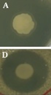
; B, <i>Staphylococcus </i>sp. WW60 (DUS56); C, B. sp. <i>cryopeg </i>(DUS59); D, <i>Paenibacillus </i>(DUL128); E, <i>S. warneri </i>(DUS130); F, B. <i>psychrodurans </i>(DUS131) against <i>C. albicans</i> KACC 30062; G, B. sp. <i>cryopeg </i>(DUS59); H, <i>Paenibacillus </i>sp. (DUL128); I, <i>S. warneri </i>(DUS130); J, (B. <i>psychrodurans </i>(DUS131) against <i>C. albicans</i> KACC 3003; K, B. sp. <i>cryopeg </i>(DUS59) against C. <i>saitoana</i> KACC 41238; L, B. <i>psychrodurans </i>(DUS131) against <i>C. glabrata </i>KBNO6P00368")
Anticandidal potential of endophytic bacteria patches against pathogenic Candida species. A, Burkholderia sp. UR 1-07 (DUS14); B, Staphylococcus sp. WW60 (DUS56); C, B. sp. cryopeg (DUS59); D, Paenibacillus (DUL128); E, S. warneri (DUS130); F, B. psychrodurans (DUS131) against C. albicans KACC 30062; G, B. sp. cryopeg (DUS59); H, Paenibacillus sp. (DUL128); I, S. warneri (DUS130); J, (B. psychrodurans (DUS131) against C. albicans KACC 3003; K, B. sp. cryopeg (DUS59) against C. saitoana KACC 41238; L, B. psychrodurans (DUS131) against C. glabrata KBNO6P00368
In the current study, the endophytic bacteria Bacillus sp. Cryopeg showed broad spectrum anticandidal activity against three Candida species, C. saitoana (KACC 41238), C. albicans (KACC 30062), and C. albicans (KACC 30003), with inhibition zone diameters of 9.55 ± 0.28 mm, 21.47 ± 0.18 mm, and 23.42 ± 0.52 mm, respectively (Table 1 and Figure 1).
However, B. psychrodurans also showed broad spectrum anticandidal activity against three Candida species (such as C. albicans KACC 30062, C. albicans KACC 30003, and C. glabrata KBNO6P00368) with inhibition zone diameters of 19.31 ± 0.02 mm, 23.49 ± 0.22 mm, and 9.29 ± 0.34 mm, respectively (Table 1 and Figure 1). Paenibacillus sp. and S. warneri both showed strong anticandidal activity against C. albicans KACC 30062 and C. albicans (KACC 30003) with inhibition zone diameters of 25.52 ± 0.10 mm and 23.06 ± 0.30 mm, and 23.59 ± 0.67 mm and 22.84 ± 0.08 mm, respectively (Figure 1 and Table 1).
Burkholderia sp., which showed a clear inhibition zone with the highest value against C. albicans (KACC 30062) among all six promising endophytic bacteria, was selected for further extraction by different solvents, including n-hexane, chloroform, ethyl acetate, as well as n-butanol. The different solvent extracts existed were verified for the anticandidal activity assay against C. albicans (KACC 30062). The n-hexane, chloroform, and ethyl acetate extracts did not show any inhibitory activity against C. albicans (KACC 30062), but the butanol fraction exerted anticandidal effect against C. albicans (KACC 30062) with an inhibition zone diameter of 21.89 ± 0.20 mm (Figure 2 and Table 2). The MIC and MFC values of the butanol extract against C. albicans (KACC 30062) were 250 µg/mL and 500 µg/mL, respectively (Table 2).

Anticandidal potential of selected endophytic bacteria Burkholderia sp. butanol fraction against C. albicans KACC 30062
| Treatment | C. albicans (KACC 30062) | MIC (µg/mL) | MFC (µg/mL) |
|---|---|---|---|
| BuOH Fr. | 21.89 ± 0.20a | 250 | 500 |
a Inhibition zone diameters are expressed as the mean ± SD in mm.
The sequence of the one endophytic bacteria identified as Burkholderia sp. UR 1-07 (DUS14) showing strong anticandidal activity against C. albicans was aligned using 16S rRNA gene sequencing and the neighbor joining tree was constructed. The phylogenetic tree of the promising endophytic bacteria DUS14 revealed that this isolate has 99% similarity with Burkholderia sp. UR 1-07 (Figure 3). Scanning electron microscopy analysis was conducted to visualize the treatment effect of the butanol extract administered at its MIC on the morphology of C. albicans. The SEM analysis results revealed variations in the morphology of tested pathogenic C. albicans KACC 30062 strain. Compared with the smooth, regular oval shape and the high density cells found in the control, the C. albicans treated with butanol extract had rough cell surfaces with burst cells and low density cells (Figure 4).
 generated from 1000 bootstrap trees and correspond to the scale bar of branch lengths (0.001)")
The phylogenetic relationship of the endophytic bacteria DUS14 based on 16S rRNA gene sequences by the maximum likelihood method was calculated with 1000 bootstrap replicates. The numbers above each node are the confidence levels (%) generated from 1000 bootstrap trees and correspond to the scale bar of branch lengths (0.001)

SEM images showing the consequence of the butanol fraction on the morphology of C. albicans KACC 30062 treated with 5% DMSO. A, C. albicans KACC 30062 treated with butanol fraction of Burkholderia sp. at the MIC
5. Discussion
Candida species are frequently isolated and may be associated with high mortality in the absence of appropriate treatment (36). Persistent candidiasis is widely documented as a reason for mortality and morbidity in hospitals and also responsible for the most common nosocomial bloodstream infection (37-40). The majority of yeast infection cases are caused by C. albicans (39). Inherent resistance to antifungal treatment has been observed in a few species, but is emerging as a prominent problem in the management of Candida infections (41). Recently, the incidence of diseases caused by drug-resistant fungi has increased. To treat contagious fungal infections Amphotericin B has been used, but it is acknowledged to have high and potentially dangerous side effects, counting the occurrence of high fever and trembling anxieties later a hours of infusion (42).
The general side effects of antifungal drugs include kidney damage, gastrointestinal discomfort, skin complaints, and hepatotoxicity (43). Hence, there is a necessity to identify novel drugs from diverse natural sources (44). Mostly, endophytic bacteria produce diverse dynamic compounds with special biological activities (45). To this end, the present study evaluated the anticandidal effects of endophytic bacteria isolates from D. uniformis against 5 Candida species, C. albicans (KACC 30062), C. albicans (KACC 30003), C. glabrata (KBNO6P00368), C. saitoana (KACC 41238), and C. geochares (KACC 30061).
Several researchers have isolated various endophytes from different parts of plants. Bacterial endophytes have been documented as storehouse of new secondary metabolites for possible remedial use (46-49). According to Strobel and Daisy (50), the study of endophytes of medicinal and widespread fern plants is required as they are anticipated to harbor unique and exciting endophytes with new bioactive metabolites. The growth of availability of antifungal compounds from natural products has encouraged search for superior efficiency strategies, like using the antifungal agents from edible medicinal plants (40).
This has directed to the detection of quite a lot of bioactive composites from bacterial as well as fungal endophytes isolated from medicinal and fern plants (48, 51-54). Several species in the Bacillus genus are economically significant for use in the construction of numerous molecules and additional products for foodstuffs, pharmaceutical, agricultural, and biological industries (55, 56). Several Bacillus species are remarkably important owing to their use in antibiotic construction and ability to induce production of anti-inflammatory cytokine in vitro (57-59). The use of Bacillus species to produce a wide range of metabolites having antimicrobial prospective is extensive in the medical and pharmaceutical industries; they can assist plant growth in adverse conditions and also capable of controlling many infections in humans, animals, and plants after applied as an organic control mediator (60-62).
Paenibacillus sp. are known for their endophytic existence (14, 63) and have been described as capable plant-growth promoting bacteria (64). Numerous Paenibacillus-based products have been patented and commercially established. The antagonistic prospective of Paenibacillus sp. against a wide range of phytopathogenic fungi has been previously acknowledged (65, 66). As a source of novel bioactive compounds, Paenibacillus sp. strains have a huge potential in biotechnology; at present, this potentially has only partially been explored (67). Burkholderia is a group of metabolically important gram negative bacteria (68). Various Burkholderia species are acknowledged for their metabolic prospective and be able to be used for biotechnological functions, for instance the biological management of plant infections (69), nodulation as well as plant development elevation (70, 71), and bioremediation (72, 73). Promising application of endophytic bacteria to the natural control of pathogenic fungi and in the plant growth improvement was reported in the previous study of Zhao et al (64).
Scanning electron microscopy analysis results revealed variations in the morphology of tested pathogenic C. albicans (KACC 30062) strain with respect to the control with the smooth, regular oval shape and the high density cells. These variations in the morphological feathers in the treated C. albicans (KACC 30062) might arise from the bioactive compounds extracted from the butanol extract from the endophytic bacteria that resulted in the destruction of cell integrity and lysis of the cell wall as evidenced from earlier findings (21).
5.1. Conclusion
In the present study, we screened a large number of endophytic bacteria from D. uniformis and confirmed that six endophytic bacteria displayed promising anticandidal effects against four Candida species. Two of these bacteria showed strong anticandidal activity against C. saitoana, C. albicans, and C. glabrata species; three showed strong anticandidal activity against two C. albicans species. One of the selected endophytic bacteria, Burkholderia sp. UR 1-07, which showed clear and highest anticandidal activity against C. albicans (KACC 30062), was used for further solvent extraction. The butanolic fraction was effective against C. albicans (KACC 30062). Thus, it was concluded that endophytic bacteria isolated from D. uniformis may serve as the potential source of natural anticandidal agents useful for the inhibition of the Candida species C. saitoana (KACC 41238), C. albicans (KACC 30062), C. albicans (KACC 30003), and C. glabrata (KBNO6P00368). It may also be a substitute for artificial fungicides to control candidiasis.
