1. Background
Cancer is regarded as a multi-stage process in which the genetic and epigenetic changes in a cell, result in a progressive alteration from normal to cancer cells (1). Several factors are involved in developing cancer (2), the internal reasons of cancer may be related to oxidative stress generation, apoptotic process disorders, and genetic mutations; hence the external reasons of cancer may be consistent with the environmental factors like viruses, bacteria, smoking, chemicals, radiation, and stress (3). Several researchers indicated that oncogenes activation and the inaction of tumor suppressors are needed for cancer cells proliferation (4). The main genes lead to cancer are included in apoptosis, tumor suppressors, oncogenes, and DNA repair genes (1). Apoptosis is a kind of gene-dependent programmed cell death identified by special morphological and biochemical properties, like chromatin density, cell shrinkage, and nuclear condensation (5). The cell death process can be induced by extracellular endogenous proteins or cytokines and oxidative stress reactions (6). In apoptotic process, caspases are among the most significant factors involved in apoptosis (7). The name of caspases is derived from their function and these enzymes coordinate the route of programmed cell death (6). Skin cancer (melanoma) is a common type of cancer, and early detection increases survival (8). Conventional treatments for melanoma involve surgery, chemotherapy, and immunotherapy (9). Nowadays, general tendency of society to utilize herbal medicines and natural products is raising, so that herbal medicines have an important effect on pharmaceutical products (10). Ficus carica (fig) belongs to Moraceae family, the name of species derived from Carica is the name of fig-growing region in the southwest of Turkey. Its major origin is Mediterranean region and it grows in multiple regions of the world (11). Figs have an essential role to reduce cholesterol, controlling respiration and strengthening the heart. Other features involve decreasing body temperature, increasing body sweating, and treating anemia (12). Fig leaf extract has anti-cancer, anti-proliferative effects (13). The most significant compounds in figs are proteins, fatty acids, enzymes, beta-carotene, ficusin, arabinose, and xanthotoxols (14). Furthermore, it involves phenolic compounds like catechin, gallic acid and syringic acid (15). Ficus carica leaf extract was indicated to involve rhein, chrysin and quercetin which can prevent proliferation of cancer cells, and rhein has anticancer impacts (16). Ficus carica leaf extract has a stimulating impact on cellular and humoral immune response; hence it can be applied for therapeutic objectives in the diseases of immune origin (13).
2. Objectives
Regarding different chemical compounds in F. carica and therapeutic features of this plant to treat various diseases, we decided to survey the impact of F. carica leaves extract on apoptosis induction in melanoma cancer cells (B16F10).
3. Methods
3.1. Chemicals
B16F10 cell line (melanoma cancer) was obtained from Pasteur Institute of Iran. Culture medium DMEM (Dulbecco’s Modified Eagle Medium), FBS (Fetal bovine serum), antibiotics (penicillin-streptomycin) and Trypsin- EDTA were provided from Bioidea (Iran). MTT (3-(4,5-Dimethylthiazol-2-yl)-2,5-diphenyltetrazolium bromide), DAPI (4′,6-Diamidino-2-phenylindole dihydrochloride), Propidium Iodide and Acridine orange were provided from Sigma (Germany), Annexin V-FITC kit was provided from Abcam (UK), as well as SYBR Green, RNA extraction kit and CDNA synthesis were provided from Pars Tous (Iran).
3.2. Plant Materials
Ficus carica leaves were provided from Mashhad (Khorasan Razavi, Iran) in May 2019, and confirmed in Islamic Azad University, Mashhad botanical lab with herbarium code 10319.
3.3. Extraction Method
In this study, the dried F. carica leaves were crushed into a powder and 100cc of methanol per 1 gram was added to it. Then, the resultant mixture was kept in the dark at room temperature for 72 h. During this time, the mixture was well stirred three times a day and the extract was filtered using Whatman paper after 72 h, and the total methanolic extraction was performed by using rotary evaporator. To evaporate methanol, the resulting extract, which was water-insoluble green precipitate was placed at 66.5°C for 72 h.
3.4. Cancer Cell Survival Analysis
Melanoma cancer cells (B16F10) and normal epithelial kidney cells (HEK-293) were cultured in DMEM medium supplemented with 10% FBS and 1% antibiotic. The cells were maintained at 37°C in a 5% CO2 atmosphere and 95% humidity. Cell viability was examined by MTT assay. Briefly, B16F10 and HEK-293 cells were cultured in 96-well plate with 1 × 10⁴ per well and incubated for 24 hours. Cells were treated by different concentrations of the methanolic extract of F. carica leaf (150, 250, 350, 450, 550, 650, 750 and 850 μg /mL) and sham (DMSO as extract diluent) for 24 and 48 hours. After incubation, MTT solution was added to culture medium which was maintained in darkness for 4 hours at 37°C. Next, the culture medium was discarded and 80 μL of DMSO was added to cells. The absorbance was estimated at 560 nm. Then, the results were compared to control groups.
3.5. DAPI Staining
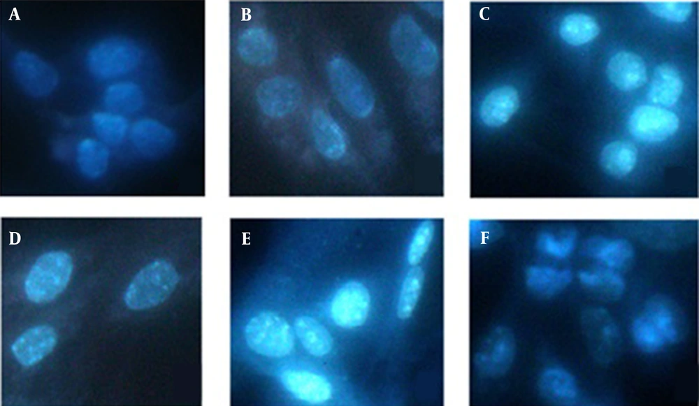
In this study, the nuclear morphology alteration was assessed with DAPI dye. B16F10 cells were treated in various concentrations of 350, 450, 550, 650 and 750 μg/mL of F. carica leaves extract which incubated for 24 hours. Then, the cells were fixed and stained with methanol and DAPI, respectively. Nucleus morphology was seen under fluorescence microscope (Olympus, Japan) via blue filter, the excitation and emission wavelength were 364 nm and 454 nm, respectively.
3.6. Acridine Orange-Propidium Iodide (AO/PI) Staining
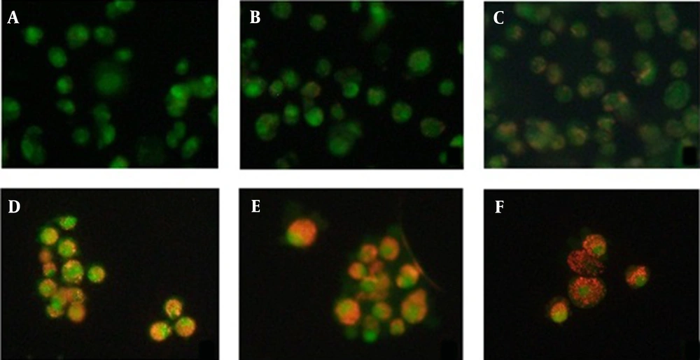
Cell viability and apoptosis were assessed by fluorescence microscopy utilizing AO/PI. In brief, B16F10 cells were incubated for 24 h at 350, 450, 550, 650 and 750 μg/mL concentration of methanolic extract of F. carica leaves. Then, the cells were trypsinized and centrifuged. The cells were stained with 10 μL of a mixture of AO and PI fluorescence. At last, the cells were seen under ultraviolet fluorescence microscope (Olympus, Japan) at 490 nm excitation and 520 nm emission wavelength.
3.7. Annexin V Fluorescence Assay
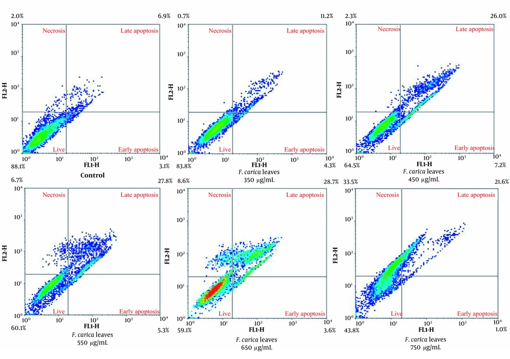
In this experiment, apoptosis and necrosis cells were quantified by flow cytometry. In brief, melanoma cells were treated by multiple concentrations of F. carica leaves extract. After 24 hours, the cells were collected and 500 μL of binding buffer 1X was added to every sample. Subsequently, 5 μL of Annexin V-FITC and 5 μL of propidium iodide were added to every sample which was estimated by FACSCalibur flow cytometer (BD Biosciences).
3.8. Real-time Quantitative Polymerase Chain Reaction
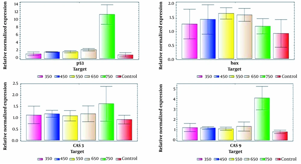
The expression levels of Bax, caspase3, caspase9, and p53 genes were assessed by real-time PCR. The total RNA was extracted from melanoma cancer cells after treatment with various concentrations of F. carica leaf extract (350, 450, 550, 650 and 750 μg/mL), due to the manufacturer’s instructions. The extracted RNA was utilized for cDNA synthesis regarding Pars Tous manufacturer instructions. The specific primer sequences for p53, Bax, caspase3, caspase9 and GAPDH genes was designed as shown in Table 1. The relative quantification was performed in real-time PCR detection system (Bio-Rad CFX96) using SYBR Green. The results were normalized to an internal reference gene GAPDH. All PCR amplifications were analyzed in triplicates.
| Gene | Sense | Anti-sense |
|---|---|---|
| GAPDH | AACTCCCACTCTTCCACCTTCG | GTCCACCACCCTGTTGCTGTAG |
| p53 | AACCGCCCGACCTATCCTTACC | TCCCAGGGCGGCACAAAC |
| Bax | TCATGGGCTGGACACTGGACTTC | GAGCGAGGCGGTGAGGACTC |
| Caspase3 | TGACTGGAAAGCCGCCGAAACTC | GCAAAGCCATCTCCTCATCAG |
| Caspase9 | GAAGAACGACCTGACTGCCAAG | GAGAGAGGATGACCACCACAAAG |
3.9. Statistical Analysis
All results were expressed as mean ± SEM. Statistical analysis was performed using GraphPad Prism 8 software, ANOVA test followed by Tukey test to compare the data and P < 0.05 level was considered significant.
4. Results
4.1. Cancer Cell Survival Analysis
The result of MTT assay showed that F. carica leaves extract had cytotoxic impacts on B16F10 cells in a dose- and time-dependent manner as compared with control or Sham (DMSO) groups (Table 2). Therefore, our data indicated the inhibitory impact of F. carica extract on skin cancer cells by increasing extract concentration. Due to our results, the concentration of 750 and 650 μg/mL of methanolic extract of F. carica leaves decreased cells viability (IC50) by 50% for 24 h and 48 h, respectively. However, these concentrations did not exert high cytotoxic activity against normal epithelial HEK-293 cells and indicated that B16F10 cancer cells were more sensitive to F. carica leaves extract than normal cells (Table. 2).
| Groups | HEK-293, 24 h | B16F10, 24 h | B16F10, 48 h |
|---|---|---|---|
| Control | 100.00 ± 1.13 | 100.00 ± 1.55 | 100.00 ± 1.23 |
| Sham | 98.16 ± 1.23 | 98.03 ± 1.63 | 98.31 ± 2.20 |
| F. carica leaves, 150 µg/mL | 97.10 ± 1.18 | 97.42 ± 2.23 | 94.78 ± 2.94 |
| F. carica leaves, 250 µg/mL | 97.55 ± 0.74 | 95.21 ± 1.35 | 90.43 ± 5.04 |
| F. carica leaves, 350 µg/mL | 95.95 ± 2.24 | 90.47 ± 5.02 | 85.42 ± 2.92 b |
| F. carica leaves, 450 µg/mL | 91.88 ± 0.39 | 82.39 ± 2.71 c | 78.36 ± 2.72 c |
| F. carica leaves, 550 µg/mL | 87.47 ± 1.49 b | 69.17 ± 4.70 c | 62.82 ± 3.35 c |
| F. carica leaves, 650 µg/mL | 75.37 ± 1.86 c | 54.38 ± 3.28 c | 49.00 ± 3.11 c |
| F. carica leaves, 750 µg/mL | 62.98 ± 1.66 c | 45.61 ± 4.60 c | 41.87 ± 3.23 c |
| F. carica leaves, 850 µg/mL | 58.71 ± 1.06 c | 41.23 ± 3.50 c | 34.84 ± 5.13 c |
aData are represented as the mean ± SD.
b P value significant at the level of P < 0.01 compared to the control.
c P value significant at the level of P < 0.001 compared to the control.
4.2. DAPI Staining
Regarding DAPI staining results, increasing concentration of the methanolic extract of F. carica leaves result in condensed nuclei and their DNA was seen as fragmented that confirmed cells underwent apoptosis. The apoptosis population was significantly higher after treatment with 650 and 750 μg/mL of extract (Figure 1). While in control group, the stained nuclei of B16F10 cells maintained the round shape and homogeneous.
4.3. AO/PI Staining
Staining with AO/PI indicated that F. carica treatment promoted apoptosis in melanoma cancer cells (B16F10). Figure 2 showed control cells were stained green with intact nucleus structure while the apoptosis was clearly obvious in cells treated with cytotoxic concentrations of methanolic extract of F. carica leaf as marked by orange nuclei and red staining represents necrosis.
4.4. Annexin V-FITC Test
Detection of apoptosis by Flow cytometry revealed that the percentage of apoptotic cells increased after methanolic extract of F. carica leaves treatment in a dose-dependent manner. Due to flow cytometry histogram, the lower concentrations of F. carica leaf extract, exhibited the least population of apoptosis and higher viable cell. In contrast, the percentage of early and late apoptosis increased in higher concentrations of F. carica extract (Figure 3 and Table 3).
| Group | Liver Cells (%) | Early Apoptotic Cells (%) | Late Apoptotic Cells (%) | Necrotic Cells (%) |
|---|---|---|---|---|
| Control | 88.1 | 3.1 | 6.9 | 2.0 |
| F. carica, 350 μg/mL | 83.8 | 4.3 | 11.2 | 0.7 |
| F. carica, 450 μg/mL | 64.5 | 7.2 | 26.0 | 2.3 |
| F. carica, 550 μg/mL | 60.1 | 5.3 | 27.8 | 6.7 |
| F. carica, 650 μg/mL | 59.1 | 3.6 | 28.7 | 8.6 |
| F. carica, 750 μg/mL | 43.8 | 1.0 | 21.6 | 33.5 |
4.5. Real-time PCR
The alteration in expression of p53, Bax, caspase 3 and caspase 9 genes in melanoma cancer cells treated at concentrations of 350 to 750 μg/mL of F. carica leaves extract were evaluated by real-time PCR. Our data revealed that increasing expression of p53, Bax, caspase 3 and caspase 9 genes in treated group compared to control which confirmed the apoptosis induction in B16F10 cells through the activation of mitochondrial pathways (Figure 4). By increasing concentrations of F. carica leaf extract, particularly at high concentrations from 650 to 750 μg/mL, genes expression involved in apoptosis has increased than reference gene in skin cancer cell line.
5. Discussion
Epidemiological studies indicate the significant prevalence of skin cancer and mortality in recent years (17). Melanoma, as serious skin cancer, develops in melanocytes. Melanocytes make melanin, or skin pigment. The strongest risk factors for melanoma are different abnormal moles and a family history of melanoma (18). Apoptosis is a very regular and precise process which is regulated by a series of signaling cascades and cellular proteins. Thus, a deep understanding of apoptosis mechanisms and related pathways is efficient not only in understanding the disease but also in disease treatment (3).
Apoptosis defects play a significant role in tumor formation and regulation disruption causes resistance to chemotherapy and radiation therapy. Moreover, this trend may increase metastasis. Any factor which can inhibit normal development of cells provides conditions for apoptosis. Thus excessive apoptosis can result in diseases like Parkinson's and Alzheimer's and ischemic injuries (19). Ficus carica have been utilized in conventional medicine to treat numerous diseases including cancer. Some of the phenolic compounds in fig leaves include furanocoumarins such as psoralen and bergapten, flavonoids such as quercetin 3-O rutinoside, and phenolic acids such as ferulic acid, 3-O-caffeolic acid and 5-O-caffeolic acid (20). Furanocoumarin and psoralen show the anticancer and anti-cholinesterase activities, and flavonoids indicate anticancer, anti-inflammatory, and antidiabetic activities (13). Purushotham et al. investigated the anti-proliferative, anti-inflammatory and anti-apoptotic activity of methanolic extract of Andrographis paniculata (AN) in A375 and B16F10 cell lines. Due to their results, methanolic extract of AN leaf and stem caused a decrease in B16F10 cell viability. In addition, leaf extract had a higher anticancer activity compared with its stem on cancer cells. These results revealed that AN decreased the proliferation and induced apoptosis in cancer cell lines (21).
Regarding obtained results, increasing the dose of methanolic extract of F. carica leaf induced apoptosis in B16F10 cancer cells, followed by preventing cancer cell proliferation. Goldberg et al. surveyed the impact aqueous extract of Hibscus rosa sinesis compounds in vitro. The results confirmed that extract contained compounds which prevented the growth of melanoma cells by dose-dependent manner (22). Our results including MTT, Annexin V/ PI, DAPI, and AO/PI assays all indicated that B16F10 cells treated with methanolic extract of F. carica leaves extract promote the apoptosis through the activation of mitochondrial pathways. Regarding increased expression of genes involved in mitochondrial pathways of caspase 3 and caspase 9, methanolic extract of F. carica leaves induces apoptosis in B16F10 cells. methanolic extract of F. carica leaf prevent proliferation of skin cancer cells (B16F10 cell line) which the results are consistent with above research. MTT test results indicated that F. carica leaves extract has a cytotoxic impact on B16F10 cell line in dose and time dependent manner and decreased the cells viability and induced apoptosis. It is followed by inhibiting melanoma cancer cell proliferation. Zhang et al. investigated the inhibition of pancreatic β-cell apoptosis by the lower concentration of F. carica leaves extract (FCL) through the AMPK/JNK/Caspase 3 signaling pathway by antioxidation properties. They demonstrated that FCL at 60 μg/mL concentration could inhibit apoptosis induced by hydrogen peroxide and reduce ROS production. Therefore, FCL could inhibit pancreatic β-cell apoptosis by inhibiting the AMPK/JNK/ Caspase 3 signaling pathway and by antioxidation properties at lower concentration of FCL (60 μg/mL) (23).
In the present study, the results of Annexin, MTT, DAPI tests indicated that in B16F10 cells treated with the methanolic extract of fig leaves at lower concentration have no apoptotic and inhibiting effect on B16F10 cells while the higher concentration of F. carica leaves extract (650 μg/mL) induced apoptosis and activated mitochondrial pathways, which inhibted the proliferation of the skin cancer cells. Turkoglu et al. investigated the effect of extract of F. carica leaf on the expression of inflammatory factor genes in HaCaT cells. The results of gene expression analysis by real-time PCR showed that extract of F. carica leaf significantly reduced interleukin-1, TNF-α compared to the untreated cells. Moreover, based on the results of cytotoxicity analysis, the extract of F. carica leaf reduced the proliferation of HaCaT cells. Therefore, this extract is useful for some inflammatory and skin disorders such as androgenic alopecia (24). In addition, the MTT test showed that the alcoholic extract of fig leaves had a cytotoxic effect on B16F10 cells, which reduced the viability of cells in low concentrations. Thus, the cells underwent apoptosis, which caused the inhibition of the proliferation of melanoma cancer cells. In 2018 Zhang et al. analyzed the extracts and compounds of F. carica leaves on cell survival, cell cycle and breast cancer cell migration (MDA - MB-231 cell line), and consistent with our results their results indicated that chemical compounds in F. carica leaves like Psoralen and Bergapten caused a decrease in breast cancer cell proliferation with a time and dose-dependent effect. Therefore, these extracts and compounds prevent survival of cells and the cell cycle of cancer cell line (25). Purnamasari et al. evaluated the anticancer activity of the alcoholic extract of F. carica leaf and fruit against proliferation, apoptosis and necrosis in Huh7it cells. Based on the results, F. carica is a rich source of polyphenols, and the results of MTT and AnnexinV assays indicated that the difference in anticancer activity is related to the various compounds in each extract (26). In this regard, the results of the present study indicated that the methanolic extract of fig leaves inhibits the proliferation of skin cancer cells.
5.1. Conclusions
Using herbal and traditional remedies in medicine and pharmacy opened a novel insight for researchers to treat various disease. Our results indicated the efficiency of F. carica leaves extract on preventing proliferation and apoptosis induction in melanoma cancer cells. It is expected that in near future, the impact of F. carica leaves, its efficient compounds and related signaling pathways which induce apoptosis in cancer cells will be addressed.


