1. Background
One of the largest genera of the Lamiaceae family, genus Nepeta, belongs to the subfamily Nepetoideae and tribe Mentheae, which comprises about 300 herbaceous perennial, rarely annual species (1). Iran, particularly, is one of the centers of origin of the genus with sixty-seven species, described by the common Persian name of ‘Pune-sa’ and about 53% of endemics (2). Several Nepeta spp. are used in folk medicine as diuretic, diaphoretic, anti-tussive, anti-inflammatory, anti-spasmodic, anti-asthmatic, febrifuge, emmenagogue and sedative agents and for antiseptic and astringent properties as a topical remedy in children with cutaneous eruptions, and for snake and scorpion bites (1). The diversity, species richness and variation, as well as chemical properties have led to much research on the genus Nepeta. Phytochemical investigations have reported nepetalactones, iridoids and their glucosides, diterpenes, triterpenes and flavonoids as major constituents of Nepeta species (3).
Nepeta depauperata Benth.is one of the endemic perennial species distributed just in south of Iran. It has beautiful flowers with a pleasant odor and grows up to a height of about 40 - 80 cm (4). N. depauperata which is locally called “Oryan Pune-sa” is extensively exploited as a medicinal plant in Iranian traditional medicine (4). It is used to relieve of snake, scorpion and wasp bites, and for wound healing. It is also believed to be very effective in the treatment of inflammation and relieve of pain and is used orally and topically by patients suffering rheumatism and inflammatory disorders. The essential oil chemical composition of this plant has been investigated and spathulenol (31.84%), beta caryophyllene (12.93%) and caryophyllene oxide (10.27%) were identified as the major components of the oil (5).
2. Objectives
Due to the widespread use of N. depauperata in Iranian folk medicine for relief and treatment of pain and inflammation, we prompted to evaluate the anti-nociceptive activities of the methanol extract of its flowering aerial parts and investigate the pharmacological basis for the folkloric use of it as a pain killer agent.
3. Materials and Methods
3.1. Plant Collection and Preparation of Extract
Fresh flowering aerial parts of N. depauperata were collected in March 2013 from the mountain areas of Genow protected region in 30 Km west north of Bandar Abbas, Hormozgan province, Iran: (27° 24' 6.62" N 56° 10' 46.57" E, 1,800 m). Specimen was identified voucher was deposited in the herbarium of pharmaceutical sciences branch, Islamic Azad University (IAUPS), Tehran under code number LNd-1030. The plant supply (500 g) was dried in exposure to air and away from sun beam, and after being crushed, was taken to the percolator where it was percolated by means of methanol (3 times per day, 20 cc solvent each time, for 10 days). The extraction was repeated for 3 times. The extract was concentrated by rotary evaporator apparatus and the solvent removed to produce a dark green gummy solid. The resultant methanol extract was preserved in closed and dark containers in refrigerator until the time of experiment.
3.2. Chemicals
Formalin, morphine sulphate and naloxone were purchased from Merck Chemical Co. (Germany), Temad Pharmaceutical Co. (Iran) and Darou Pakhsh Pharmaceutical Co.(Iran) respectively.
3.3. Experimental Animals
In this experimental study, Male albino mice (20 - 25 g) were housed in groups of 6 - 8 and were allowed free access to food and water except for the short time that the animals were removed from their cages for testing. All experiments were conducted during the period between 10 a.m. and 13 p.m. with normal room light (12 hours regular light/dark cycle) and temperature (22 ± 1°C). The procedures were carried out in accordance with the institutional guidelines for animal care and use (ethical approval number: 22510303922088). Each mouse was used only once.
3.4. Formalin Test
The analgesic effects of N. depauperata extract were investigated by formalin test. 15 minutes after separately injection of different doses of the extract (25, 50, 100 and 200 mg/kg), morphine (2 mg/kg, positive control) and the vehicle, 50 μL of 2.5% formalin was injected into the right hind paw of the mice. The animals were immediately placed in the formalin test container scoring of nociceptive behaviors began immediately after formalin injection and continued for 60 minutes. The two of main formalin related behavioral responses, flexing and licking were scored. To score the pain responses, the one hour recording session was divided into 5 minutes intervals during which the duration (in seconds) spent licking and flexing the injected paw was scored. Plots of the mean duration time of licking and flexing were generated for each concentration group. Behavioral responses in formalin test were converted to first phase values, 0 - 10 minutes after formalin injection and second phase values, 15 - 60 minutes after formalin injection.
Naloxone (2 mg/kg), an opioid antagonist, was administrated subcutaneously 15 minutes prior to treatment with the extract. The extract and morphine were administered 15 minutes before formalin.
3.5. Hot-Plate Test
Male albino mice (20 - 25 g) were divided into 7 groups (6 - 8 animals in each group).
Control animals received normal saline (10 mL/kg, IP); while the positive control group received morphine (5 mg/kg, IP) and the test groups received different doses of the extract (50 - 80 - 160 and 250 mg/kg, IP). Briefly, the animals were placed on the hot-plate apparatus and the time between placement of the mouse on the platform and shaking or licking of the paws or jumping was recorded as the reaction time or latency of the pain response. In order to avoid the damage to the paws of the animals, the time standing on the plate was limited to 15 seconds (cut-off time). Hot plate test was performed on all animals individually in 15th, 30th, 45th and 60th minutes after the treatment.
3.6. Statistical Analysis
One-way ANOVA test was used to compare mean duration time of licking and flexing between the groups. In order to assess the difference in mean time of licking and flexing in first and second phase between each test and control group, Tukey test was used. Data obtained for pain behavior in hot plate test were scrutinized using one way analysis of variance (ANOVA) followed by the Tukey post-hoc test. The results are expressed as mean ± S.E.M and P < 0.05 was considered as significant difference of means. The data were analyzed using SPSS statistical software.
4. Results
4.1. Formalin Test
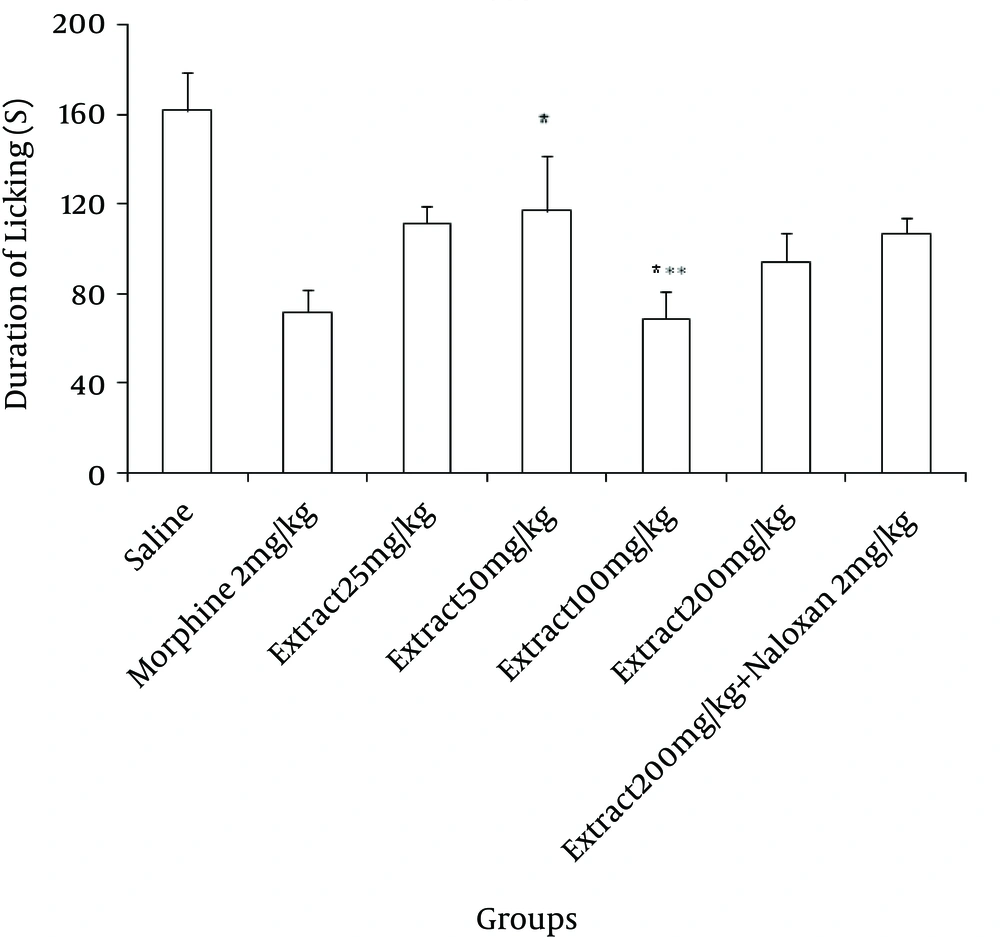
The effects of systemic IP administration of different doses of the extract on the behavioral responses in the first and second phases of formalin test were observed. In both phases of the pain behaviors were separately analyzed as total licking and flexing duration time. As it is mentioned in Figure 1, in the first phase of formalin test 25, 50, 100 and 200 mg/kg doses of the extract reduced the licking duration (P < 0.001, in 100 mg/kg dose) and morphine as a standard analgesic drug significantly (P < 0.001) reduced pain behavior.
Different doses of ND (25, 50, 100 and 200 mg/kg) and saline were administered IP, 15 minutes prior to sub-plantar injection of formalin and time spent for licking was measured during a 0 - 10 minutes period after formalin injection. Morphine (2 mg/kg, IP) was used as reference drug. Data are mean ± SEM of 6 - 8 animals in each group.*P < 0.05, ***P < 0.001 compared with control group.
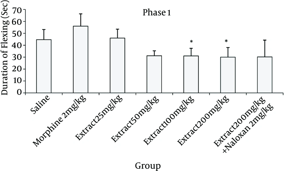
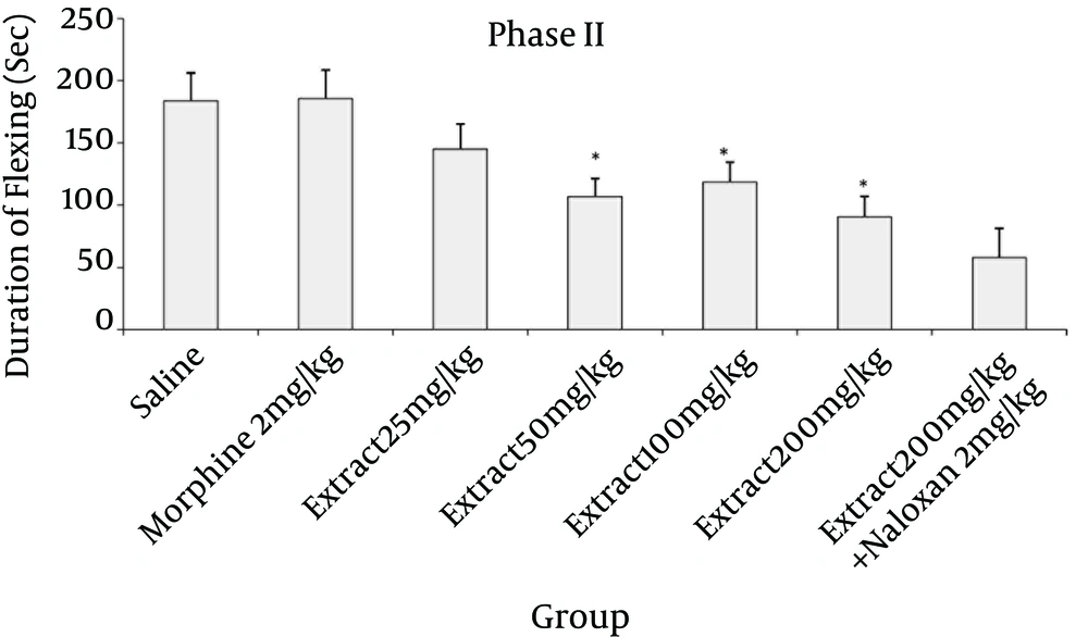
Doses of 50, 100 and 200 mg/kg of the extract reduced the flexing time in formalin test in compared to saline (P < 0.05 in both phases; Figures 2 and 4).
Different doses of ND (25, 50, 100 and 200 mg/kg) and saline were administered IP, 15 minutes prior to sub-plantar injection of formalin and time spent for flexing was measured during a 0 - 10 minutes period after formalin injection. Morphine (2 mg/kg, IP) was used as reference drug. Data are mean ± SEM of 6 - 8 animals in each group. *P < 0.05 compared with control group.
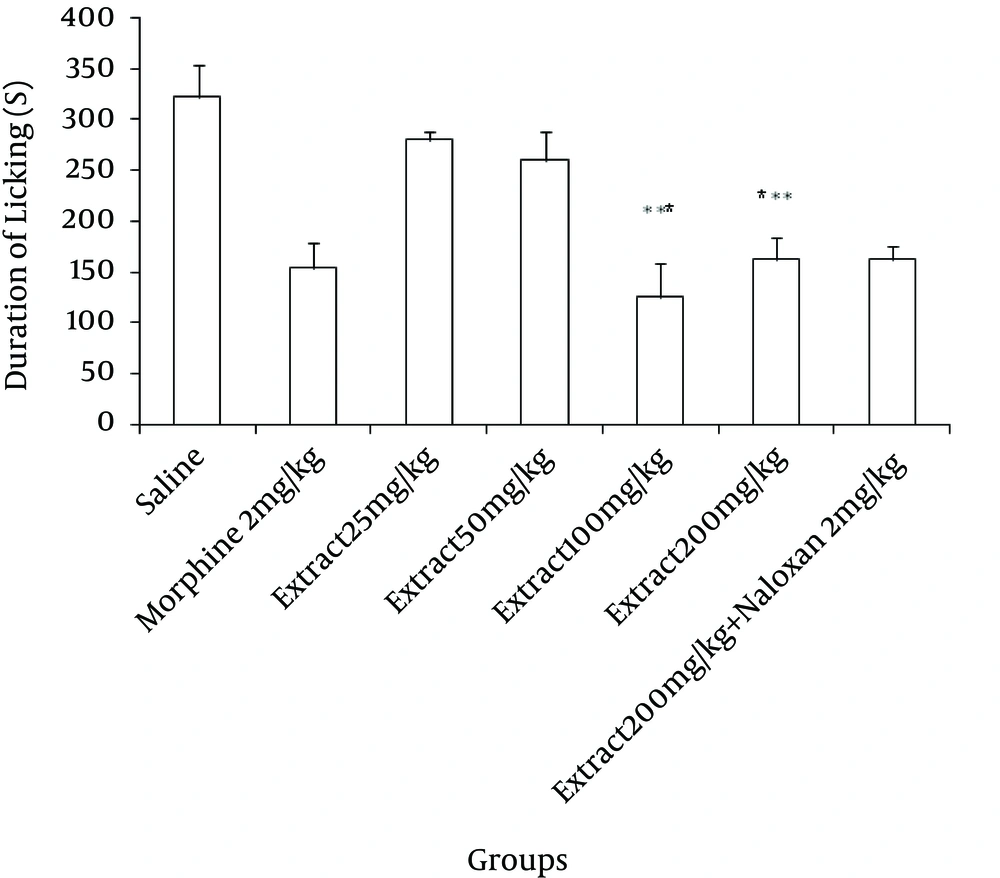
Again in the second phase of formalin test, as it is mentioned in Figure 3, all the studied doses of the extract reduced the licking duration (P < 0.001, in 100 and 200 mg/kg in compared to saline) and morphine as a standard analgesic drug significantly (P < 0.001) reduced pain behavior.
Different doses of ND (25, 50, 100 and 200 mg/kg) and saline were administered IP, 15 minutes prior to sub-plantar injection of formalin and time spent for licking was measured during a 15 - 60 minutes period after formalin injection. Morphine (2 mg/kg, IP) was used as reference drug. Data are mean ± SEM of 6 - 8 animals in each group. ***P < 0.001 compared with control group.
Different doses of ND (25, 50, 100 and 200 mg/kg) and saline were administered IP, 15 minutes prior to sub-plantar injection of formalin and time spent for flexing was measured during a 15 - 60 minutes period after formalin injection. Morphine (2 mg/kg, IP) was used as reference drug. Data are mean ± SEM of 6 - 8 animals in each group. *P < 0.5 compared with control group.
4.2. Hot-Plate Test
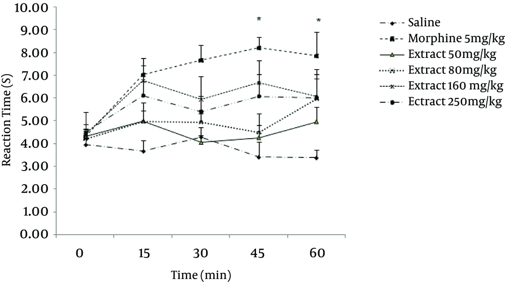
The anti-nociceptive effect of N. depauperata methanol extract (50, 80, 160 and 250 mg/kg, IP) were investigated in an acute thermal model of nociception in mice. In hot plate test, morphine as the standard drug produced significant analgesia 15 minutes after injection which remained until 60 minutes.
As evaluated at 15 minutes post injection, 50 and 80 mg/kg doses of N. depauperata extract did not produce any significant analgesia, but the doses of 160 and 250 mg/kg reduced the acute thermal pain significantly (P < 0.05). The time course assessment demonstrated that the extract began to show anti-nociceptive action in the hot-plate paradigm only 15 minutes after intra peritoneal administration, an effect that lasted up to 1 hour post-treatment (Figure 5). Values were found to be significant (P < 0.05) at 15, 45 minutes after treatment with 160 mg/kg of N. depauperata extract in compared to saline group. Moreover, in this dose exhibited anti-nociceptive effect similar to morphine.
Saline and extract (50, 80, 160 and 250 mg/kg, IP) were administered 15 minutes prior to placement of the animal in hot plate and reaction time of mice was measured at 15 minutes intervals until 1 hour. Morphine (5 mg/kg, IP) was used as reference drug. Data are mean ± SEM of 6 - 8 animals in each group. *P < 0.05 compared with control group.
5. Discussion
The management of pain is probably one of the most common and yet most difficult aspects in medical practice. Many improved analgesics and anti-inflammatory agents have been developed, but there is considerable opportunity for conceptual innovation.
We used heat induced and formalin induced pain model for evaluating anti-nociceptive of Nepeta depauperata methanol extract in experimental mice. Our data demonstrated that N. depauperata extract elicited anti-nociceptive effects in mice subjected to both the acute thermal (hot plate) and chronic or persistent formalin pain stimuli.
In the hot plate test which consists of a thermal stimulus, an increase in the reaction time is generally considered as an important parameter for evaluating central anti-nociceptive activity (6, 7). Relative to controls, the studied extract increased significantly the mice hot plat reaction time. The anti-nociceptive activity of the extract was to be similar to that of morphine, despite less potent. This reduced response could be a consequence of N. depauperata extract dose employed. In addition, this plant anti-nociceptive effects started in 15 minutes after the treatment and remained up to 60 minutes after administration of the extract.
In comparison, the formalin test is a model of acute and persistent pain and involves an inflammatory response with release of neurogenic molecules such as substance P, glutamate and TNFa in the spinal cord (8, 9). The level of pain in this model is sensitive to both centrally and peripherally acting analgesics. In the present study, as the IP administration of different doses of N. depauperata extract inhibited both phases of pain response relative to controls especially in second phase, it may be suggested that it has both the central and peripheral anti-nociceptive effects. In all tests the relative potencies of N. depauperata and the positive controls, morphine, were compared and different doses of the extract were revealed to be similar to that of morphine.
The anti-nociceptive activity induced by the plant in formalin test was not altered by naloxone, demonstrating that their actions did not depend on opioid receptors (Williamson et al., 1996) (10). Phytobiological evaluation of Nepeta species displayed that the essential oil contained in these plants have marked anti-inflammatory and antinociceptive properties. Previous studies on N. caesarea essential oil showed significant analgesic activity in the tail flick and tail immersion tests. This effect was accompanied by a marked sedation, which was also blocked by naloxone, indicating the involvement of opioid receptors (Aydin et al., 1998) (11).
Ricci et al., reported anti-nociceptive properties of N. cataria essential oil. The treated animals exhibited an increased latency of tail withdrawal and so peripheral anti-inflammatory analgesic effects in the acetic acid writhing reflex and the carrageenan-induced edema tests (12).
Similar to the previous report (Sefidkon and Akbarinia, 2003) (13), N. pogonospermaoil contained 1, 8-cineole and 4aα, 7α, 7a β-Nepetalactone as two major constituent. It has been suggested by many investigators that 1, 8-cineole has potent antinociceptive (Martinez et al., 2009) (14) and anti-inflammatory activity (Santos and Rao, 2000) (15). In addition, Aydin and colleagues (11) suggested that 4aα, 7α, 7aβ- Nepetalactone, might be responsible for the significant analgesic activity and marked sedation in a rat model. They also recommended this nepetalactone might have specific opioid receptor subtype agonist activity (Aydin et al., 1998) (11). Again in a behavioral study on rats a significant decrease in performance was observed following IP administration of nepetalactone enriched fraction (Harney et al., 1977) (16). Furthermore the nepetalactone isomers were suggested to be the responsible component of the anti-nociceptive and anti-inflammatory actions of N. cataria (Ricci et al., 2010) (12). Moreover, the essential oil of N. crispa which contained 20.3% 4aα, 7α, 7aβ-Nepetalactone and 47.9% 1, 8-cineole, showed strong antibacterial, antifungal, antinociceptive and anti-inflammatory activity (Ali et al., 2012) (17). Also, it was showed the analgesic activity of the essential oil of N. italica to be correlated with the amount of 1, 8-cineole (Aydin et al., 1999) (18).
Literature survey revealed that the extracts of other Nepeta species had significant anti-inflammatory activities. In previous study the anti-inflammatory activity of N.sibthorpii methanol extract has been reported to have significant anti-inflammatory properties due to its ursolic acid and polyphenol contents (Miceli et al., 2005) (19).
5.1. Conclusion
The present study indicates that the methanol extract of N. depauperata exhibited central and peripheral antinociceptive effects. However, the mechanisms behind the central and peripheral antinociceptive activity of it are not completely understood and may need further studies with different antagonists (such as serotonergic, adrenergic, etc.). Taking findings into account, it seems quite possible that N. depauperata contains constituents with antinociceptive activity, which may lead for the development of new natural products having analgesic effect. In further investigations, the different fractions of N. depauperata will be evaluated and the structural characterization of responsible components will be clarified.
