1. Background
Nanofibers are strings of fibers with a diameter of less than one micron. Their high surface area-to-volume ratio, high porosity, and high elasticity make their application possible in different fields (1, 2). Today, the unique properties of nanofiber membranes that are easily prepared by electrospinning have attracted a great deal of attention in drug delivery and tissue engineering (3). Due to their high porosity and high surface-to-volume ratio, nanofibers have become effective drug delivery systems. Taking advantage of a variety of materials in this way provides multiple structural forms containing drug molecules from integrated nanofibers for a variety of combined systems. For example, the use of different drugs to produce nanofibers using this method has been studied; such drugs include ibuprofen (4), cefazolin (5), rifampin (6), itraconazole (7), mefoxin (8), tetracycline (9), ciprofloxacin (10), and ketoprofen (11). The efficiency of this method has been evaluated by placing high doses of drugs in the fibers and facilitating the solubility of some insoluble drugs (12, 13).
Fast-dissolving delivery systems (FDDSs) are drug delivery systems that have become widespread; several drugs have been prepared in this manner or are under preparation. FDDSs provide a solution for patients who have problem with swallowing solid oral dosage forms; these systems also can be used to increase solubility, increase the half-life of the drug in the body, and escape the hepatic first-pass effect (14). In this dosage form, the formulation dissolves quickly in the mouth and is absorbed within a few minutes. This causes increased drug bioavailability and accelerates the effect of the drug; moreover, it may bypass the hepatic first-pass effect.
Yu et al. used the electrospinning method to prepare the FDDS of ibuprofen. The results of this work indicated that more than 80% of the drug was released from the nanofibers within 20 seconds (15). In another study, electrospinning and polyvinyl alcohol were used to prepare caffeine and riboflavin in SD form. The results showed that 100% of the caffeine and 40% of the riboflavin were released from the nanofibers within 60 seconds (16).
Loratadine is an antagonist of the H1 receptor and is classified as class II BCS drugs. It has low water solubility and bioavailability.
2. Objectives
In this study, our goal was to prepare loratadine FDDS nanofibers by electrospinning using polyvinylpyrrolidone (PVP). Furthermore, evaluation of various important factors, such as the applied voltage, feed rate, and the concentrations of the polymer and the drug, was used for factorial design to determine the effects of these parameters on the different properties of the resulting nanofibers. In addition, in terms of drug-release characterization, the nanofibers are compared to the solid dispersion preparation obtained from the freeze-drying technique.
3. Materials and Methods
3.1. Materials
Loratadine (Abidi Ind., Iran), PVP K30 with a molecular weight of 58,000 (Sigma, USA), hydrochloric acid (Merck, Germany), and ethanol (Merck, Germany) were purchased from the indicated sources.
3.2. Experimental Design
In this study, the effect of formulation parameters on the characteristics of the resulting film was assessed using full factorial design with four variables at two levels. The independent variables studied in this research were the concentration of polyvinylpyrrolidone in ethanol, drug-to-polymer ratio, voltage, and feed rate. The independent variables and their corresponding levels are shown in Table 1. Based on these variables and their levels, 16 formulations were designed (Table 2). Responses (dependent factors) were the mean dissolution time (Y1), fiber diameter (Y2), and disappearance time (Y3).
| Variables | Levels | |
|---|---|---|
| +1 | -1 | |
| PVP concentration in ethanol, % | 35 | 30 |
| Ratio of drug to polymer | 1/2 | 1/4 |
| Voltage, kV | 20 | 10 |
| Feed rate, mL/h | 6 | 1 |
The Independent Variables and Corresponding Levels
| Run | X1 | X2 | X3 | X4 |
|---|---|---|---|---|
| F1 | 30 | ¼ | 10 | 1 |
| F2 | 30 | ¼ | 10 | 6 |
| F3 | 30 | ¼ | 20 | 1 |
| F4 | 30 | ¼ | 20 | 6 |
| F5 | 30 | ½ | 10 | 1 |
| F6 | 30 | ½ | 10 | 6 |
| F7 | 30 | ½ | 20 | 1 |
| F8 | 30 | ½ | 20 | 6 |
| F9 | 35 | ¼ | 10 | 1 |
| F10 | 35 | ¼ | 10 | 6 |
| F11 | 35 | ¼ | 20 | 1 |
| F12 | 35 | ¼ | 20 | 6 |
| F13 | 35 | ½ | 10 | 1 |
| F14 | 35 | ½ | 10 | 6 |
| F15 | 35 | ½ | 20 | 1 |
| F16 | 35 | ½ | 20 | 6 |
Components of the Formulations
3.3. Preparation of Electrospinning Solution
To prepare the initial solution, 30 and 35 g of PVP were dissolved in 100 mL of ethanol. The polymeric solutions were placed on a magnetic stirrer for 15 minutes at room temperature; subsequently, loratadine was added to this solution at ratios of 1: 2 and 1: 4 relative to the weight of the polymer and mixed until the loratadine was completely dissolved.
3.4. The Electrospinning Method
After preparing various polymer–drug solutions, they were loaded into a 10 mL syringe. The electrospinning process was carried out at voltages of 10 and 20 kV and feed rates of 1 and 6 mL/h. The nozzle distance to the collector was considered as fixed and equal to 5 cm. Electrospinning was carried out at room temperature (25°C) under a relative humidity of 40 - 50%. After production, the nanofibers that formed on the collector were carefully removed and cut into 1 cm square pieces; they were kept in dry, hermetically sealed containers until further tests were carried out.
3.5. The Solid Dispersion Method
To prepare the solid dispersion, 30 g of PVP was dissolved in 100 mL; loratadine was then added to this solution at ratios of 1: 2 and 1: 4 relative to the weight of the polymer and mixed until the loratadine had completely dissolved. The two prepared solutions were placed in a freeze-dryer (model FDCF-12012, OPERON Co., South Korea). The freeze-drying process was performed for 24 h at a temperature of -130°C. After collection of lyophilized powder, it was kept in tightly capped containers at room temperature until the tests were conducted.
3.6. Scanning Electron Microscopy (SEM)
The surface properties of electrospun nanofibers were verified using a 1455vp model SEM (Leo Co., Germany). The samples were sputter coated with a thin layer of gold under a nitrogen atmosphere. Average diameters of 30 fibers were measured from SEM images using the Microstructure measurement software (Nahamin Pardazan Asia).
3.7. Differential Scanning Calorimetry (DSC)
The DSC thermogram of the drug powder, PVP, the physical mixture of drug, and PVP and nanofibers of PVP with and without the drug were obtained. Five milligrams of the sample were placed in an aluminum pan and then inserted into the DSC device (model CH 8907, Mettler Toledo, Switzerland). The target samples were heated in the range of 25 - 300°C at a speed of 10°C/minute, and their thermograms were recorded.
3.8. Fourier Transform Infrared Spectroscopy (FTIR)
To study the possible physical interference between the drug and polymer, FTIR was carried out on the loratadine powder, PVP, and nanofibers obtained from PVP with and without the drug. Two milligrams of the samples were weighed and homogenized using a mortar and mixed with 10 mg of potassium bromide. Subsequently, the samples were compressed using a hydraulic press. The resultant disc was placed in an infrared spectrometer (Broker, Germany) with a scanning range of 4,000 to 450 cm−1.
3.9. Dissolution Studies
Five milligram samples of each nanofiber (equivalent to 1 mg and 1.7 mg loratadine for F1 - F8 and F9 - F16, respectively) was carefully weighed; following this, while mixing, the samples were immersed in 20 mL of dissolution medium of 0.1 N hydrochloric acid at 37°C. At 15, 30, 45, 60, and 120 s, 1 mL of the medium was determined. The samples were passed through the filter, and the concentration of the released drug was obtained using the ultraviolet-visible spectrophotometry method at a wavelength of 280 nm. A graph of the percentage of the drug released from the nanofiber versus the time is shown. Each test was carried out in triplicate and the mean and standard deviation were recorded. The mean dissolution time (MDT) for each formulation was assessed according to the following equations:


Where ti is the midpoint of the time period during which the fraction ΔMi of the drug was released from the dosage form. A high MDT value for a drug delivery system means that it has a slow in vitro drug release.
Dissolution tests were carried out on 5 mg of lyophilized powder similar to the procedure performed on nanofiber.
3.10. Determination of the Disappearing Time of Nanofibers
To determine the dissolution time of the fibers and check the characteristics of their rapid solubility, a 1 cm2 piece of each nanofiber formulation was carefully cut and immersed in 20 mL of a dissolution medium of 0.1 N hydrochloric acid at 37°C with simultaneous mixing. Photographs of the samples were taken using a Canon camera (A4000) at 0, 10, 20, 30, 40, 50, and 60 s. The time was recorded as the disappearing time when the whole sample disappeared from the medium.
3.11. Statistical Analysis of the Data
Using the backward, stepwise linear regression method and significant terms (P < 0.05), SPSS software was used to perform statistical analysis and find significant relationships between variables and responses. The overall pattern of the regression models is given in the following equation:
Y = C + b1X1 + b2X2 + b3X3 + b4X4 + b5X12 + b6X22 + b7X32 + b8X42 + b9X1X2 + b10X1X3 + b11X1X4 + b12X2X3 + b13X2X4 + b14X3X4+ b15X1X2X3+ b16X1X2X4+ b17X2X3X4+ b18X1X3X4 + b19X1X2X3X4.
The three-dimensional graphs related to the effect of variables on response according to the above equation were plotted using Statgraphics version 16.1.
4. Results
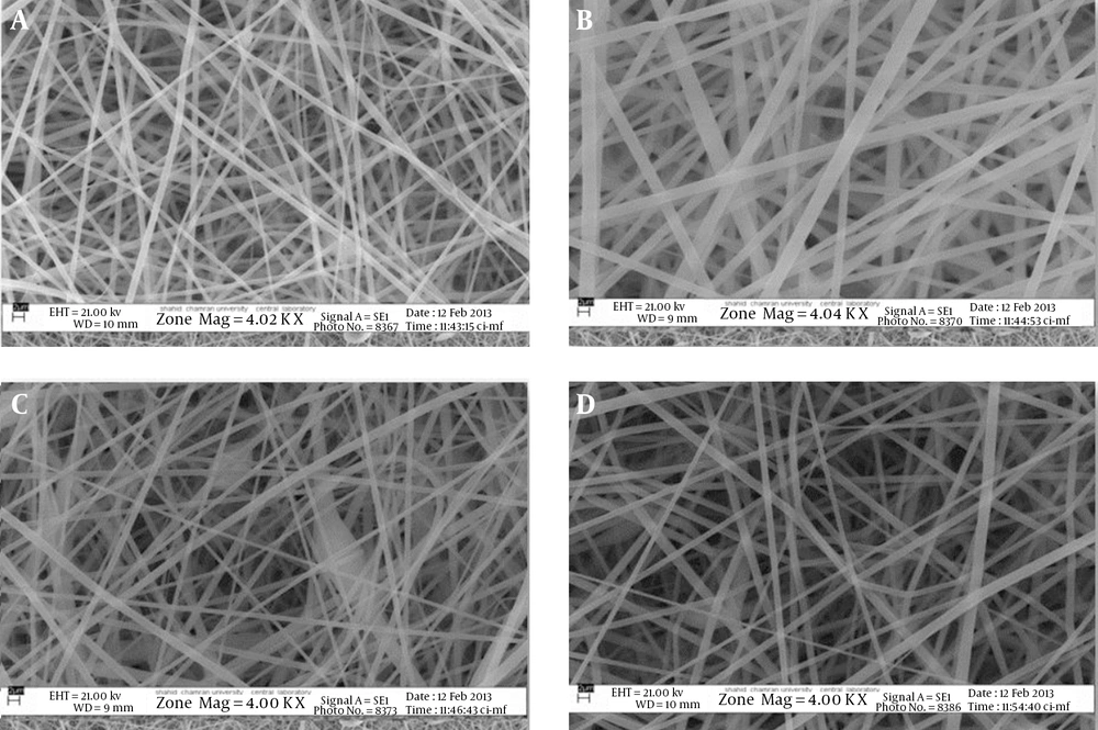
The SEM images of some nanofibers are shown in Figure 1. Figure 1A, 1B shows that the feed rate had the maximum effect on mean diameter of nanofibers such that an increase in the feed rate from 1 to 6 mL/h, substantially increased the diameter of the nanofibers. In addition, a complete structure of the nanofiber was not formed at a feed rate of less than 1 mL/h. An increase in the diameter of the nanofiber according to an increased feed rate was also observed in other studies (17, 18). Reducing the concentration of the drug and PVP somewhat decreased the diameter of the nanofibers. The results of preformulation also indicated that if the concentration of PVP in ethanol and the ratio of drug to polymer was reduced to beneath than a certain limit, nanofibers were formed in the cut pieces; a greater decrease in the concentration of PVP in ethanol and the ratio of drug to polymer led to the formation of micro- and nanoparticles. Compared with F1, the bead was seen in the F3 formulation at a voltage of 20 kV. The voltage could be increased, but only up to a certain limit; if increased further after this point, this led to ragged and incomplete fiber formation, as the electrostatic repulsive force on fibers was too high (19, 20).
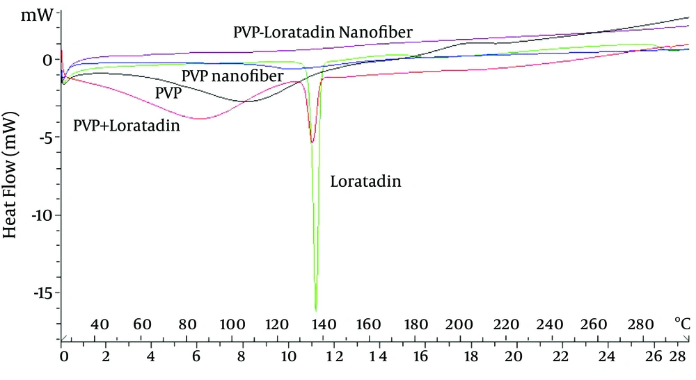
DSC thermograms are shown in Figure 2; a sharp endothermic peak can be observed at 135°C, which is attributed to the drug melting (21). The thermogram of PVP shows a broad endotherm peak at 80 - 140°C due to the evaporation of absorbed water; this indicates the hygroscopic nature of this polymer (22). The melting point of the drug and polymer are observed in the thermogram of the physical mixture of loratadine and PVP powder, which shows that no physical change occurred in the drug and polymer powder mixture. In PVP nanofibers containing loratadine, the peak showing the melting of loratadine disappeared, representing a loss of the crystal structure of loratadine and its conversion to the amorphous form during the electrospinning process. Moreover, two small endothermic peaks were observed at 60 and 110°C, which can be attributed to the water absorbed by the nanofiber and the glass transition (Tg) of PVP, respectively. Regarding the smaller peak, which shows the bond water compared to the powdered polymer, it can be concluded that the hygroscopic nature of PVP in a powdered state is much greater than in nanofibers. This is probably related to the presence of loratadine, which is a hydrophobic component in the structure of the nanofiber and therefore reduces the water absorption effect of nanofibers compared with PVP powder alone.
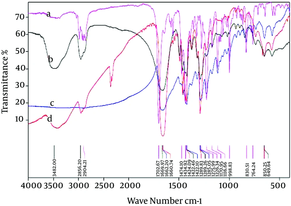
FTIR testing was conducted to detect possible interactions of loratadine and PVP in the nanofibers. The results of FTIR are shown in Figure 3. The loratadine spectrum showed a series of bands at 2,904 cm-1 (C-H stretch) and in the range of 3,000 to 2,850 cm-1, which was associated with C-H and H stretch (23). A very strong band at 1,702 cm-1 associated with an ester C=O was seen in the loratadine spectrum. Bands of 1,474 and 1,227 cm-1 were also related to the stretching vibrations of the benzene ring and C-H stretching (24). Loratadine also showed bands in wave numbers 830, 996, and 1116 cm-1. The PVP spectrum exhibited a broad band in the region of 3,480 cm-1 that was associated with the presence of water in the polymer, thereby demonstrating the hydrophilic properties of PVP (25). In addition, bands of areas of 2,955 cm-1 and 1,669 cm-1 were associated with C-H stretch and C=O bond, respectively (22), thus confirming the DSC results.
Similar bands of nanofiber containing PVP showed that the hygroscopic property of the polymer did not change in combination with the nanofiber (Figure 3). The thermogram of nanofibers containing drug and PVP exhibited the same bands as loratadine and PVP in the area below 1,700 cm-1. Nevertheless, the band at 3,480 cm-1 was related to PVP. Moreover, the area at the 2,900 cm-1 band was replaced with a wider band caused by the amorphous deformation of the drug, as well as the hydrophobic nature of loratadine; the removal of the band is associated with moisture. In a similar study, the possible interferences of loratadine and PVP were examined in the form of solid dispersion by the FTIR method. The results of the study did not show clear interference between the drug and polymer (23), and the low percentage of the drug was mentioned as a possible reason for the lack of interference. In the present study, the ratio of drug to polymer was much greater, and so the effect of the drug on the PVP FTIR spectrum was clearly evident. In another research, the displacement of bands related to PVP and the creation of a broader band when mixed with carbamazepine has been attributed to the establishment of hydrogen bonds between functional groups (26).
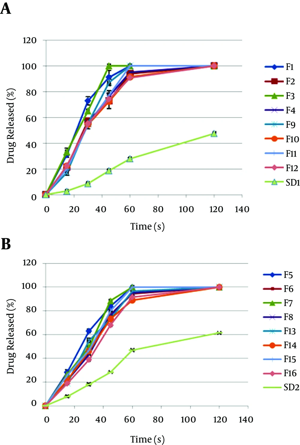
Considering that the aim of the study was to obtain a fast-dissolving formulation of loratadine nanofibers, drug release from the nanofibers was compared with the solid dispersion form to identify which one exhibits a faster release. Figure 4 corresponds to the drug release from 16 different nanofiber formulations containing loratadine, as well as two solid dispersion formulations with different ratios of drug to polymer (1: 2 and 1: 4). As depicted in Figure 4, drug releases from solid dispersion with drug: polymer ratios of 1:4 and 1:2 at 120 s were equal to 45 and 60%, respectively. Meanwhile, the 16 loratadine nanofibers formulations exhibited 100% release at this time. Among the formulations, F1 and F3 showed the fastest drug release. The ratio of drug to polymer in both formulations was equal to 1: 4, and the initial concentration of PVP and feed rate remained the same (30% and 1 mL/h, respectively).
To determine the effect of independent variables on the responses, mathematical models were generated between the dependent and independent variables using the SPSS software. The equations of the responses are as follows:
Y1 = -43.412-0.039X1X1 + 12.088X1X2 + 0.208X1X3- 506.857X2X2 +25.354X2X3+ 19.314X2X4-0.240X3X3 + 0.297X4X4 - 0.756X1X2X3 - 0.614X1X2X4 + 0.002X1X3X4-0.849X2X3X4 + 0.022X1X2X3X4.
Y2 = 309.558 + 0.939X1X1 - 17.326X1X4 + 1057.524X2X2 - 1235.027X2X4 + 81.500X4X4 - 1.052X1X2X3 + 45.092X1X2X4.
Y3 = -56.096 + 13.566X1X2 + 0.241X1X3 - 0.713X1X4 - 488.698X2X2 + 32.996X2X3 - 0.255X3X3 + 3.973X4X4 - 1.094X1X2X3 - 2.467X2X3X4 + 0.079X1X2X3X4.
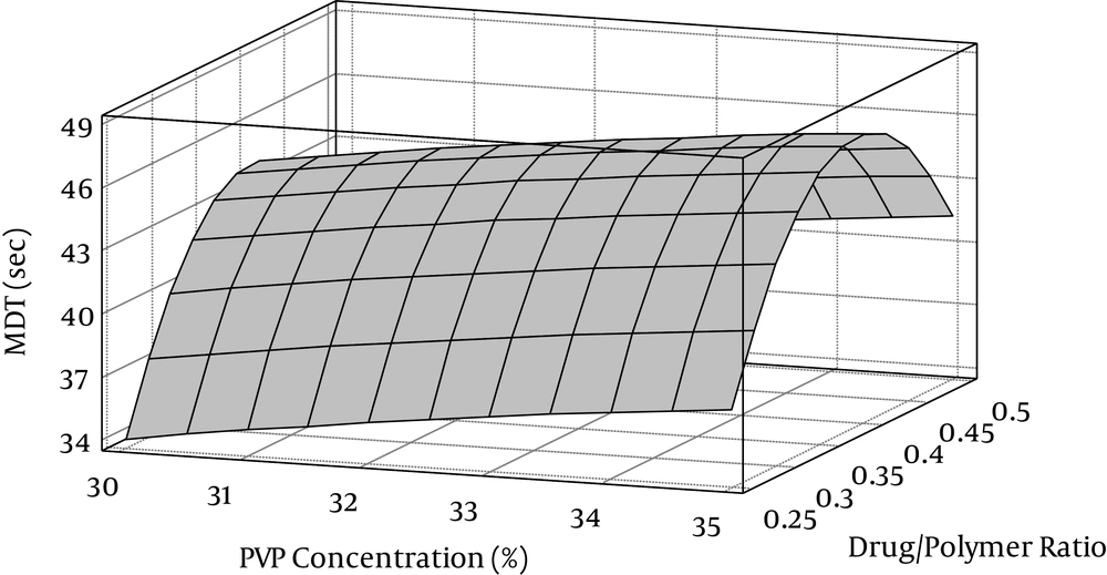
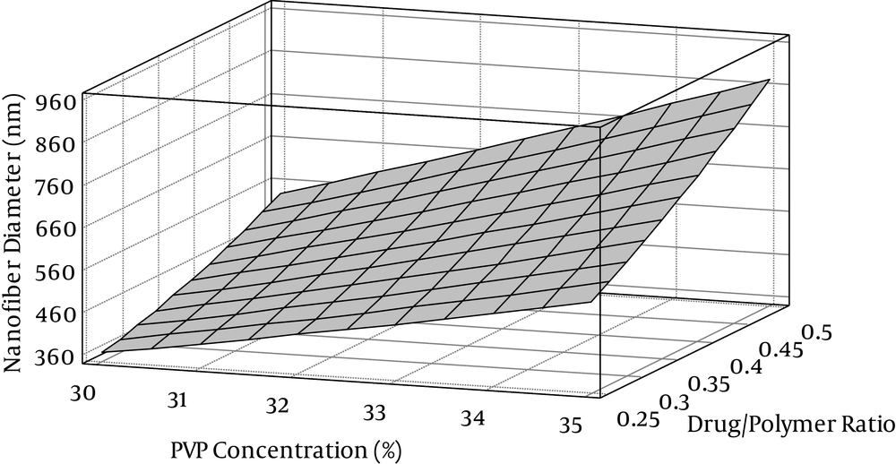
Where Y1, is the MDT (s), Y2 is the nanofiber diameter (nm), Y3 is the disappearance time (s), X1 is the PVP concentration in the electrospinning solution, X2 is the drug to polymer ratio, X3 is the voltage applied, and X4 is the feed rate. The three-dimensional response surfaces were drawn to estimate the effects of the independent variables on each response. Figure 5 shows the effect of the polymer solution’s concentration and the ratio of drug to polymer on MDT (Y1). As can be observed, reducing the concentration of PVP decreased MDT and a faster drug release from nanofibers occurred. This effect can be explained by the effect of the polymer’s concentration on the diameter of nanofibers. As can be seen in Figure 6, an increase in the concentration of the polymer increased the diameter of the nanofibers; this could be attributed to increasing polymer solution viscosity. At low concentration and viscosity, the time it takes for droplets to dry is limited until they can reach the collector. The effect of a high concentration of the polymer solution on the increasing diameter of nanofibers has been established in other research (27-30). In the present study, the increased concentration of the polymer solution by more than 35% did not result in suitable nanofibers; therefore, this was regarded as the maximum concentration. By reducing the diameter of the nanofibers, a higher amount of the drug was exposed on the surface, and consequently, the drug release was faster. As shown in Figure 5, increasing the ratio of drug to PVP by up to 0.4 enhanced MDT, while at higher ratios, MDT was reduced and drug release was faster. In addition, as Figure 6 illustrates, increasing the ratio of drug to polymer enlarged the diameter of the nanofibers. Therefore, the initial increase in MDT due to the increase in drug-to-polymer ratio can be caused by increasing the diameter of the nanofibers. Nevertheless, because of the hydrophobic characteristic of loratadine, increasing the drug-to-polymer ratio above a specific limit enhanced migration of the drug molecules to the surface of nanofibers during the electrospinning process, and as a result, the drug release rate from the nanofibers became faster (31).
5. Discussion
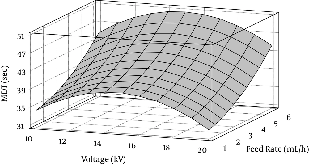
The results of drug release indicated that formulations with a feed rate of 1 mL/h exhibited a more rapid release compared to a feed rate of 6 mL/h (Figure 4). Figure 7 shows graphs of the response surfaces related to the effect of feed rate and voltage on the MDT. As illustrated in this illustration, an increase in feed rate increases the MDT. This effect can be caused by increasing the diameter of the nanofibers due to the increased feed rate. Other investigations have shown similar effects of feed rate on the diameter of nanofibers (17, 32). As can be seen in Figure 7, the effect of the voltage used for the electrospinning process on drug release from nanofibers was variable. With a voltage range of up to 15 kV, increased voltage caused an increase in MDT and slow drug release. However, increasing voltage above 15 kV reduced MDT. Similar results were also found concerning the effects of voltage on the diameter of fibers (data not shown). In a similar study on retinoic acid nanofibers, Puppi et al. (32). investigated the effect of voltage on the diameter and morphological properties of nanofibers; they identified an optimum range of voltage for the production of fibers with appropriate characteristics. It was determined that at a critical voltage, a stable fiber jet can be produced by polymer solution (30). In the present study, the minimum and maximum MDT values were observed at 14 and 20 kV voltages, respectively. Above 20 kV, nanofibers were incomplete due to the lack of the formation of a stable jet and irregular spinning of polymer solution (33). Therefore, the voltage of 20 kV was considered a critical voltage.
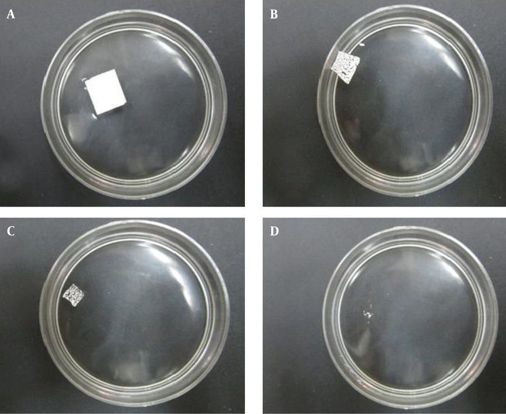
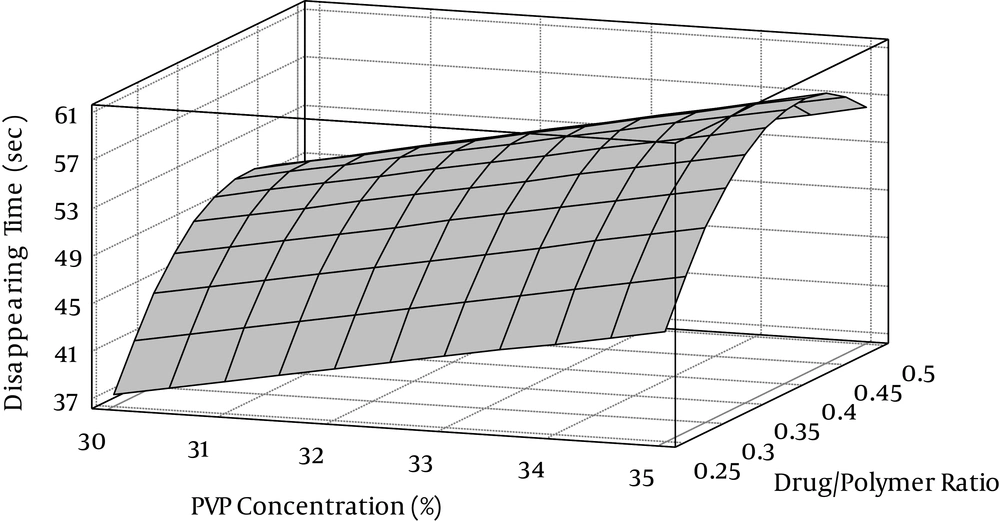
The disappearance time of nanofiber formulations was less than 60 seconds (data not shown), while the formulations of solid dispersion were still visible after 60 seconds. Figure 8 shows the disintegration process of F1 formulation at a time of less than 40 seconds. The quick disintegration of nanofibers compared to SD formulations can be attributed to a higher surface to volume ratio of nanofibers and full distribution of loratadine in this dosage form, as well as the high porosity of nanofibers (16, 34). According to Figure 9, the effect of the polymer solution concentration and the amount of drug on the disappearance time was similar to the effect of variables on MDT; by increasing the concentration of the polymer solution, the disappearance time was also increased. Moreover, increasing the drug-to-polymer ratio by up to 0.4 increased the disappearance time, while above this ratio, nanofibers disappeared quickly. Therefore, the disappearance time of nanofibers was concordant with drug release.
Generally, given that a proper formulation of oral FDDS must release the drug at a high speed in addition to having physical stability, due to their high porosity and surface-to-volume ratio, electrospun nanofibers are suitable as a candidate dosage form for loratadine. Among the formulations, the lowest MDT and time of disappearance were attained with a solution of 30% PVP in ethanol, a drug-to-polymer ratio of 1:4, 10 - 20 kV of voltage, and a feed rate of 1 mL/h (F1 and F3 formulations). Among the formulations, F1 had more uniform and bead-free nanofibers. Therefore, this formulation can be a perfect choice for an oral, fast dissolving system for loratadine.
5.1. Conclusion
The results of this research showed that reducing the concentration of polymer, reducing the feed rate, and increasing the voltage up to a sufficient level make it feasible to generate acceptable nanofibers with a smaller diameter and more uniform structure. Furthermore, the nanofiber diameter and ratio of loratadine had an effect on the drug release and disappearance time of fibers, so that a decrease in the fiber diameter and amount of the drug enhanced the nanofibers’ drug release.
The F1 formulation (30% PVP concentrations in ethanol, drug-to-polymer ratio of 1: 4, 10 kV of voltage, and a feed rate of 1 mL/h) resulted suitable and uniform nanofibers which exhibited fast drug release. Therefore, this formulation is a good candidate as a fast-dissolving oral delivery system for loratadine. It was also shown that electrospinning is an appropriate procedure for preparing the fast-dissolving form of loratadine compared with solid dispersion.
