1. Background
Diabetes mellitus, a multi-factorial chronic disease, is characterized by persistent hyperglycemia, resulting from complete or relative lack of insulin production, action, or both (1). The current medications for diabetes mellitus are included diet, exercise, several oral anti-diabetic drugs, insulin therapy or even combination therapies (2). The modern drugs, insulin and other oral hypoglycemic agents such as sulphonylureas, biguanides, and inhibitors of glycosidase have several typical undesirable effects, which include diarrhea, hypoglycemia, hepatotoxicity, lactic acidosis, dyslipidemia, and hypertension (3). In view of the alarming increase in the worldwide diabetic population, there is a need for novel therapies, which are effective with fewer undesirable outcomes (4). Plants have always been an excellent source of medications and many of the presently available drugs have been derived directly or indirectly from them (5). This has led to an increasing trend for research on anti-diabetic herbal products, which produce the smallest or no side-effects.
Calendula officinalis Linn (C. officinalis), belonging to the family of Asteraceae, known as “gole-hamishe-bahar” in Persian, is an aromatic herb used in Iranian traditional medicine. It is also commonly known as Pot Marigold and its flowers have been used for the treatment of inflammatory conditions in internal organs, gastrointestinal ulcers, dysmenorrhea and as a diuretic and diaphoretic in convulsions (6). C. officinalis has been reported to possess several pharmacological activities, which include antioxidant (7, 8) anti-inflammatory (9), antidiabetic (10, 11), antibacterial (12), and anti-pyretic, cytotoxic as well as tumor reducing (13). This plant is rich in several classes of chemical compounds including terpenoids, flavonoids, coumarins, flavoxanthin, glycosides, volatile oil, sterols, and steroids (8).
There are several reports about the antidiabetic effect of the leaf extract of Calendula officinalis (10, 11) and a study conducted by Preethi et al. has shown an extract of Calendula officinalis flower has significant antioxidant activity in vitro and in vivo (7). However, to the best of our knowledge, there is no report about the effect of its flower’s extract on diabetes and insulin secretion from isolated islets in the animal model. Thus, we assessed the effect of C. officinalis hydroalcoholic extract on diabetic complications and insulin secretion in an animal model.
2. Objectives
In the current study, we have done a systematic investigation on the anti-diabetic potential of hydro-alcoholic flower extract of Calendula officinalis in vivo, as well as its direct effects on glucose-stimulated insulin secretion from the rat’s isolated pancreatic islets in vitro.
3. Methods
3.1. Plant Collection and Preparation of Hydroalcoholic Extract
Calendula officinalis flowers were prepared from Ahvaz, southwest of Iran and its hydroalcoholic extract was obtained as previously described elsewhere (14). A voucher specimen of the plant was stored in the herbarium of medicinal plants and natural products research center, Ahvaz Jundishapur University of Medical Sciences, Ahvaz, Iran (Herbarium No. A150200400FP).
3.2. Animals and Induction of Diabetes
Thirty six adult male Wistar rats with weight range of 220 - 280 g were purchased from the research center and experimental animal house, Jundishapur University of Medical Sciences, Ahvaz, Iran. Animals were divided into four groups with nine rats in each group and treated as follows: group I: healthy control rats treated with normal saline (HC) (15); group II: diabetic control rats treated with normal saline (DC); group III: diabetic rats treated with the extract (250 mg/kg body weight, orally); group IV: diabetic rats treated with the extract (500 mg/kg body weight, orally). Diabetes was induced by a single intraperitoneal injection of 60 mg/kg streptozotocin (Sigma-Aldrich, USA) dissolved in 0.1 M citrate buffer (pH: 4.5). Fasted rats with blood glucose level ≥ 350 mg/dL were considered as diabetic.
3.3. Analysis of Serum Biochemical Parameters and Histological Examination
After 4 weeks of intervention, all animals were anesthetized by using the combination of 40 mg/kg ketamine and 5 mg/kg xylazine intraperitoneally. Then, blood samples were collected from the heart and serum separated. All serum biochemical parameters including glucose, total protein, alanine aminotransferase (ALT), aspartate aminotransferase (AST), albumin, and total bilirubin determined with the use of commercially available kits (Pars Azmoon, Tehran, Iran) and using an automatic analyzer (Biotecnica BT-3000 Plus Chemistry Analyzer, Italy). Total lactate dehydrogenase (LDH) activity was measured using a kinetic spectrophotometry method to measure the conversion of reduced coenzyme NADH to oxidized NAD+ at 340 nm produced in the pyruvate-to-lactate (P → L) reaction. The serum insulin level was determined by an enzyme-linked immunosorbent assay (ELISA) method using a rat insulin kit (Mercodia, Uppsala, Sweden). We used homeostasis model assessment (HOMA-β) for evaluation of beta cell function by the following formula: β-cell function (%) = (20 × insulin µIU/mL)/(glucose mg/dL - 3.5): Moreover, histological assessment was performed as previously described elsewhere (16).
3.4. Islets Isolation and Incubation with Hydro-Alcoholic Extract
The pancreas of male healthy control rats was isolated by collagenase digestion method (17). After anesthetization of rats, the pancreas was isolated and well washed using normal saline. Then, it was washed with 20 mL of cold Hank’s balanced salt solution (HBSS) three times. In the next stage, HBSS was discarded and isolated pancreas well pipetted using 1.5 - 2 mg collagenase type IV/mL HBSS. Using cold HBSS, enzymatic digestion was stopped and ultimately digested pancreas centrifuged at 1500 RPM for 1 minute. For final approval, isolated islets were stained with dissolved dithizone at 5 mL dimethyl sulfoxide (DMSO). Then, 10 µmol of this solution was added to isolated islets and placed for 15 minutes in an incubator, subsequently washed with HBSS. Due to the high presence of zinc, the beta cells were observed as red color under light microscopic (18). Extracted islets were placed into the medium (2 cc RPMI, 10% FBS) and simultaneously treated with different doses of hydro-alcoholic extract (0.01, 0.1, 1 mg/mL HBSS) overnight at 37°C. The islets were pre-incubated with 1 mL HBSS with 2.8 mM glucose during 30 minutes, subsequently, the pre-incubation liquid was discarded. Finally, the wells were treated under two conditions including basal (1 mL HBSS with 2.8 mM glucose for 30 minutes) and stimulatory (1 mL HBSS with 16.7 mM glucose for 30 minutes). The supernatant was collected and then stored at -20°C for evaluation of insulin level.
3.5. Statistical Analysis
All statistical analyses were performed using SPSS software (SPSS, Inc., Chicago IL, USA). Continuous variables are expressed as mean ± SD and compared by one-way analysis of variance (ANOVA) followed by Tukey’s post hoc test. The significant level was set at P < 0.05.
4. Results
4.1. Effects of the Extract on the Body Weight and Blood Glucose Levels
Four weeks after induction of diabetes, a significant weight loss was observed in diabetic controls (Table 1). Treatment of STZ-diabetic rats for 4 weeks with daily doses of the extract induced a significant recovery in the body weight (Table 1). As shown in Table 1, STZ diabetic rats showed a significant increase in fasting blood glucose compared to healthy controls (P = 0.001). After four weeks intervention, in rats treated with 250 mg/kg of the extract, a mild reduction of blood glucose levels was observed (P = 0.017). However, the reduction of blood glucose level was more significant in rats treated with 500 mg/kg of the extract (P = 0.001).
| Groups (N = 9) | Body Weight, g | Fasting Blood Glucose, mg/dL | ||
|---|---|---|---|---|
| Initial | After Intervention | Initial | After Intervention | |
| HC | 266.78 ± 10.89 | 253.33 ± 18.43 | 93.11 ± 12.23 | 93.22 ± 11.51 |
| DC | 240.00 ± 20.31 | 209.78 ± 50.97c | 331.42 ± 77.75c | 389.00 ± 132.93c |
| DT250 | 267.22 ± 30.32 | 235.00 ± 26.92d | 462.67 ± 90.17 | 158.00 ± 106.54d |
| DT500 | 255.44 ± 26.71 | 244.44 ± 44.47d | 364.89 ± 129.52 | 93.78 ± 12.56d |
Abbreviations: DC, diabetic control; HC, healthy control.
aValues are expressed as mean ± SD.
bComparisons were made using one-way ANOVA followed by Tukey’s post hoc test.
cSignificant difference with HC (P < 0.05).
dSignificant difference with DC (P < 0.001).
4.2. Effects of the Extract on the Parameters Related to Insulin
As shown in Table 2, intraperitoneal injection of streptozotocin resulted in an obvious reduction of serum insulin level in diabetic rats compared to healthy controls (P = 0.001). The increase of insulin level in diabetic rats treated with the extract (250 or 500 mg/kg) was 39.9% and 44.9%, respectively. This increase was significant for 250 and 500 mg/kg of the extract (P = 0.001). Moreover, the serum insulin level in diabetic rats treated with 500 mg/kg of the extract was higher than diabetic rats treated with 250 mg/kg (Table 2). The HOMA-β calculation as an index of beta cell function confirmed that insulin secretion from beta cells improved after treatment with the extract. Although, a significant improvement in insulin secretion was observed in both groups treated with the extract, however, this improvement in rats treated with 500 mg/kg was higher than those treated with 250 mg/kg of the extract (P < 0.05).
Abbreviations: DC, diabetic control; HC, healthy control; HOMA-β, the homeostasis model assessment of β cell function; LDH, lactate dehydrogenase.
aValues are expressed as mean ± SD.
bSignificant difference with DC (P < 0.001).
cSignificant difference with DC (P < 0.05).
4.3. Effects of the Extract on the Biochemical Parameters
As shown in Table 3, STZ-induced diabetes resulted in increased serum concentration of liver biomarkers including AST and ALT. Moreover, these liver biomarkers were not significantly decreased in diabetic rats treated with 250 mg/kg of the extract, while oral administration of 500 mg/kg of the extract significantly decreased serum ALT compared to diabetic controls (P < 0.05). The mean value of serum LDH activity in diabetic controls was dramatically increased compared to healthy controls. While, treatment with the extract (250 and 500 mg/kg) could significantly decrease LDH activity (P < 0.05). The mean value of total protein in diabetic rats treated with 250 and 500 mg/kg of the extract was not changed compared to diabetic controls, however, serum albumin was significantly decreased in diabetic rats treated with the extract (P < 0.05). Also, administration of 500 mg/kg of the extract significantly decreased total bilirubin concentration compared to diabetic controls (P < 0.05). While, after treatment with the extract direct bilirubin concentration was not dramatically changed in both groups. Table 4 shows the effect of marigold flower extract on the insulin secretion from isolated islets under glucose stimulatory condition. Results indicated the mean value of insulin level from the isolated islets treated with different concentrations (0.01, 0.1 and 1.0 mg/mL) of the extract in HBSS were 6.87, 8.60 and 10.56 µIU/islets/24 h, respectively.
| Groups (N = 9) | AST, U/L | ALT, U/L | Total Protein, g/dL | Albumin, g/dL | Total Bilirubin, mg/dL |
|---|---|---|---|---|---|
| HC | 143 ± 32.44 | 64 ± 13.56 | 8.04 ± 0.68 | 3.76 ± 0.24 | 0.23 ± 0.52 |
| DC | 162 ± 39.30 | 115 ± 37.08 | 7.36 ± 0.65 | 3.20 ± 0.54 | 0.44 ± 0.18 |
| DT250 | 152 ± 46.78 | 99 ± 41.97 | 7.87 ± 0.36 | 3.55 ± 0.25b | 0.41 ± 0.16 |
| DT500 | 146 ± 26.86 | 70 ± 19.41b | 7.81 ± 0.52 | 3.80 ± 0.18b | 0.31 ± 0.12b |
Abbreviations: AST, aspartate aminotransferase; ALT, alanine aminotransferase; DC, diabetic control; HC, healthy control.
aValues are expressed as mean ± SD.
bSignificant difference with DC (P < 0.05).
Abbreviation: HC, healthy control.
aValues are expressed as mean ± SD.
bInsulin level (µIU/islets/24 h).
cT0.01, the islets treated with 0.01 mg/mL of the extract.
dSignificant difference compared to basal glucose (P < 0.05).
eT0.1, the islets treated with 0.1 mg/mL of the extract.
fT1.0, the islets treated with 1.0 mg/mL of the extract.
4.4. Effects of the Extract on the Pancreas, Liver, and Kidney Histopathology
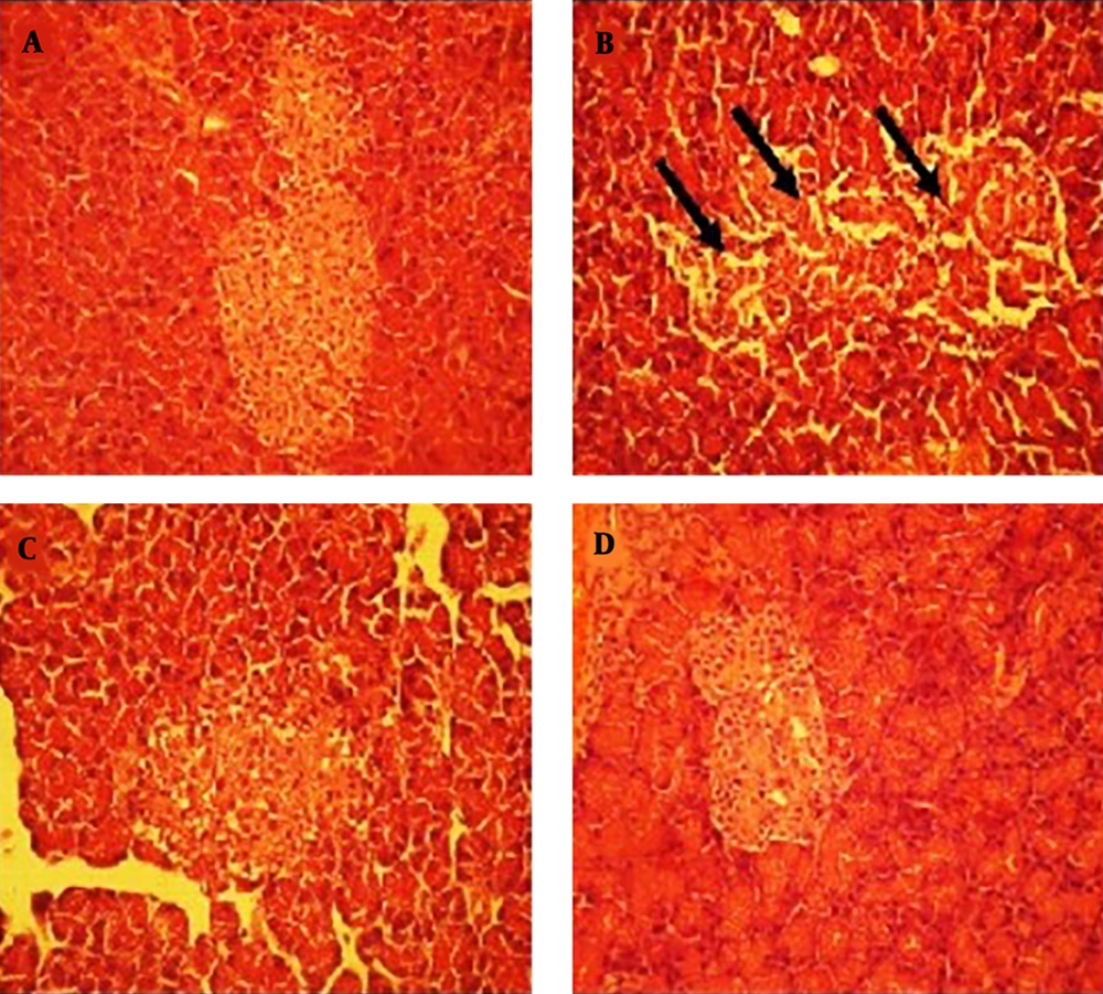
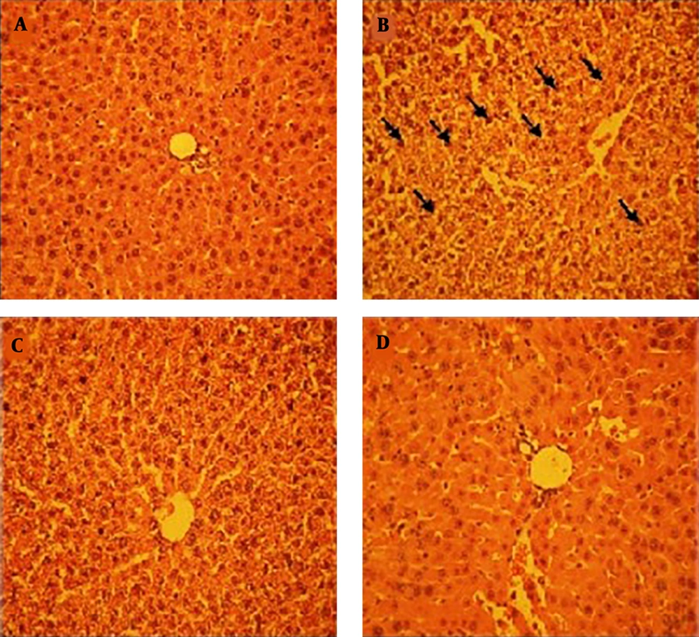
Histological examinations indicated that the pancreas structure was normal in healthy control (Figure 1A). While the pancreatic tissue in diabetic control showed a relative reduction of islets size with marked loss of its cells and normal cellular cord arrangement (Figure 1B). The pancreatic tissue of diabetic rats treated with 250 mg/kg of the extract showed a reduction of degenerative changes of Langerhans islets, however, destruction of cells and disturbance of cells arrangement are still present (Figure 1C). However, the pancreatic tissue of rats treated with 500 mg/kg of the extract demonstrated a partial amelioration in the histological structure of Langerhans islets with an obvious decrease in cells destruction (Figure 1D). Liver sections in healthy control showed there were no histopathological changes (Figure 2A). In contrast, STZ-induced diabetic rats showed obvious histopathological changes such as lack of radial arrangement, dilated sinusoid, clear cytoplasm and condensed nuclei (Figure 2B). Treatment with the extract with both doses showed marked amelioration in liver structure. Hepatocyte arrangement and enlargement of sinusoids dramatically returned to normal state. Also, nuclei and cytoplasm had a normal structure as compared with the diabetic control (Figure 2C and D). However, this improvement was more obvious in the group receiving 500 mg/kg of the extract.
Histopathological evaluation of liver. Healthy control (A); diabetic control (B); diabetic rats treated with 250 mg/kg of the extract (C); diabetic rats treated with 500 mg/kg of the extract (D); (H&E X300); notice to the condensed nuclei (arrows) and cytoplasm surrounding of nucleus in the diabetic control in (B).
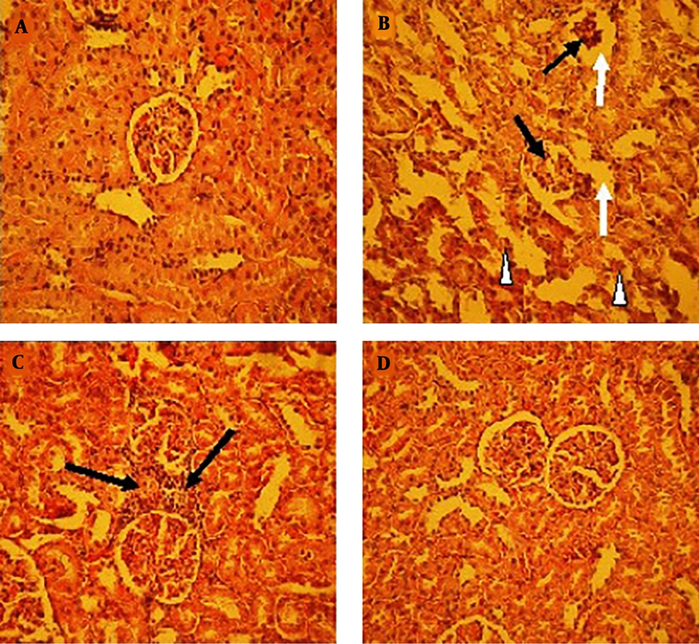
The prepared tissue sections of the kidney in healthy controls indicated that there was a normal histological structure without any pathological changes (Figure 3A). The kidney of the diabetic rats showed deterioration in some tubular epithelial cells, and also, some glomeruli demonstrated atrophy or deterioration with wide urinary space (Figure 3B). The kidney of diabetic rats treated with the extract (250 and 500 mg/kg) showed a decline in glomerular and tubular epithelial cells deterioration as compared to diabetic controls. While mild cellular infiltration in interstitial tissue in the kidney of rats treated with 250 mg/kg was also seen (Figure 3C and D).
Histopathological evaluation of kidney. Healthy control (A); diabetic control (B); diabetic treated with 250 mg/kg of the extract (C); diabetic treated with 500 mg/kg of the extract (D); (H&E X300); notice to the destruction of some renal tubules (rectangle) and glomeruli (black arrows) also, increase in urinary space (white arrows) in (B), and cellular infiltration into interstitial tissue (black arrows in (C)).
5. Discussion
Increased blood glucose level is expected in STZ-induced diabetic animals, since STZ causes a significant reduction of insulin release, by selectively destroying pancreatic insulin-secreting β-cells and inducing persistence of hyperglycemia (19). Oral administration of C. officinalis (250 and 500 mg/kg body weight) resulted in a significant reduction in the blood glucose level and improvement in body weight. The decrease in body weight in diabetic rats clearly confirms a loss or degradation of structural proteins due to diabetes. The structural proteins are known to contribute to the body weight (20). Protein biosynthesis is decreased in all tissues due to absolute or relative deficiency of insulin (the most anabolic hormone) in STZ-induced diabetic rats. This result is consistent with the results reported by Ramesh and Pugalendi (21) and Erenmemisoglu et al (22). Moreover, our results indicated oral administration of both doses of the extract resulted in a significant reduction of blood glucose level which is consistent with the results reported previously (10, 11).
The measurement of aminotransferases (AST and ALT) and LDH activities have clinical and toxicological significance, since, changes in their activities may be reflecting tissue toxicity damage or disease conditions (23). Our results indicated, serum levels of AST, ALT, and LDH were significantly increased in diabetic control, however, diabetic rats treated with the extract showed improvement. Recovery of AST, ALT, and LDH activities in diabetic rats towards normal range shows that the C. officinalis flower extract has no harmful effect on the liver functions. A significant increase in serum activity of AST, ALT, and LDH possibly resulted from the leak of these enzymes of liver into the bloodstream, which provides a hepatotoxic effect of streptozotocin (24). In addition, the histological study confirmed these biochemical findings and liver structure in rats treated with 500 mg/kg of the extract was similar to healthy controls. Moreover, the improvement of the liver damage by the extract could be confirmed through assessing its impact on the plasma bilirubin level. Our finding showed the experimentally induced diabetes markedly increased plasma bilirubin levels. However, after treatment with the extract a great reduction of plasma bilirubin was observed. Rana et al. (25) reported that increase in plasma bilirubin possibly resulted from a reduction of liver uptake, conjugation, or an increase of bilirubin production.
The STZ- induced hyperglycemia resulted in an increase in the serum levels of urea and creatinine, which are reflecting of symptoms for renal dysfunction (26). After treatment with the extract, the levels of urea and creatinine were significantly decreased in comparison to the diabetic controls. This further confirms the utility of C. officinalis in diabetes prospective complications. Moreover, renal histology demonstrated tubular, glomerular, and interstitial alterations in diabetic rats. These results are consistent with the results reported by Hamada and Fukagawa (27), and Teoh et al. (28). Our results showed that treatment with the C. officinalis extract can ameliorate the STZ-induced diabetic alterations in the kidney.
Our results showed that islet and beta cells destruction were significant in diabetic controls. These findings are agreement with the previous results which found that STZ is a destructive molecule for the pancreatic beta cell (19, 29). Structurally, STZ is similar to glucose so that it enters into beta cells, and by producing reactive oxygen species results in diabetes (30, 31).
Previous studies showed treatment with the C. officinalis extract resulted in a proliferation of beta cells and recovery of islet injuries. And, suggested this plant may comprise pharmaceutical compounds with anti-oxidant effects and capability to stimulate regeneration of beta cells (32, 33). Also, this result is consistent with the previous result which reported the anti-oxidant properties of the C. officinalis flower extract (7). Moreover, our results showed treatment with the extract significantly increased insulin secretion in a dose-dependent manner (P < 0.05). It has been demonstrated phenolic compounds including flavonoids are effectiveness on the beta cell function and insulin secretion. Moreover, another phenolic substances such as quercetin can be beneficial in the improvement of the oxidative stress in diabetic rats (34). Youl et al. 2010 demonstrated that quercetin is able to induce insulin secretion in INS-1 β-cell line and protects the beta cell against oxidative stress (35). Although, increased blood glucose level is the most stimulating factor for insulin secretion, however, accompanied by progression of diabetes, this effect will be gradually reduced due to the disorder for glucose entry into the beta cells, so that, reduction of glucose-induced insulin secretion is a common feature of diabetic patients (36). Thus, clinically, maintenance of insulin secretion can delay several side effects of diabetes, and studies in this direction can be invaluable for the treatment of diabetes. Although, the promising effects of Calendula officinalis in the treatment of diabetes particularly its leaf extract on reducing blood glucose have been reported (9, 10). Also, our results showed that the extract can induce the insulin secretion in a dose-dependent manner. However, to date, no study assessing the effect of the extract on insulin secretion in STZ-induced diabetic rats has been reported. Therefore, we were not able to compare our results in this regard with the others.
5.1. Conclusions
Our findings indicate that hydro-alcoholic extract of the marigold flower has a potential protective effect on the main organs including liver, kidney, and pancreas against STZ-induced diabetic complications. In addition, it has a direct effect on the glucose-stimulated insulin secretion from the rat’s isolated pancreatic islets in vitro condition.


